Edelstein-Keshet L. Mathematical Models in Biology
Подождите немного. Документ загружается.

306
Continuous Processes
and
Ordinary
Differential
Equations
This system
is
called
a
substrate-inhibited reaction since
the
chemical
X can
deactivate
the
complex
Mi
which
is
required
in
forming
the
products.
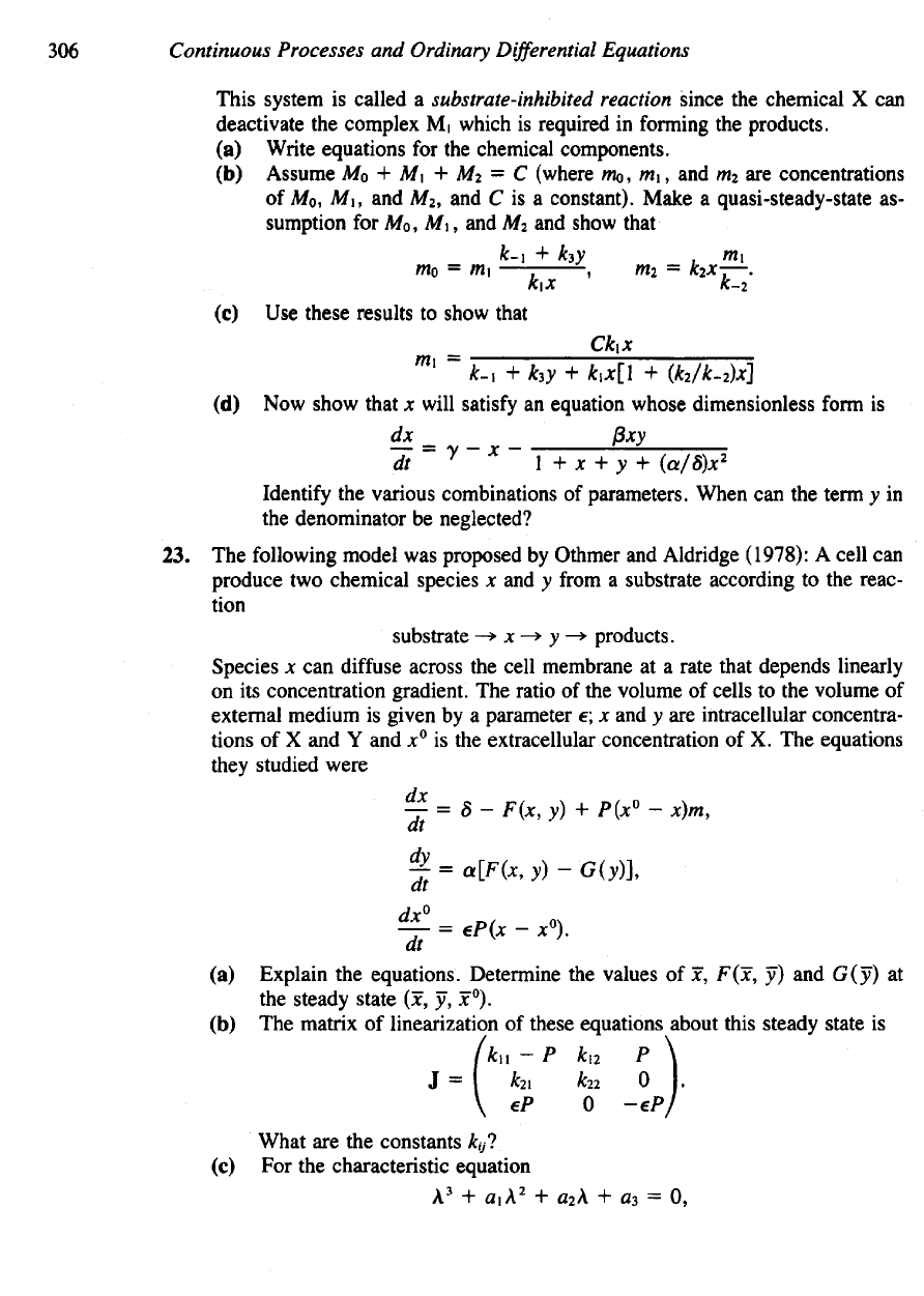
(a)
Write equations
for the
chemical components.
(b)
Assume
Mo + M\ + M
2
= C
(where
mo, m\, and m
2
are
concentrations
of
A/o,
A/I,
and
A/
2
,
and C is a
constant). Make
a
quasi-steady-state
as-
sumption
for
A/o, A/i,
and A/
2
and
show that
(c) Use
these results
to
show
that
(d) Now
show that
x
will
satisfy
an
equation whose dimensionless
form
is
Identify
the
various combinations
of
parameters. When
can the
term
y in
the
denominator
be
neglected?
23. The
following model
was
proposed
by
Othmer
and
Aldridge (1978):
A
cell
can
produce
two
chemical species
x and y
from
a
substrate according
to the
reac-
tion
Species
x can
diffuse
across
the
cell membrane
at a
rate that depends linearly
on its
concentration gradient.
The
ratio
of the
volume
of
cells
to the
volume
of
external medium
is
given
by a
parameter
e; x and y are
intracellular concentra-
tions
of X and Y and x° is the
extracellular concentration
of X. The
equations
they
studied were
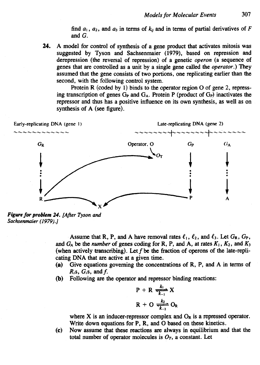
(a)
Explain
the
equations. Determine
the
values
of jc,
F(x,
y) and
G(y)
at
the
steady
state
(x, y,
x°).
(h) The
matrix
of
linearization
of
these eauations about this
steadv
state
is
What
are the
constants
&//?
(c)
For the
characteristic
equation
Models
for
Molecular Events
307
find a\, a
2
, and a
3
in
terms
of
Jk,y
and in
terms
of
partial derivatives
of F
and G.
24. A
model
for
control
of
synthesis
of a
gene product that activates mitosis
was
suggested
by
Tyson
and
Sachsenmaier (1979), based
on
repression
and
derepression (the reversal
of
repression)
of a
genetic
operon
(a
sequence
of
genes that
are
controlled
as a
unit
by a
single gene called
the
operator.) They
assumed that
the
gene consists
of two
portions,
one
replicating earlier than
the
second,
with
the
following
control system.
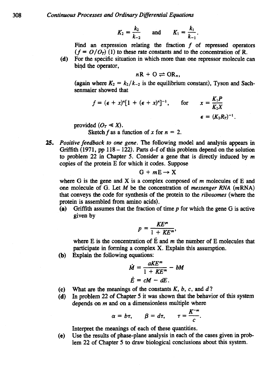
Protein
R
(coded
by 1)
binds
to the
operator region
O of
gene
2,
repress-
ing
transcription
of
genes
G
P
and G
A
.
Protein
P
(product
of G
P
)
inactivates
the
represser
and
thus
has a
positive
influence
on its own
synthesis,
as
well
as on
synthesis
of A
(see
figure).
Figure
for
problem
24.
[After
Tyson
and
Sachsenmaier
(1979).]
Assume that
R, P, and A
have removal rates
i\, €
2
, and €
3
Let
GR,
G
P
,
and
G
A
be the
number
of
genes coding
for R, P, and A, at
rates
K\, K
2
, and £
3
(when actively transcribing).
Let/be
the
fraction
of
operons
of the
late-repli-
cating
DNA
that
are
active
at a
given time.
(a)
Give equations governing
the
concentrations
of R, P, and A in
terms
of
Rt&,
G,s, and/.
(b)
Following
are the
operator
and
represser binding reactions:
where
X is an
inducer-repressor complex
and OR is a
repressed
operator.
Write down equations
for P, R, and O
based
on
these kinetics,
(c) Now
assume
mat
these reactions
are
always
in
equilibrium
and
that
the
total number
of
operator molecules
is O
T
, a
constant.
Let
308
Continuous Processes
and
Ordinary
Differential
Equations
Find
an
expression relating
the
fraction
/ of
repressed operators
(/ =
O/Or)
(1) to
these
rate constants
and to the
concentration
of R.
(d) For the
specific situation
in
which-more than
one
represser
molecule
can
bind
the
operator,
(again where
AT
2
=
k
2
/k-
2
is the
equilibrium constant), Tyson
and
Sach-
senmaier showed that
provided
(O
T
<X).
Sketch/as
a
function
of x for n = 2.
25.
Positive feedback
to one
gene.
The
following model
and
analysis appears
in
Griffith
(1971,
pp 118 -
122). Parts
d-f of
this problem depend
on the
solution
to
problem
22 in
Chapter
5.
Consider
a
gene
that
is
directly induced
by m
copies
of the
protein
E for
which
it
codes. Suppose
where
G is the
gene
and X is a
complex composed
of m
molecules
of E and
one
molecule
of G. Let M be the
concentration
of
messenger
RNA
(mRNA)
that conveys
the
code
for
synthesis
of the
protein
to the
ribosomes (where
the
protein
is
assembled
from
amino acids).
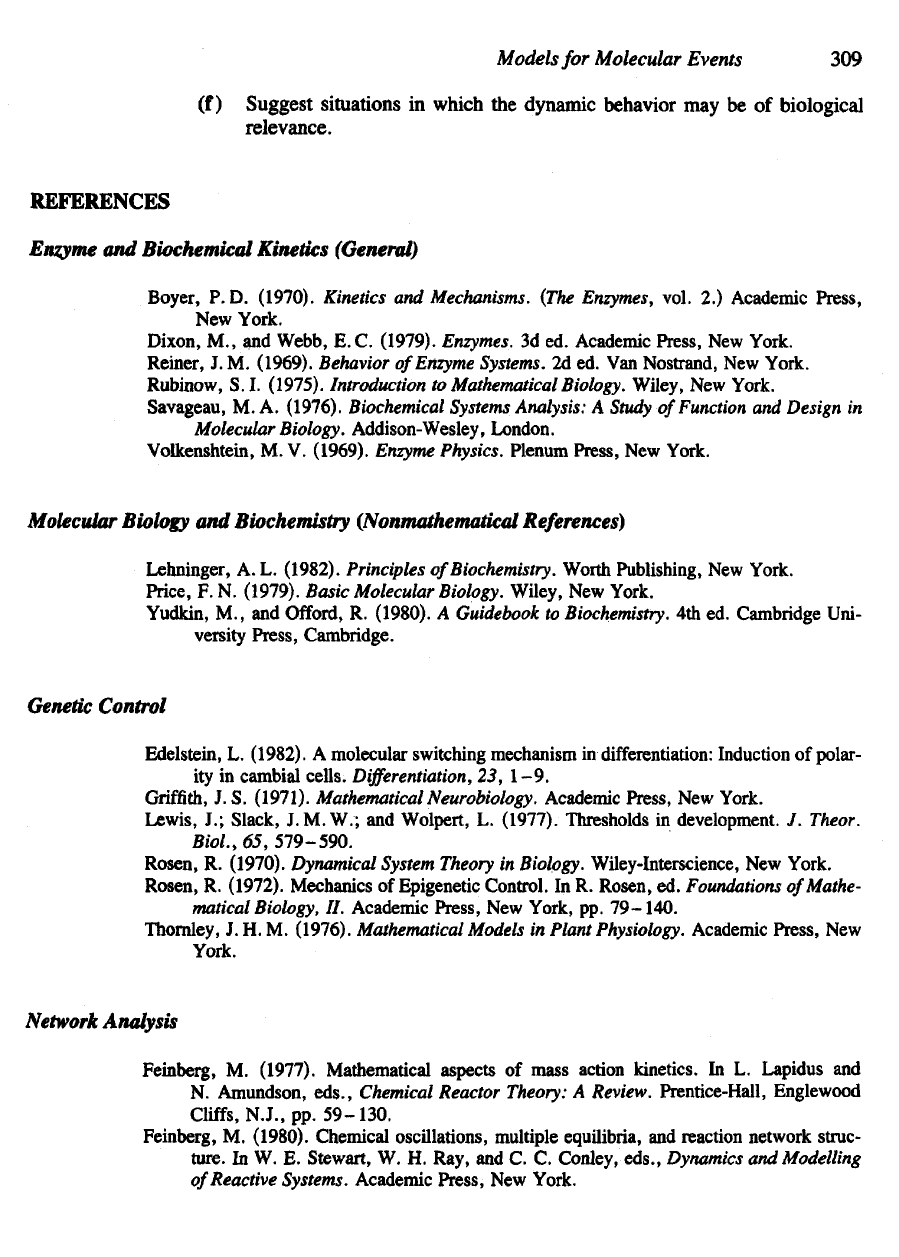
(a)
Griffith
assumes that
the
fraction
of
time
p for
which
the
gene
G is
active
given
by
where
E is the
concentration
of E and m the
number
of E
molecules that
participate
in
forming
a
complex
X.
Explain this assumption.
(b)
Explain
the
following equations:
(c)
What
are the
meanings
of the
constants
K, b, c, and d ?
(d) In
problem
22 of
Chapter
5 it was
shown that
the
behavior
of
this system
depends
on m and on a
dimensionless multiple where
Interpret
the
meanings
of
each
of
these quantities.
(e) Use the
results
of
phase-plane analysis
in
each
of the
cases given
in
prob-
lem
22 of
Chapter
5 to
draw biological conclusions about this system.
Models
for
Molecular
Events
309
(f)
Suggest situations
in
which
the
dynamic behavior
may be of
biological
relevance.
REFERENCES
Enzyme
and
Biochemical Kinetics
(General)
Boyer,
P. D.
(1970). Kinetics
and
Mechanisms. (The Enzymes,
vol.
2.)
Academic Press,
New
York.
Dixon,
M, and
Webb,
E. C.
(1979).
Enzymes.
3d ed.
Academic Press,
New
York.
Reiner,
J. M.
(1969).
Behavior
of
Enzyme
Systems.
2d ed. Van
Nostrand,
New
York.
Rubinow,
S. I.
(1975).
Introduction
to
Mathematical
Biology. Wiley,
New
York.
Savageau,
M. A.
(1976). Biochemical
Systems
Analysis:
A
Study
of
Function
and
Design
in
Molecular
Biology. Addison-Wesley, London.
Volkenshtein,
M. V.
(1969). Enzyme Physics. Plenum Press,
New
York.
Molecular
Biology
and
Biochemistry
(Nonmathematical
References)
Lehninger,
A. L.
(1982). Principles
of
Biochemistry.
Worth Publishing,
New
York.
Price,
F. N.
(1979).
Basic Molecular Biology. Wiley,
New
York.
Yudkin,
M., and
Offord,
R.
(1980).
A
Guidebook
to
Biochemistry.
4th ed.
Cambridge Uni-
versity
Press,
Cambridge.
Genetic
Control
Edelstein,
L.
(1982).
A
molecular switching mechanism
in
differentiation:
Induction
of
polar-
ity
in
cambial
cells.
Differentiation,
23,
1-9.
Griffith,
J.S.
(1971).
Mathematical
Neurobiology.
Academic
Press,
New
York.
Lewis,
J.;
Slack,
J.
M.W.;
and
Wolpert,
L.
(1977). Thresholds
in
development.
J.
Theor.
Bio/.,
65,
579-590.
Rosen,
R.
(1970).
Dynamical
System
Theory
in
Biology. Wiley-Interscience,
New
York.
Rosen,
R.
(1972). Mechanics
of
Epigenetic Control.
In R.
Rosen,
ed.
Foundations
of
Mathe-
matical
Biology,
II.
Academic Press,
New
York,
pp.
79-140.
Thornley,
J. H. M.
(1976).
Mathematical
Models
in
Plant
Physiology.
Academic
Press,
New
York.
Network
Analysis
Feinberg,
M.
(1977). Mathematical aspects
of
mass action kinetics.
In L.
Lapidus
and
N.
Amundson,
eds.,
Chemical Reactor
Theory:
A
Review. Prentice-Hall, Englewood
Cliffs,
N.J.,
pp.
59-130.
Feinberg,
M.
(1980). Chemical oscillations, multiple equilibria,
and reaction
network struc-
ture.
In W. E.
Stewart,
W. H.
Ray,
and C. C.
Conley,
eds.,
Dynamics
and
Modelling
of
Reactive Systems. Academic Press,
New
York.
310
Continuous Processes
and
Ordinary
Differential
Equations
Feinberg,
M. (in
press).
Chemical reaction network structure
and the
stability
of
complex
isothermal reactors.
I. The
deficiency zero
and
deficiency
one
theorems;
n.
Multiple
steady states
for
networks
of
deficiency one. Chemical Engineering Science.
Other
References:
Banks,
H. T., and
Mahaffy,
J.M. (1979). Mathematical
Models
for Protein Biosynthesis.
LCDS Report. Brown University, Providence, Rhode Island.
Banks,
H. T.;
Miech,
R. P.; and
Zinberg,
D. S.
(1975). Nonlinear systems
in
models
for en-
zyme
cascades.
In A.
Ruberti
and R.
Mohler, eds.
Variable
Structure
Systems
with
Ap-
plications
to
Economy
and
Biology.
(Lecture
Notes
in
Economy
and
Mathematical
Sys-
tems, vol.
3.)
Springer-Verlag,
New
York.
Kadas,
Z. M.
(1982).
A
piecewise linear activator-inhibitor model, (preprint
of
unpublished
paper).
Lin,
C. C., and
Segel,
L. A.
(1974).
Mathematics
Applied
to
Deterministic Problems
in the
Natural
Sciences. Macmillan,
New
York.
Murray,
J. D.
(1977). Lectures
on
Nonlinear
Differential
Equation
Models
in
Biology.
Clarendon
Press,
Oxford.
Othmer,
H., and
Aldridge,
J.
(1978).
The
effects
of
cell density
and
metabolite
flux on
cellu-
lar
dynamics.
J.
Math.
Biol.,
5,
169-200.
Prigogine,
I. and
Lefever,
R.
(1968). Symmetry-breaking instabilities
in
dissipative systems.
117. Chem.
Phys.,
48,
1695-1700.
Prigogine,
I., and
Nicolis,
G.
(1967).
On
symmetry-breaking instabilities
in
dissipative sys-
tems.
J.
Chem. Phys.,
46,
3542-3550.
Rapp,
P. E.
(1979): Bifurcation theory, control theory,
and
metabolic regulation,
in D. A.
Linkens,
ed.
Biological Systems, Modelling
and
Control. Peregrinus,
New
York.
Segel,
L. A., ed.
(1980). Mathematical
Models
in
Molecular
and
Cellular Biology. Cam-
bridge University Press, Cambridge.
Tyson,
J., and
Sachsenmaier,
W.
(1979). Derepression
as a
model
for
control
of the
DNA-
division cycle
in
eukaryotes.
/.
Theor.
Biol.,
79,
275-280.
O
Limit Cycles,
Oscillations,
and
Excitable Systems
One
ring
to
rule them all,
one
ring
to find
them,
One
ring
to
bring
them all,
and in the
darkness
bind
them
J.R.R.
Tolkien
(1954)
The
Lord
of
the
Rings, Part
1
Ballantine Books,
NY
(1965)
Periodicity
is an
inherent phenomenon
in
living things. From
the
cell cycle, which
governs
the
rate
and timing of
mitosis (cell division),
to the
diurnal
(circadian) cycle
that
results
in
sleep-wake patterns,
to the ebb and flow of
populations
in
their natural
environment—life
proceeds
hi a
rhythmic
and
periodic style.
The
stability
of
these
periodic phenomena,
the
fact
that they
are not
easily disrupted
or
changed
by a
noisy
and
random environment, leads
us to
believe that
the
pattern
is a
ubiquitous part
of
the
process
of
growth,
of
biochemical
and
metabolic control systems,
and of
popula-
tion
fluctuations.
Oscillations
are
easily
found
in
such physical examples
as
spring-mass systems
and
electrical
circuits,
which
ideally
are
linear
in
behavior. Nonlinear equations such
as the
Lotka-Volterra predation model yield oscillations too. However,
as
previously
suggested,
the
Lotka-Volterra equations
are not
sufficiently
descriptive
of the
oscil-
lations encountered
in
natural population cycles.
For one
thing,
the
Lotka-Volterra
cycles
are
neutrally stable. This means that
the
amplitude
of
oscillations depends
on
the
initial population level.
(We
shall
see hi
Section
8.8
that
all
conservative
systems
share
this property.) Another unrealistic
feature
is
that
the
Lotka-Volterra model
is
structurally
unstable: very slight modifications
of the
equations will disrupt
the cy-
cling behavior (see comments
in
Chapter
6).
For
this combination
of
reasons,
we are led to
consider more suitable descrip-
tions
of the
stable
biological
cycles.
It
transpires that
the
notion
of a
limit cycle
312
Continuous
Processes
and
Ordinary
Differential
Equations
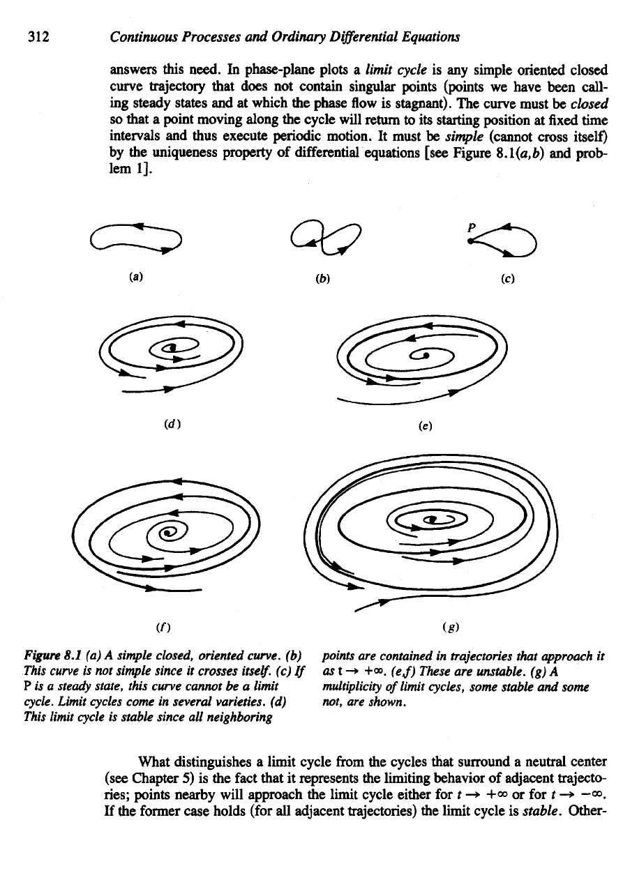
answers this
need.
In
phase-plane plots
a
limit
cycle
is any
simple oriented
closed
curve trajectory that
does
not
contain singular points (points
we
have been
call-
ing
steady states
and at
which
the
phase
flow is
stagnant).
The
curve must
be
closed
so
that
a
point moving along
the
cycle will return
to its
starting position
at fixed time
intervals
and
thus execute
periodic
motion.
It
must
be
simple
(cannot cross itself)
by
the
uniqueness property
of
differential equations [see Figure
8.1(a,fc)
and
prob-
lem
1].
Figure
8.1 (a) A
simple
closed, oriented curve,
(b)
points
are
contained
in
trajectories
that
approach
it
This
curve
is not
simple
since
it
crosses
itself,
(c) If
ast-*
+<». (
e
,f)
These
are
unstable,
(g) A
P is a
steady
state, this curve cannot
be a
limit
multiplicity
of
limit cycles, some stable
and
some
cycle.
Limit
cycles
come
in
several varieties,
(d)
not,
are
shown.
This
limit
cycle
is
stable since
all
neighboring
What
distinguishes
a
limit cycle
from
the
cycles that surround
a
neutral center
(see Chapter
5) is the
fact
that
it
represents
the
limiting behavior
of
adjacent trajecto-
ries;
points nearby will approach
the
limit cycle either
for t -* +» or for t ->
-<».
If
the
former case holds (for
all
adjacent trajectories)
the
limit cycle
is
stable. Other-
About
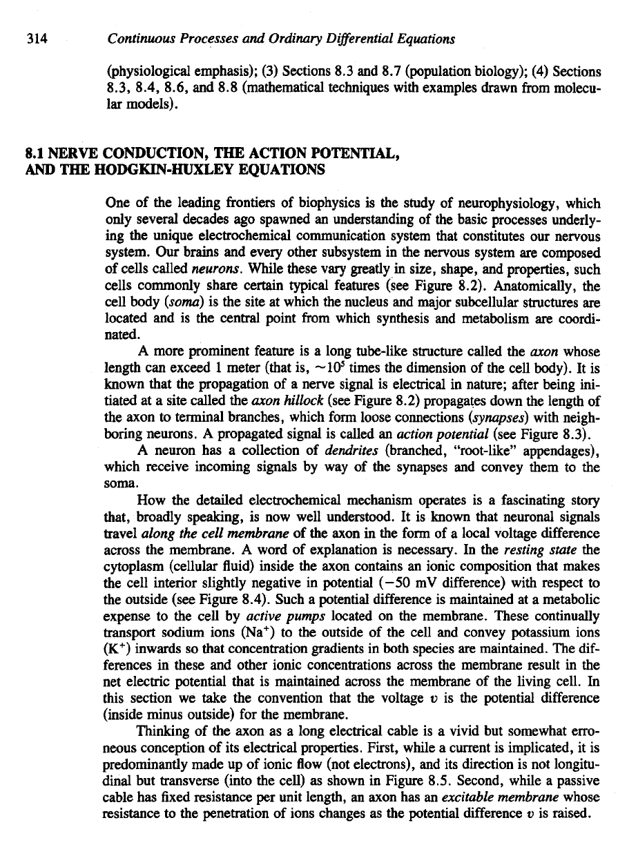
F and G we
shall assume that these
are
continuous
functions
with con-
tinuous
partial derivatives
with
respect
to x and v so
that
a
unique solution
to
(la,b)
will
exist
for a
given
set of
initial values
(xo,y
0
).
Before
launching into
the
mathematical techniques that
are
useful
in
determin-
ing
whether
limit
cycles
to a
system such
as
equations
(la,b)
exist,
we
discuss neural
excitation
by way of a
motivating example. Some
of the
basic concepts
and
physio-
logical
detail
are
described
in
Section 8.1.
A
model
due to
Hodgkin
and
Huxley
is
then
derived
and
partially analyzed
in
Section 8.2,
to be
followed later
by a
simpler
set of
equations
due to
Fitzhugh
and
Nagumo
in
Section 8.5.
We find
that both mod-
els
have
the
potential
for
exhibiting sustained oscillations typical
of a
limit cycle
solution,
as
well
as
excitable
behavior:
by
this
we
mean that
a
stimulus larger than
some threshold will provoke
a
very large response. (This type
of
behavior
is
depicted
by a
large excursion away
from
some steady state
and
then back
to it as
excitation subsides.) While phase-plane analysis (described
in
Chapter
5)
suffices
for
understanding
Sections
8.1 and
8.2,
we find it
useful
to
draw
on new
results
in
Section 8.5.
The
Poincare-Bendixson theory
is a
cornerstone
on
which much
of the
theory
of
limit cycles
rests.
Section
8.3
provides
an
informal
introduction,
and the
results
are
then applied
in
Section
8.4 to a
system
of
equations known
as the van der Pol
oscillator.
This prototype serves
to
illustrate
how a
nullcline
configuration
in
which
one
or
both nullclines
is
S-shaped tends
to
produce oscillatory
or
excitable behavior.
Many
examples drawn
from
the
literature
are
based
on
similar principles. Among
these
is
Fitzhugh's model
for
neural excitation described
in
Section 8.5.
Another
mathematical method commonly encountered
in the
quest
for
limit-cy-
cle
solutions
is the
Hopf bifurcation theorem (Section 8.6). Here
we are
more
specific
about
the
spectrum
of
possible
effects
that
are
encountered close
to a
steady
state
of a
nonlinear system
at
which linear stability calculations predict
a
transition
from
a
stable
focus-through
a
neutral center
to an
unstable
focus
as
some parameter
is
varied. (The comments
at the end of
Section
5.9
were deliberately vague
in
antici-
pation
of the
upcoming discussion.)
In
Sections
8.7 and 8.8 we
apply
the new
mathematical techniques
to
problems
stemming
from
population
fluctuations and
oscillations
in
chemical systems. These
sections
are
extensions
of
material covered
in
Chapters
6 and 7
respectively.
For a
shorter course,
any one of the
following sequences
is
suitable:
(1)
Sec-
tions 8.1 and 8.2
(based only
on
previous material);
(2)
Sections
8.1 to 8.5
Limit
Cycles,
Oscillations,
and
Excitable
Systems
313
wise
it is
unstable. Figure
8.1
provides several examples
of
stable
and
unstable limit
cycles
in a
two-dimensional phase plane.
In
this chapter
we are
concerned with identifying criteria that point
to the
exis-
tence
(or
nonexistence)
of
limit
cycles.
We
shall address this issue primarily within
the
context
of the
system
of the two
equations
314
Continuous Processes
and
Ordinary
Differential
Equations
(physiological emphasis);
(3)
Sections
8.3 and 8.7
(population biology);
(4)
Sections
8.3, 8.4, 8.6,
and 8.8
(mathematical techniques with examples drawn
from
molecu-
lar
models).
8.1
NERVE CONDUCTION,
THE
ACTION POTENTIAL,
AND
THE
HODGKIN-HUXLEY EQUATIONS
One of the
leading
frontiers of
biophysics
is the
study
of
neurophysiology, which
only several decades
ago
spawned
an
understanding
of the
basic processes underly-
ing the
unique electrochemical communication system that constitutes
our
nervous
system.
Our
brains
and
every other subsystem
in the
nervous system
are
composed
of
cells
called neurons. While these vary greatly
in
size, shape,
and
properties, such
cells
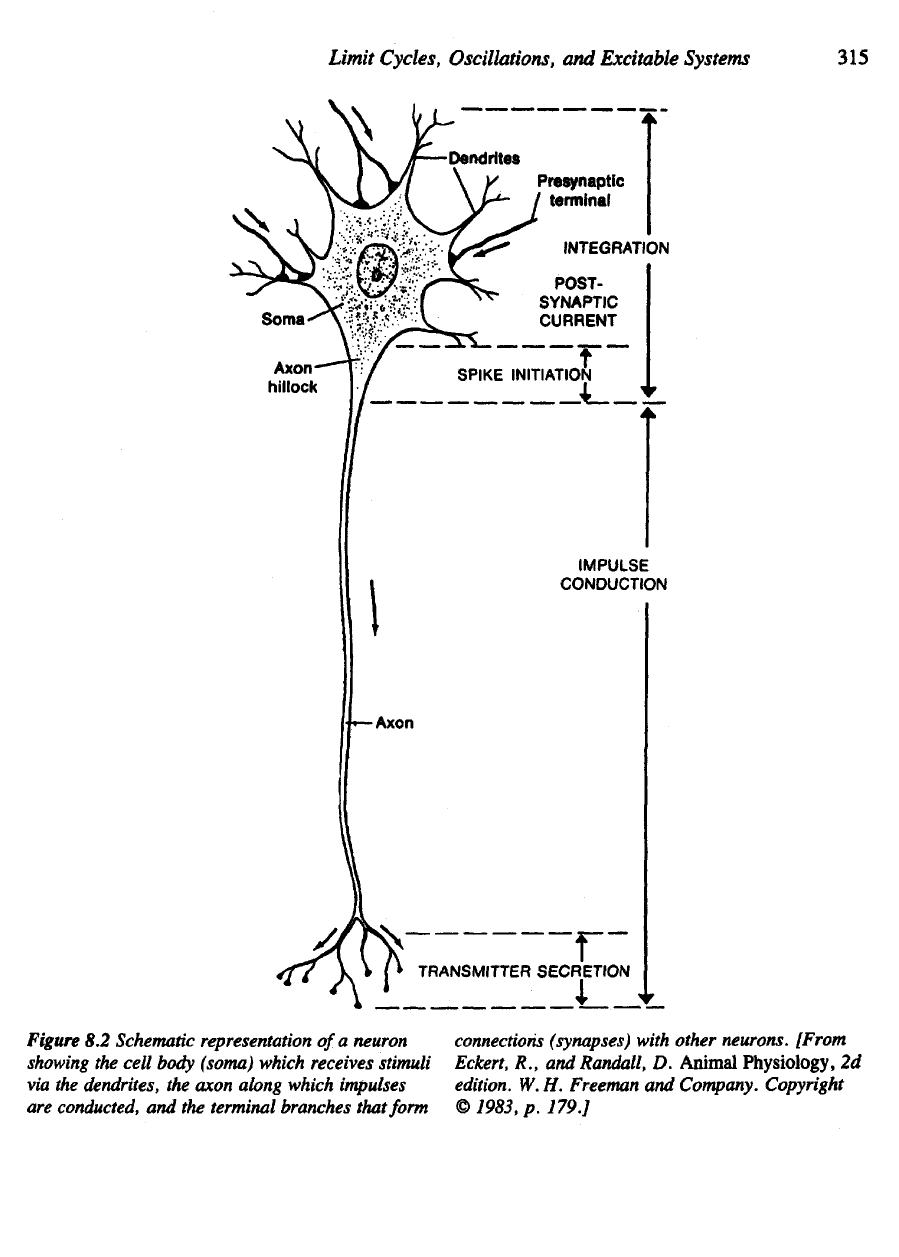
commonly share certain typical features (see Figure 8.2). Anatomically,
the
cell body
(soma)
is the
site
at
which
the
nucleus
and
major
subcellular structures
are
located
and is the
central point
from
which synthesis
and
metabolism
are
coordi-
nated.
A
more prominent feature
is a
long tube-like structure called
the
axon whose
length
can
exceed
1
meter (that
is,
~10
5
times
the
dimension
of the
cell body).
It is
known
that
the
propagation
of a
nerve signal
is
electrical
in
nature;
after
being ini-
tiated
at a
site
called
the
axon hillock (see Figure 8.2) propagates down
the
length
of
the
axon
to
terminal branches, which
form
loose connections
(synapses)
with neigh-
boring neurons.
A
propagated signal
is
called
an
action potential (see Figure 8.3).
A
neuron
has a
collection
of
dendrites
(branched,
"root-like"
appendages),
which
receive incoming signals
by way of the
synapses
and
convey them
to the
soma.
How
the
detailed
electrochemical mechanism operates
is a
fascinating story
that, broadly speaking,
is now
well understood.
It is
known that neuronal signals
travel along
the
cell membrane
of the
axon
in the
form
of a
local voltage
difference
across
the
membrane.
A
word
of
explanation
is
necessary.
In the
resting
state
the
cytoplasm (cellular
fluid)
inside
the
axon contains
an
ionic composition that makes
the
cell
interior slightly negative
in
potential (-50
mV
difference)
with respect
to
the
outside (see Figure 8.4). Such
a
potential
difference
is
maintained
at a
metabolic
expense
to the
cell
by
active pumps located
on the
membrane. These continually
transport sodium ions (Na
+
)
to the
outside
of the
cell
and
convey potassium ions
(K
+
) inwards
so
that concentration gradients
in
both species
are
maintained.
The
dif-
ferences
in
these
and
other ionic concentrations across
the
membrane result
in the
net
electric potential that
is
maintained across
the
membrane
of the
living cell.
In
this
section
we
take
the
convention that
the
voltage
v is the
potential difference
(inside minus outside)
for the
membrane.
Thinking
of the
axon
as a
long electrical cable
is a
vivid
but
somewhat erro-
neous conception
of its
electrical
properties. First, while
a
current
is
implicated,
it is
predominantly made
up of
ionic
flow
(not electrons),
and its
direction
is not
longitu-
dinal
but
transverse (into
the
cell)
as
shown
in
Figure 8.5. Second, while
a
passive
cable
has fixed
resistance
per
unit length,
an
axon
has an
excitable membrane whose
resistance
to the
penetration
of
ions changes
as the
potential difference
v is
raised.
Limit
Cycles,
Oscillations,
and
Excitable
Systems
315
Figure
8.2
Schematic representation
of
a
neuron
showing
the
cell
body
(soma) which receives stimuli
via
the
dendrites,
the
axon along which impulses
are
conducted,
and the
terminal branches that form
connections
(synapses)
with other neurons. [From
Eckert,
R., and
Randall,
D.
Animal
Physiology,
2d
edition.
W. H.
Freeman
and
Company. Copyright
©
1983,
p.
179.]
