Dennison С. A Guide to Protein Isolation
Подождите немного. Документ загружается.

168 Chapter 6
6.5 Amplification methods
At low levels of Ab and Ag, no visible precipitation may be formed.
In modem methods of immunological analysis, therefore, use is made of
amplification methods which enable the Ab/Ag reactions to be visualised
and quantitated. Amplification methods will be discussed here in more
-
or
-
less historical order. the enzyme and immunogold methods being more
recent.
6.5.1 Complement fixation
ìComplementî is a name given to a group of serum proteins which
bind Ag/Ab complexes and cause lysis of cells displaying surface antigens.
Complement thus functions in vivo as part of the immune response,
aimed at the lysis of foreign cells.
This activity is exploited in the
complement fixation assay, which is a sensitive means of detecting
Ag/Ab complexes. The complement fixation assay is about 100
-
fold
more sensitive than the precipitin reaction.
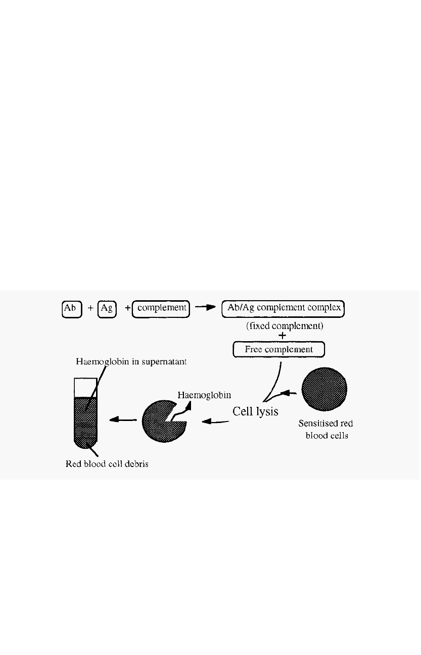
Figure 113. The complement fixation assay.
The complement fixation assay depends on the fact that complement
is consumed (fixed) by Ag/Ab complexes, making less complement
available. The amount of complement left (i.e. not fixed by the Ag/Ab
complexes) can be measured by its ability to lyse red blood cells sensitised
by antibodies binding to surface antigens. The amount of lysis can be
quantitated by measuring the haemoglobin released into the supernatant,
by its absorbance at 541 nm.
Immunological methods 169
If all of the complement was bound by Ag/Ab complexes, none would
be available to bind to the red blood cells and no lysis, and consequently
no release of haemoglobin, would occur. On the other hand, if no Ag/Ab
complexes were formed, no complement would be fixed, so all of it would
be available to lyse red blood cells and the absorbance of the released
haemoglobin would be at a maximum.
In order to measure the amount of a specific Ag present in a complex
mixture, it is necessary to first establish a standard curve by measuring
the complement fixed when different amounts of Ag are added to a fixed
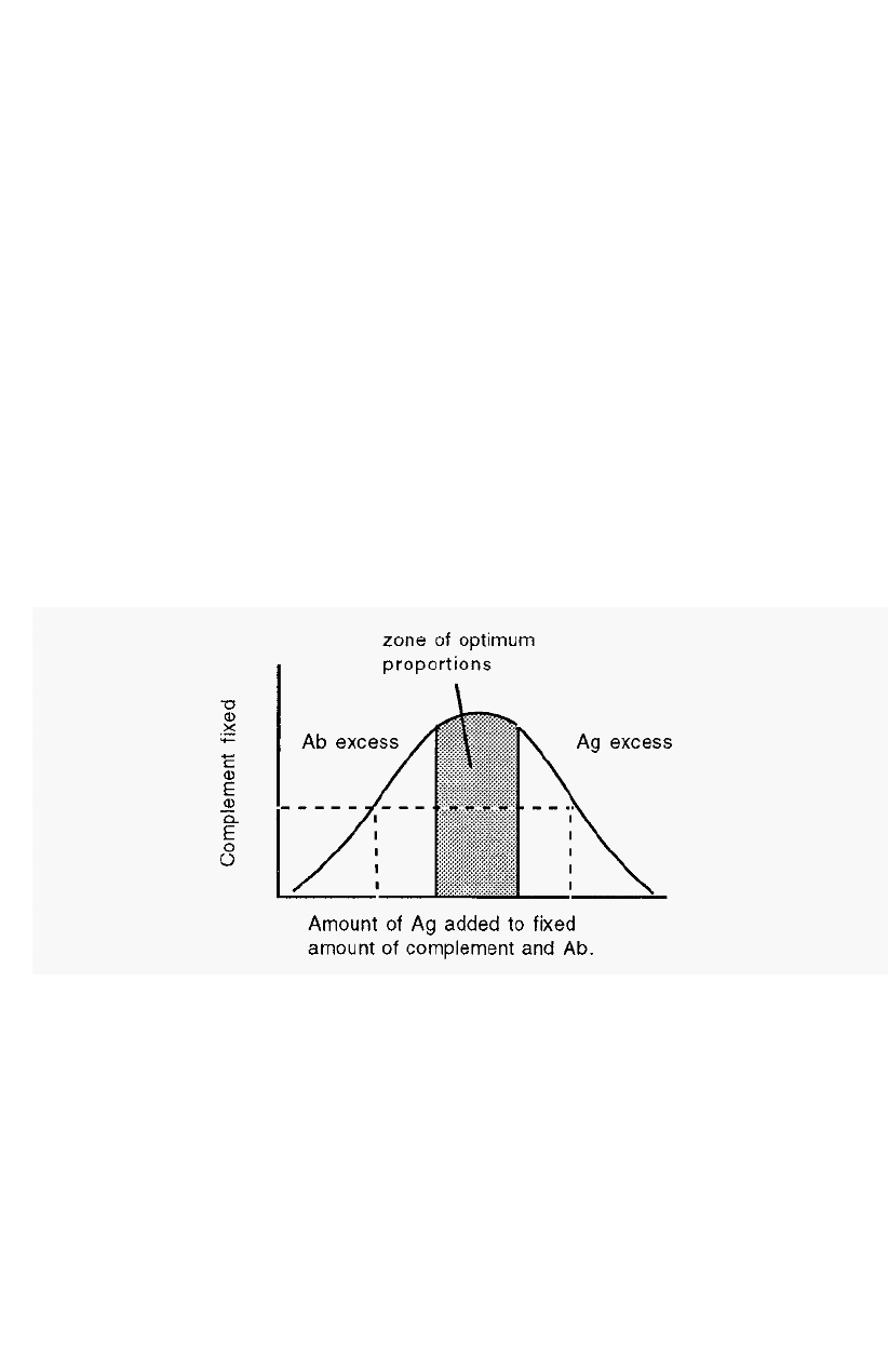
amount of complement and Ab. The standard curve (Fig. 114) has a
shape similar to that of an immunoprecipitation curve (Fig. 101).
Because of the shape of the standard curve, two different Ag
concentrations can give the same degree of complement fixation (dotted
lines in Fig. 114). In measuring an unknown, therefore, it is necessary to
test several dilutions of the Ag to establish on which side of the curve the
Ag concentration falls. The [Ag] can then be read off from the standard
curve.
Figure 114. Standard curve for complement fixation.
The method has two principal disadvantages:
-
• Certain crude mixtures cause haemolysis by mechanisms unrelated to
complement, and,
•
Some crude mixtures inactivate complement in the absence of the
appropriate Ag/Ab complex.
I70 Chapter 6
6.5.2 Radioimmunoassay (RIA)
Radioimmunoassays
(RIAs)
combine the sensitivity of radioisotope
detection with the selectivity of immunoassays.
RIAs
can be used to
detect molecules that do not
fix complement when combined with a
specific antibody, for example haptens (see p152), and
RIAs
are mostly
used for the assay of small molecules. Examples of compounds that can
be assayed by
RIA
are peptide hormones, steroids (such as testosterone),
cyclic AMP, morphine, digitalis etc.
The principle of the
RIA
is the same as that of the chemical assay
technique known as an isotope dilution assay. In an isotope dilution
assay a known amount of a radioisotope, of known specific activity (i.e
radioactivity per unit mass), is added to a sample and a pure sample of the
element of interest is subsequently extracted
and its specific activity
determined. The decrease in the specific activity of the isolated element
is due to the presence of the non
-
radioactive isotope which dilutes the
radioactive isotope. From the extent of this dilution, the concentration
of the endogenous, non
-
radioactive isotope can be determined.
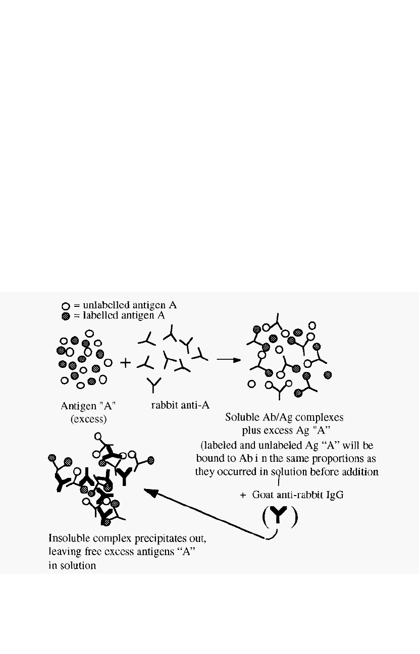
Figure I 15. Radioimmunoassay.

Immunological methods 171
A RIA is illustrated in Fig. 115. In a RIA, a radio
-
labelled antigen is
used to dilute an unknown amount of an unlabelled antigen, present in the
sample. A non
-
saturating amount of, say, rabbit antibody to the antigen
is added (i.e. the antigen must be in excess). The labelled and unlabelled
antigens will bind to the antibodies in the same ratio as they are present
in the sample. The Ab/Ag complexes can then be precipitated, for
example by addition of a goat anti
-
rabbit IgG antibody. The radioactivity
in the precipitate will be inversely related to the amount of the antigen
originally present in the sample. The concentration of the unknown
antigen in the sample can thus be measured by reference to a straight line
standard curve in which the % inhibition is plotted against log[non
-
radioactive Ag].
Radioimmunoassay has the following disadvantages:
• A radioactively labelled Ag may not be available, especially for an
antigen which has not been extensively studied.
• Associated with the radioimmunoassay are all of the hazards of
radioisotopes, which means that specially equipped laboratories and
special licences are necessary.
6.5.3 Enzyme amplification
The advantage of enzyme methods over radioisotope methods of
amplification is that, because enzymes are safe and biodegradable, no
special licences or safety facilities are required, and disposal after use is no
problem.
6.5.3.1 Enzyme linked immunosorbent assay (ELISA)
The ELISA method was introduced in its modern form by Engvall and
Perlmann
14
and van Weemen and Schuurs
15
. The principle of an ELISA
is that an enzyme, linked to an immunoreactive molecule (an antibody or
protein A) can be used to detect the presence of an antigen with great
sensitivity, due to the amplification achieved by the enzyme catalysed
reaction. The method can also be turned
about and used to detect Ab.
Several different formats of ELISA are possible. Only some concepts
pertaining to ELISAs are discussed here. For details on how to conduct
an ELISA, a specialist text
16,17
should be consulted.
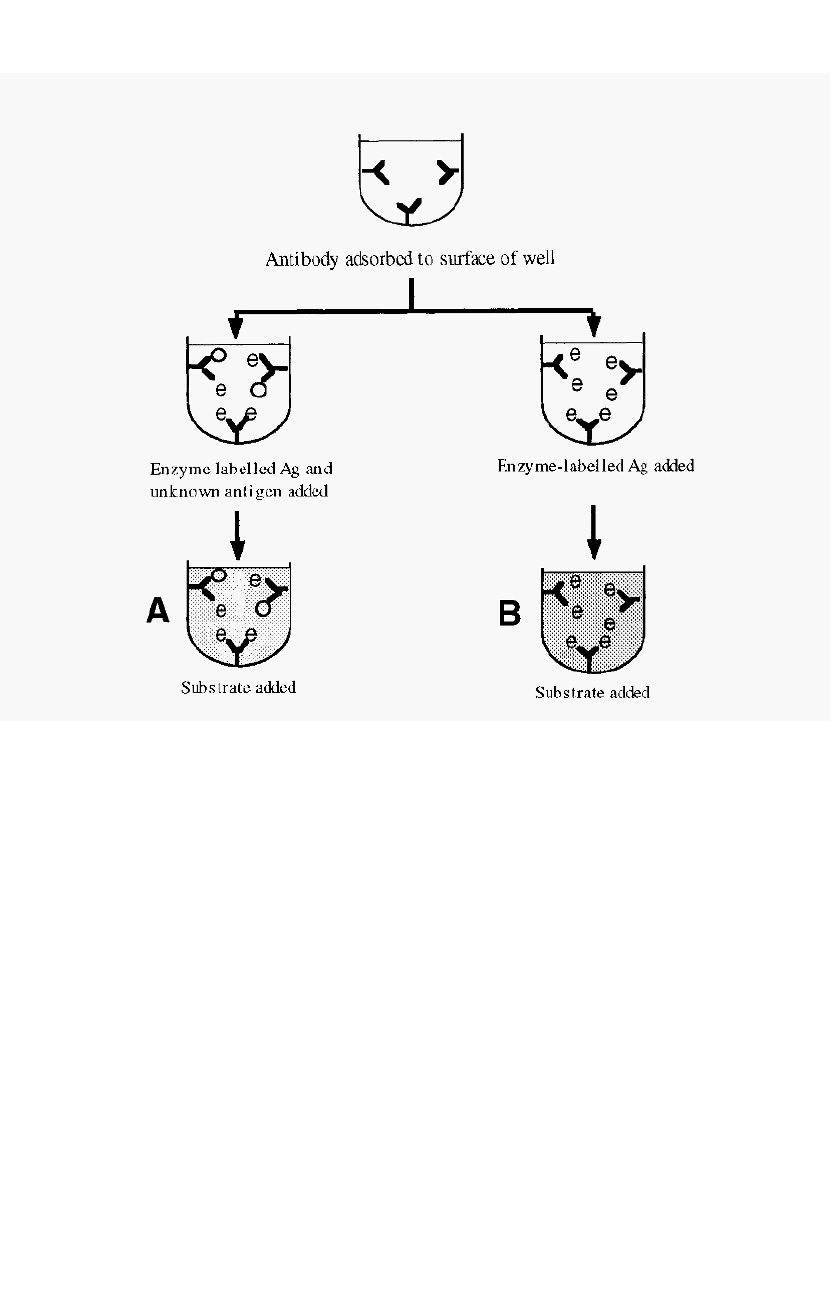
The competitive ELISA for measurement of Ag is conceptually similar
to a RIA (Section 6.5.2), in that a labelled Ag competes with an
unlabelled Ag for binding to the antibody, but in this case the label is an
enzyme (Fig. 116).
172
Chapter 6
Figure 116. A competitive ELISA for measuring [Ag].
Unknown [Ag] is proportional to the absorbance difference
between the wells A and B.
Antibody is inmobilised by adsorption onto the walls of a plastic
microtitre plate. Excess antibody is washed off and the enzyme
-
linked
Ag is added to one set of wells while the unknown sample, mixed with
enzyme
-
linked Ag is added to another set. The unknown sample Ag thus
serves to dilute the amount of labelled Ag which reacts with the
immobilised Ab After incubation, excess antigen is washed off and a
solution of the enzyme substrate is added. The enzyme catalysed
reaction generates a colour which can be measured. The intensity of this
colour is inversely proportional to the amount of Ag originally present.
The microtitre plate contains 96 wells in an 8 x 12 array, which permits
the exploration of a number of different combinations of coating and
Ab/Ag concentrations, permitting optimisation of the reaction
16,17
.
Immunological methods 173
In another format, related to immunoblotting (Section 6.5.3.2), the
Ag (usually a solution containing a mixture of proteins, including the
protein of interest) may be adsorbed onto the walls of a microtitre plate
and the Ag of interest subsequently detected with an enzyme
-
labelled Ab.
For increased sensitivity, further amplification may be obtained by using
two antibodies, an Ag
-
specific primary Ab and an enzyme
-
labeled
secondary Ab that is specific for the type of primary Ab. This has the
advantage that the secondary enzyme
-
labeled secondary Ab can be a
universal reagent, targeting primary Abs of the same type but with
different specificities.
6.5.3.2 Immunoblotting
In one ELISA format the principle of amplified detection of an
immobilised Ag using an enzyme labeled Ab is used. The Ag, in this case,
may be coated onto the walls of a microtitre plate. A similar principle
applies to immunoblotting, whereby Ag immobilised on for e.g. a
nitrocellulose filter, can be detected using an enzyme
-
linked antibody
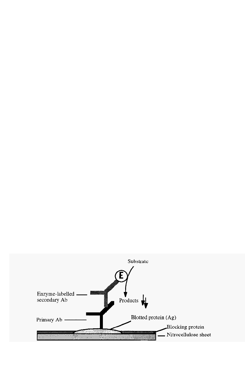
system (Fig. 117). An example is dot blotting, in which a small volume
of the antigen of interest is dotted onto a nitrocellulose filter
18
.
The
surrounding protein
-
binding sites may be blocked with milk proteins,
which do not tend to bind proteins non
-
specifically. The dot can then be
probed with a primary antibody, followed by an enzyme
-
labelled
secondary antibody, as in an ELISA. The only difference is that in an
ELISA, the enzyme product is soluble, whereas in an immunoblot an
insoluble product is necessary. A commonly used combination is
horseradish peroxidase as the enzyme label, with 4
-
chloro
-
l
-
naphthol as
the substrate. The blue/grey product is insoluble in water
19
.
Alternatively, a gold
-
labelled Ab or protein A may be used
18
.
Figure 117.
A schematic sketch of immunoblotting.
174 Chapter 6
The curious name
of
this
technique is due to a pun. A technique for the blotting of DNA
fragments, and probing these with labelled RNA, was named ìSouthern
blottingî after its originator, E. M. Southern. The reverse process:
blotting RNA and probing this with DNA fragments, was then named
ìnorthern blottingî, to indicate that it was an opposite process. The
blotting of proteins was subsequently called “western blotting” to indicate
a similar process but with different molecules, i.e. in a different direction.
In western blotting, proteins are first separated by SDS
-
PAGE (p 133).
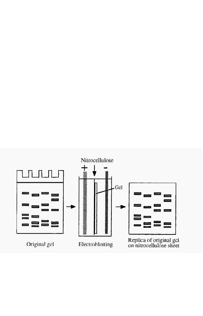
The separated proteins are then drawn out of the gel, and blotted onto
nitrocellulose, by transverse electrophoresis, forming a replica of the gel
separation (Fig. 11 8). In this process the nitrocellulose sheet must be on
the anodic side of the gel, as protein/SDS complexes are negatively
charged and have an anodic migration.
Within the gel the proteins are not accessible to probing by
antibodies, but they become accessible after electroblotting onto the
surface of a nitrocellulose sheet. Antibodies bound to the blotted proteins
can be detected with an enzyme
-
labelled secondary antibody, as in a dot
blot.
A related technique is western blotting
20
.
Figure 118. Electroblotting.
Treatment with SDS tends to denature proteins, although the effects
can be minimised if the sample is not boiled in SDS (see Section 5.8.1).
The denatured protein might not be recognised by the antibodies used to

Immunological methods 175
probe the blot. In this case a renaturing blot system
21
, can be used to
advantage. In this technique the transfer buffer does not contain SDS and
the SDS may be removed from the gel before the transfer step. The
nitrocellulose sheet must be placed on the appropriate side of the gel,
depending on the direction of migration of the protein at the pH of the
transfer buffer. Normally a high pH is used, giving an anodic migration,
but this is not appropriate if the protein is not stable at a high pH.
In a gel of constant composition, proteins separate by virtue of their
differential rates of migration, smaller proteins migrating faster than
large ones. This same differential will apply to the migration of proteins
during the transfer step, i.e. smaller proteins will transfer to the
nitrocellulose more rapidly. If insufficient time is allowed for the
transfer, the blot will be biased in favour of smaller proteins. This effect
can be overcome by running the first electrophoresis in a gradient gel
(Section 5.9). In this case the proteins will each reach a point in the
gradient where they are about equally impeded by the gel and during the
lateral transfer step they will all migrate out of the gel at about the same
rate.
Western blotting is useful for determining the presence or absence of a
specific Ag in a complex mixture. It is also useful for testing the
specificity of an Ab, before this is used in immunocytochemistry, for
example. Blotting (not immunoblotting) is also used as a step in the
sequencing of a protein band purified by gel electrophoresis. For this
purpose the protein is electroblotted onto a polyvinylidene difluoride
(PVDF) membrane, the blot excised and transferred into the sequenator.
6.5.4 Immunogold labeling with silver amplification
Colloidal gold particles, ranging in diameter from 1 to 30 nm, can be
prepared by reducing dissolved gold chlorides with various reducing
agents
2
2
. Proteins bind readily to such particles and stabilise the colloids
against a salt challenge. Colloidal gold particles can thus be used as labels,
attached to either antibodies or protein A, to form immunogold probes.
Such immunogold probes find their greatest use in electron microscopy
immunocytochemistry, whereby the subcellular distribution of an Ag of
interest may be determined. Colloidal gold particles are very electron
dense and show up readily in electron micrographs.
With silver amplification
23
, immunogold probes may also be used at
the light microscopy level and for staining immunoblots
18
. In the
immunogold
-
with
-
silver
-
amplification (IGSS) technique, the colloidal gold
label serves as a nucleation centre for the deposition of metallic silver.
This yields a black stain which marks the position of the Ag of interest.

176 Chapter 6
6.5.5
C oll oi d aggl u t in at ion
As mentioned above (Section 6.5.4), proteins can stabilise colloids.
The proteins bind to the colloidal particles and similar charge repulsion
between the bound proteins keeps the colloidal particles apart, thus
preventing flocculaton. Latex beads are commonly used as colloidal
suspensions for analysis. A natural system, which is virtually colloidal, is
blood, in which the red cells are prevented from aggregating by virtue of
their similar surface charges.
Antibodies are divalent and are thus able to simultaneously bind to two
similar antigens on two different colloidal particles. Such cross
-
linking of
the colloidal particles causes them to flocculate out of suspension, and
this provides a very sensitive method for the detection of Abs specific
for the colloid
-
bound Ag. The method is particularly useful for medical
diagnosis in the field. Since flocculation can easily be detected by eye, no
sophisticated instrumentation is required. The method gives a simple
yes
-
or
-
no answer, but it can be made semi
-
quantitative by dilution of the
Ab, until the “definite yes” becomes a “maybe”. The dilution at which
this happens is inversely related to the initial antibody concentration.
An elegant diagnostic method uses agglutination of endogenous red
blood cells as the reporter system
24,25
. Monoclonal Abs are raised against
glycophorin, a glycoprotein present on the surface of all red blood cells.
These Abs are species specific, i.e. they only recognise glycophorin from
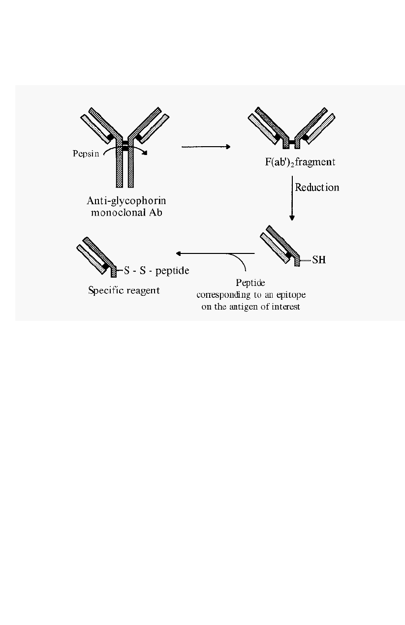
a particular species. From these monoclonal Abs, F(ab’), fragments can
be made by proteolysis with pepsin. F(ab’)
2
fragments consist of the two
Fab arms of the Ab, bound together by disulfide bridges. The presence of
the disulfide bridges is useful as these can be reduced and subsequently used
to conjugate a peptide epitope to the free -SH groups of the two separate
Fabí fragments. This generates a specific diagnostic reagent (Fig. 119).
Addition of this reagent to a drop of blood will cause
haemagglutination, if Abs targeting the peptide epitope are present.
For
example, a person infected with the AIDS virus will, in the early stages,
have anti
-
AIDS virus Abs present in their blood. These Abs will target
specific epitopes on the AIDS virus proteins. These epitopes can be
identified and corresponding peptides can be synthesised. Conjugation of
one such peptide to an anti
-
human glycophorin monoclonal Ab half
-
F(ab’)
2
fragment will generate a specific diagnostic reagent.
Addition of
an appropriate dilution of this reagent to a drop of a patient’s blood will
give a yes/no indication of the presence of anti
-
AIDS virus Abs in the
patientís blood. A positive answer is given by the agglutination of the
red blood cells. Such diagnostic analyses are useful for screening in the
Immunological methods 177
field but they should be followed by confirmatory laboratory
-
based tests,
such as ELISA
Figure 11 9. Generation of a reagent for detecting the presence of specific antibodies,
using endogenous red blood cells as the reporter system.
References
1.
Branden, C. and Tooze, J. (1991) in Introduction to Protein Structure. Garland
Publishing, New York, pp186
-
191.
2. Hopp, T. P. (1989) Use of hydrophilicity plotting procedures to identify protein
antigenic segments and other interaction sites. Methods Enzymol. 178, 571
-
585.
3. Westhof, E., Altschuh, D., Moras, D., Bloomer, A. C., Mondragon, A., Klug, A., and
van Regenmortel, M. H. V. (1984) Correlation between segmental mobility and
location of antigenic determinants in proteins. Nature 31 1, 123
-
126.
4. Polson, A., Potgieter, G. M., Largier, J. F. Mears, E. G. F. and Joubert, F. J. (1964) The
fractionation of protein mixtures by linear polymers of high molecular weight.
Biochim. Biophys. Acta 82, 463
-
475.
5. Polson, A., von Wechmar, M. B. and Van Regenmortel, M. H. V. (1980) Isolation of
viral IgY antibodies from yolks of immunized liens. Immunol. Commun. 9, 475
-
493.
6. Mancini, G., Carbonara. A. O. and Heremans, J. F. (1965) Immunochemical
quantitation of antigens by single radial immunodiffusion. Immunochemistry 2, 235
-
254.
7. Ouchterlony, O. (1948) In vitro method for testing the toxin
-
producing capacity of
diphtheria bacteria. Acta Path. Microbiol. Scad. 25, 186
-
191.
