Chandrasekaran A. (ed.) Current Trends in X-Ray Crystallography
Подождите немного. Документ загружается.


Complementary Use of NMR to X-Ray Crystallography
for the Analysis of Protein Morphological Change in Solution
417
The
15
N CSA tensor is known to be significantly dependent on the local structure,
particularly backbone torsion angle (Yao et al. 2010). Experimentally determined
15
N CSA
tensors are reported with a various method (Fushman, Tjandra and Cowburn 1999,
Cornilescu and Bax 2000, Boyd and Redfield 1999, Hiyama et al. 1988, Kurita et al. 2003).
These data give consensus
15
N CSA tensor values for the residue in each type of secondary
structure,
-helix, -sheet and others. Practically, the use of the secondary structure specific
15
N CSA tensor values can determine the alignment tensor within experimental errors.
Experimental determination of
15
N CSA tensor for protein in solution is possible using the
weak alignment technique. We previously proposed the method using magic-angle sample
spinning to determine the accurate secondary structure specific
15
N CSA tensor, in which
the bicellar media was used for a weak alignment (Kurita et al. 2003). In this experiment, we
used only one aligned state, thus, only determined the
15
N CSA tensors in a secondary
structure specific manner.
Recently, Bax and co-workers have applied this approach to determine the residue specific
15
N CSA tensors for a protein, where they used five more different aligning states to solve
the Saupe order matrix for each residue (Yao et al. 2010). The residue specific
15
N CSA
tensor determination that requires multiple aligned states of protein is rather demanding
experiments, which require a various loop mutant to change aligning angle (Yao et al. 2010).
However, the continuous effort to collect the residue specific
15
N CSA tensors in the similar
way by Bax and co-workers will establish a clearer correlation between the
15
N CSA tensor
and backbone torsion angles and also local interactions like hydrogen bonding, which may
allow the prediction of the appropriate
15
N CSA tensor values from the structure. The
refined
15
N CSA tensors will further improve the quality of alignment tensor analysis with
the RCSA, although the present RCSA based approach gives an acceptable result.
3. Achieving weak alignment
In applying the residual anisotropic spin interactions described above, it is required to make
a protein in a weakly aligned state. The aligning protein has to be carefully tuned to make
the anisotropic interactions observable in a spectrum with keeping the spectral resolution
and intensity. Alignment order is practically tuned to approximately 10
-3
, giving about 20
Hz in maximum absolute magnitude for amide
1
H-
15
N RDC. To achieve a weak alignment,
some artificial medium has to be used, because the inherent magnetic susceptibility of a
globular protein is too small to align to the desired extent, except for some heme-containing
proteins having substantial magnetic susceptibility associated with a heme group. In this
section, we will review some media for weak alignment.
3.1 Magnetically aligning liquid crystalline media
Magnetically ordering liquid crystalline media are commonly used. Discoidal phospholipid
assembly, bicelle, is one of the prevailingly used materials for a weak alignment of protein
(Ottiger and Bax 1999, Ottiger, Delaglio and Bax 1998, Tjandra and Bax 1997).. The bicelle is
composed of a mixer of dimyristoylphosphatidylcholin (DMPC) and dihexynoyl-
phosphatidylcholine (DHPC) in a ratio of 3:1. This phospholipid binary mixture forms lipid
bilayers disks 30 nm – 40 nm in diameter. Bicelle has substantial magnetic susceptibility, and
it spontaneously aligns under magnetic field with the normal of the bicelle surface staying
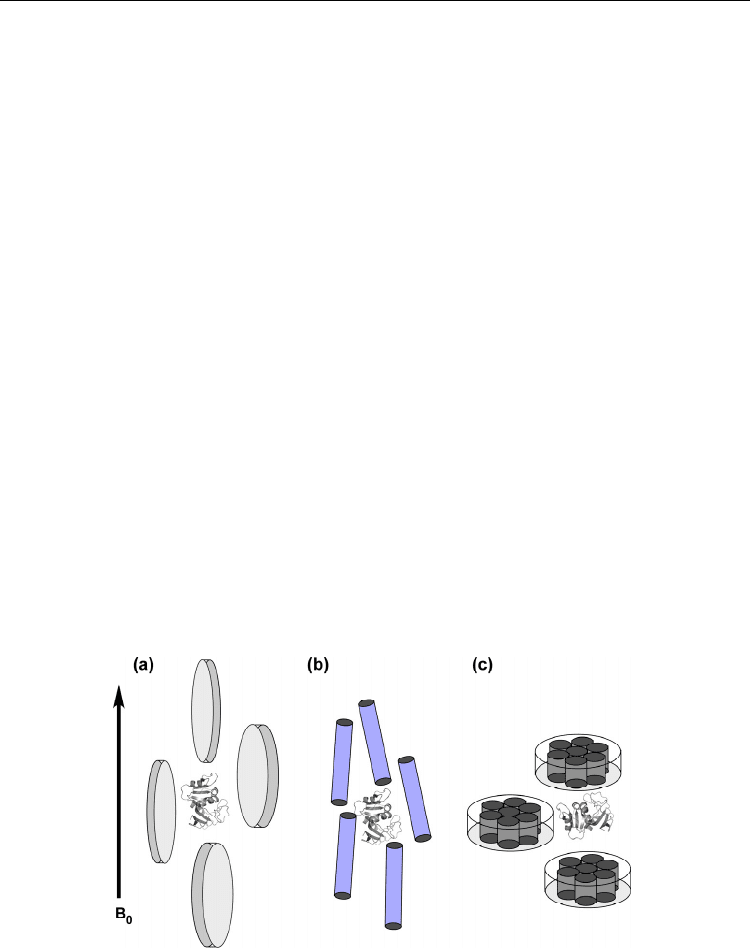
perpendicular to the magnetic field (Fig. 6a).
In the experiments to measure the anisotropic spin interactions, an appropriate amount of
bicelle is put into protein solution. In a high magnetic field, bicelles align and the aligned

Current Trends in X-Ray Crystallography
418
discoidal liquid crystalline molecules limit the space for protein tumbling. Bicelle has flat
surface, thus, the protein involved in the aligned bicellar media will be surrounded by flat
walls. Because of the steric clash, protein does not rotate freely near the bicelle wall, which
will hider some orientations of the protein. This hindrance on some orientations for the
protein in the bicellar medium causes incomplete rotational averaging of the anisotropic
interactions, which thus makes the residual anisotropic interactions observables (Berlin,
O'Leary and Fushman 2009, Vijayan and Zweckstetter 2005, Zweckstetter 2008).
The aligning magnitude is readily tuned by the bicelle concentration; higher bicelle
concentration induces the stronger order of alignment. The bicelle made of the binary
phospholipid mixture involving DMPC and DHPC has neutral charge on its surface.
Therefore, most proteins do not stick to bicelles. Protein aligns through collisional
interaction described above.
Surface charge doping to the bicellar surface is possible. Incorporation of CTAB, cetyl
trimethyl ammonium bromide, to DMPC/DHPC binary phospholipid bicelle generates the
positively charged surface, while the addition of SDS, sodium dodecyl sulphate, makes it
negatively charged. The surface charge doping to bicelle changes the aligning property from
that by neutral bicelle. In charged bicellar solutoin, electrostatic interactions between the
medium and protein become apparent. This makes it sometimes difficult to use the charged
bicelle to acidic or basic proteins such as nucleic acid binding proteins.
There are some limitations in the bicelle application as aligning media. One is in the limited
temperature range to keep bicelle in a liquid crystalline phase; it is typically 27°C – 45°C.
Some proteins precipitate in this temperature range, and the bicelle is not used for such
proteins. In addition, the bicelle is only stable around neutral pH; in acidic or basic solution,
the bicellar medium tends to make phase-separation and loses aligning ability. To avoid the
limitations associated with the physicochemical properties of the bicelle medium, other
liquid crystalline media have been reported. By properly selecting the medium, we can now
achieve weak alignment for rather various types of proteins, each of which requires its own
optimal temperature, pH, and ionic strength (Prestegard, Bougault and Kishore 2004).
Fig. 6. Weak alignment made by using various media. (a) Discoidal shape phospholipid
bicelle medium, (b) rod-like filamentous phage and (c) Purple membrane. Because of
significant anisotropic magnetic susceptibility, the molecules spontaneously align in a
magnetic field. Proteins within the aligning media are aligned through the collisional
interaction or electrostatic interactions with the aligning molecules.

Complementary Use of NMR to X-Ray Crystallography
for the Analysis of Protein Morphological Change in Solution
419
3.2 Naturally occurring materials that spontaneously align in a magnetic field
Some naturally available molecules that spontaneously align in a magnetic field are also
used for weak alignment. Purple membrane is one example, which forms two-dimensional
crystal lattice structure of bacteriorhodopsin (bR) that is rich in
-helices (Fig. 6b). Purple
membrane constitutes of ordered
-helices and thus it has intrinsic high anisotropic
magnetic susceptibility. Because of the structural characteristics, purple membrane can
spontaneously align in a high magnetic field.
Suspension of purple membrane is added into protein solution to achieve a weak protein
alignment. The alignment magnitude is tuned by the purple membrane concentration in a
sample solution, as was the case for the bicelle medium (Sass et al. 1999). Purple membrane
is the discoidal protein lipid complex, and it aligns with its normal parallel to the magnetic
field. Purple membrane is rich in negative charge on its surface. Therefore, protein is aligned
through the electrostatic interaction not collisional interaction.
Filamentous phage, which is made of a rod-like coat protein, is another example of the
naturally occurring molecule used for weak alignment. Because of the highly anisotropic
shape of the filamentous phage, it spontaneously aligns in a high magnetic field, with its rod
axis parallel to a magnetic field (Fig. 6c). Filamentous phage has also negatively charged
surface, thus it induces protein alignment through electrostatic interactions with protein as
in the case of purple membrane. The alignment order can be tuned by adjusting the
concentration of the phage suspension in protein solution.
The use of purple membrane and filamentous phage cannot be applied to basic proteins that
are positively charged. The basic proteins tightly adsorb onto the negatively charged
surfaces of the aligning molecules through the electrostatic interaction, which result in
extreme ordering of proteins and thus prohibit observing high resolution NMR signals.
3.3 Compressed acryl amide gel
The other method uses anisotropically compressed acrylamide gel (Tycko, Blanco and Ishii
2000, Ishii, Markus and Tycko 2001, Sass et al. 1999). Acrylamide hydrogel has cavities to
capture proteins within, whose cavity size is changed by altering
the ratios of the
composing chemicals, acrylamide and bis-acrylamide. Acrylamide forms a linear polymer
chain and bis-acrylamide makes bridge to link acrylamide liner polymers, thus, the
increased ratio of bis-acrylamide generates smaller cavity.
The acrylamide gel has a spatially isotropic cavity. Protein in the gel does not show any
preferential orientation, and thus it shows no residual anisotropic spin interaction on its
NMR spectrum. Protein alignment by the gel is achieved by inducing the spatial anisotropy
to the cavities in the gel.
In a weakly alignment experiment using the gel, a cast gel including a protein solution is
inserted into a NMR sample tube. There are two ways to make anisotropically compressed
gel; one is to press the gel along the NMR tube (compressed gel) and the other is squeeze it
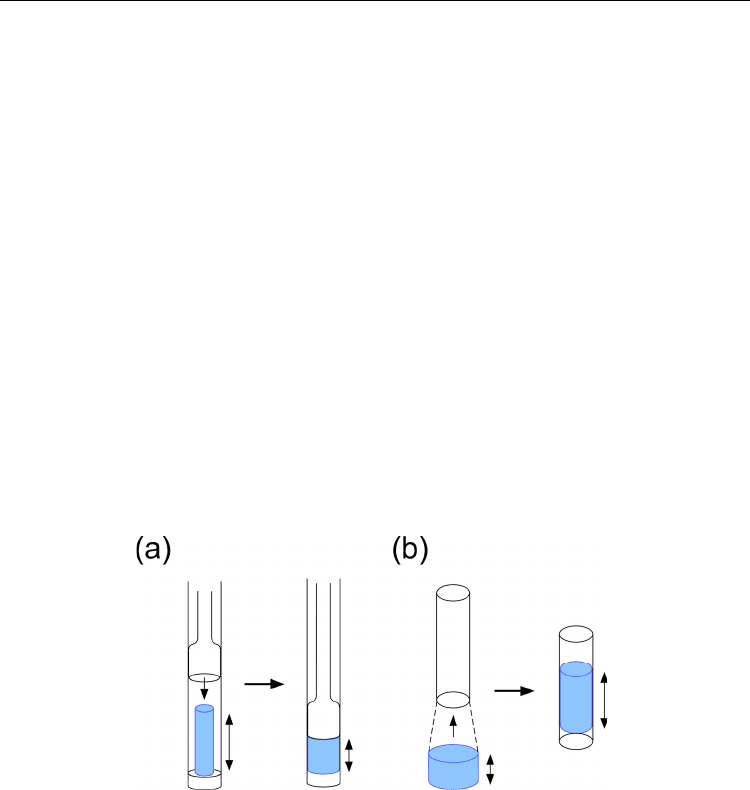
in the lateral dimension, thus it becomes stretched vertically (stretched gel) (Fig. 7).
In vertical compression, gel is cast to have a shorter diameter than the inner diameter for
NMR sample tube. After inserting the gel, vertically compressed with glass rod in the tube
(Fig. 7a). On the other hand, stretched gel is made from the cast gel having a slightly larger
diameter than that the inner diameter of the tube; the gel is inserted into the tube by using
tapered device (Chou et al. 2001) (Fig. 7b). Because of the different pressing
direction, these
two preparations change the
protein orientation to each other.

Current Trends in X-Ray Crystallography
420
The charge doping in the anisotropically compressed gel is also possible. Negative charge is
doped by replacing a part of acrylamide with acrylate, while a positive charge is introduced
by adding DADMAC, diallyldimethylammonium chloride, in casting gel chip. These charge
doping changes aligning property, because electrostatic interaction between protein and the
gel becomes active.
Acrylamide gel is neutral and thus it induces protein alignment through collisional
interaction between protein and walls having spatial anisotropy made by the compression.
The non-charged acrylamide gel is readily used for any types of protein, independent on its
pKa value. On the other hand, the charged gel is sometimes troublesome in achieving weak
alignment of basic or acidic proteins, due to their adsorption onto the gel.
The aligning order magnitude is also tuned by changing the concentration of acrylamide
and/or the ratio of the composing chemicals and also the diameter of the cased gel chip. In
addition, the extent of the charge doping may have to be considered in some cases. There
are more parameters to adjust than that for the liquid crystalline media. We, therefore, need
some trial experiments to gain the optimal gel condition to have the anisotropic spin
interactions observable in enough spectral resolution.
The great advantage in using the compressed gel is in the chemical stability of acrylamide
gel. In using acrylamide gel for a weak alignment, we do not worry about the sample
conditions including temperature, salt concentration, and solution pH. Under any sample
conditions, we can align protein by the compressed gel. It is a keen difference from the
limitation in applying liquid crystalline media and also in using the naturally occurring
materials. The compressed gel is used as a universal media for weakly aligning protein.
Fig. 7. Anisotropically compressed gels to achieve weak alignment. (a) Vertically compressed
gel. (b) Laterally compressed, thus, stretched gel. Cast gel chip (blue) is inserted into a NMR
glass tube. In the vertical compression, the cast gel is pressed by a glass rod in NMR tube.
These compressions induce spatially anisotropic cavities within the acrylamide gel.
3.4 Protein structure in aligned media – Is it natural?
A weak alignment of protein is prerequisite for the analysis of protein morphology by NMR,
because it relies on the anisotropic spin interactions that happen only under a weak aligned
condition. Because protein is dissolved in an artificial medium like magnetically ordering
liquid crystalline solution, one should worry about the structure under the condition; the
domain orientation in such a medium represents the real state of the protein in an isotropic
solution?

Complementary Use of NMR to X-Ray Crystallography
for the Analysis of Protein Morphological Change in Solution
421
The mechanisms for aligning protein in magnetically aligned bicelle solution and rod-like
filamentous phage suspension are well understood. The observed RDCs can be reproduced
with using the protein crystal structure by the simulation based on the mechanism. The
successful reproduction of the RDCs for the protein aligned by bicelle or filamentous phage
supports that the proposed theory properly describes the state of protein in the aligning media.
According to the theory, the observed residual anisotropic spin interactions can be
established by a small fraction of protein experiencing the rotational restriction through the
collision between the protein and the aligning molecule. In using the charged filamentous
phage, the electrostatic interactions are also active in inducing the rotational restriction on
the protein near the medium. The fraction for that is around 0.1% of the total number of
protein molecules in the sample. The interactions have to be weak and transient. The inter-
molecule interactions, therefore, do not actively align the protein but just prohibit some
fraction of the protein orientation near the medium. Under the condition, the aligning media
do not induce artificial protein conformation or domain arrangement, because the
prohibited orientation angles neat the medium is solely determined by the intrinsic
molecular shape of protein in the bulk solution.
The structural perturbation by the medium is easily monitored by the NMR spectral
changes. If the medium tightly interacts with protein to change the structure, some of the
chemical shifts should change from those found in an isotropic state.
The bicellar medium is in the liquid crystalline state over its phase-transition temperature.
Under the temperature, it becomes a micelle solution that does not align in a magnetic field,
thus does not induce protein alignment. If protein in this micelle solution does not show
apparent spectral changes from the solution, including no micelle, we exclude the possibility
that the bicelle induces a structural change to the protein.
In the case of the charged filamentous phage, the increased concentration of cations, which
shield the surface charge on the medium, impairs the ordering of the media in a magnetic
field. The magnitudes of the anisotropic spin interactions for the protein in the phage
suspension, therefore, are proportion to the cation concentration. If the spectral changes
show linear dependency on the cation concentration, we could rule out the structural
changes caused by the interaction with the medium.
Using compressed acrylamide gel allows the direct spectral comparison between the
samples in the reference gel (non-compressed gel) and the isotropic solution without gel to
see if any structure change happens through the interaction with the gel.
In general, under weakly aligning conditions, the spectral changes caused by the interaction
with the media are rather small, ensuring that no apparent structural changes happen to
protein in the media, even in the cases of multiple-domain proteins.
4. RDC-based domain orientation analysis – Basics and limitation
In this section, we will describe the experimental procedure to determine the domain
orientation of a multiple-domain protein, from the RDC data collection to structure
determination. In addition, the limitations in the RDC based analysis will be discussed to
emphasize the necessity of our TROSY based DIORITE approach that follows.
4.1 Collecting the RDC data
RDC is measured on a pair of
1
H coupled HSQC spectra for the samples in isotropic and
anisotropic states. The
1
H coupled HSQC spectrum gives a pair of split peaks along
15
N axis

Current Trends in X-Ray Crystallography
422
for each
1
H-
15
N correlation. The doubled number of peaks on a
1
H coupled HSQC spectrum
may increase signal overlaps that obstacle the accurate reading of peak positions. To avoid
this drawback, a particular NMR technique is used to separate the up- and down-field
components into different 2D spectra, IPAP-HSQC. IPAP-HSQC gives two separate spectra
that have in-phase and anti-phase doubles, respectively. Addition or subtraction of the
spectra will give two separate 2D spectra displaying only up-field or down-filed
components of each doublet. This signal separation reduces signal overlap on a
1
H coupled
HSQC spectrum, and keeps the spectral resolution to the same extent as in the original
HSQC spectrum (Fig. 3).
The separation width between the up- and down-field components measured from IPAP-
HSQC spectra gives
1
NH
J
for isotropic sample and
1
res
NH NH
JD for aligned sample.
Therefore, the RDC,
res
NH
D , is obtained by their difference.
4.2 Domain orientation analysis based on the RDC data
Here, we describe the domain orientation analysis based on the RDCs. The domain
orientation analysis should be done for the protein whose structure is already known by X-
ray. The primal interest of the analysis is in exploring the domain reorientation upon
interaction with the other protein or ligand. In the cases, each domain is assumed to retain
the same structure as in crystal.
As described in the theory section, the alignment tensor for a weakly aligned protein is
determined based by the RDCs and its structure coordinate, Eq. (5). In Eq. (5), direction
cosines are calculated from the structure coordinate. Because the Saupe order matrix
consists of five independent elements, we need more than five RDC data to determine the
Saupe order matrix for the corresponding part. The singular value decomposition (SVD) to
the matrix comprising of the relations for the observed RDCs will give the Saupe order
matrix (Losonczi et al. 1999). Diagonalizing the Saupe order matrix gives the alignment
tensor frame orientation relative to the molecular coordinate system and the magnate of the
orders along each principal axis. As described in the theory part, the alignment tensor frame
orientation is defined by the Euler angles (
,,
).
We consider a two-domain protein here. And we assume that the high resolution structure
of each domain is available, and each domain structure is the same as in the crystal. Based
on the collected RDCs, the alignment tensor for each domain is independently determined
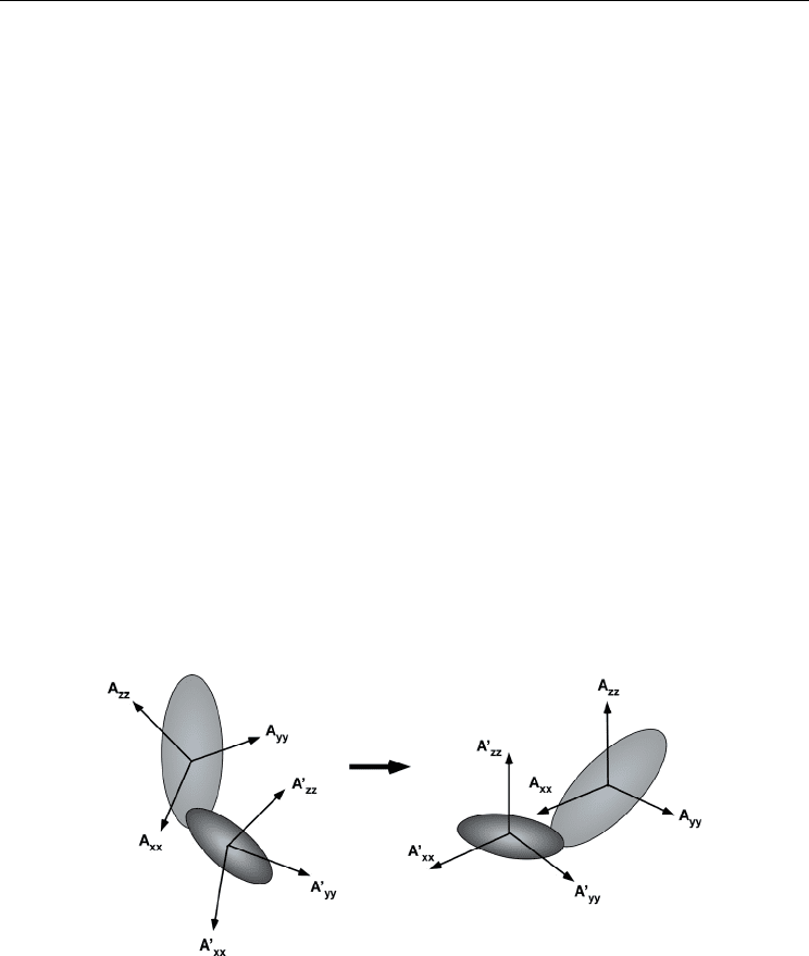
according to the above procedure. As schematically drawn in Fig. 8, the determined tensor
frames for each domain are used as a guide to define solution domain orientation; one
domain coordinate is rotated to make an overlay its tensor frame onto the other (Fig. 8). It is
noted here, the RDCs do not provide any distance information between the domains. If the
inter-domain segment has high flexibility, the additional distance restraints may be required
to build the entire structure, which should come from the other experiments like
paramagnetic relaxation enhancement (Clore and Schwieters 2002).
Alignment tensor determined by the RDCs has four possible orientations. Inversion around
each principal axis gives the same RDCs values. Therefore, the inversion is not discriminated
experimentally. To alleviate this ambiguity in orientation angle, additional alignment states
using different aligning media, including charged bicelle, or charged acrylamide gel, will be
used. In domain orientation analysis, however, the structural restrictions, which include the
length of the inter-domain linker or possible inter-domain steric clash, may allow to define one
inter-domain orientation even using a single aligning experiment.

Complementary Use of NMR to X-Ray Crystallography
for the Analysis of Protein Morphological Change in Solution
423
The alignment tensor magnitudes along each principal axis represent the extent of the
aligning order. If they differ between the domains, which may indicate that each rotates
differently to each other. The aligning orders give the insight into the domain dynamics in a
protein.
4.3 Significance of the domain orientation analysis by RDCs
The domain orientation analysis for protein in solution gives an invaluable outcome, even
its high-resolution structure is available. There are some cases to show the different domain
arrangements between solution and crystalline structures. The RDC analysis on maltose
binding protein (MBP) in the complex with
-cyclodextrin has shown that the relative
domain orientation in solution was different from that in the crystal (Skrynnikov et al.
2000b). This may indicate the crystal contact causes a subtle change in domain orientation.
Bacteriophage T4 lysozyme in solution was shown to have a more open conformation
relative to the crystal structure, which was also analyzed by the RDCs (Goto et al. 2001).
This observation appears compatible with steric requirements for the ligand bindings.
These examples illustrate how the RDC based domain analysis complements X-ray
crystallography in determining the relative domain orientation or protein morphology. The
complementary role of the RDC based analysis is considerably emphasized in exploring the
domain rearrangement upon binding to the other protein or ligand. Even if the complex
structure cannot be solved by X-ray, the complex structure is determined by the RDCs in a
solution state when the structure in a ligand-free form is available. This approach does not
require tedious and time-consuming NOE analysis as required in conventional NMR
structure determination, but just needs the backbone resonance assignments and a set of
IPAP-HSQC spectra. The structure determination is, therefore, much more efficient over the
conventional NMR structure determination.
Fig. 8. Schematic representation for the procedure to determine the relative domain orientation
based on the alignment tensors for each domain. Sets of RDC data determine the alignment
tensor for each domain, independently. The domain orientation of a protein is established by
rotating one domain coordinate to make an overlay of its tensor frame on the other.
4.4 Molecular size limitation in the RDC based approach
The domain orientation analysis with the RDCs is now recognized as a useful technique to
elucidate overall protein morphology in solution, complementing the X-ray structure

Current Trends in X-Ray Crystallography
424
analysis. This approach has, however, severe size limitation. Here, this obstacle in the RDC
application is discussed.
The size limitation comes from the rapid transverse relaxation rate of one of the split
components observed on a 2D IPAP-HSQC spectrum. The high-filed component shows
faster transverse relaxation rate than that of the other. This component has even faster
relaxation rate than that of HSQC counterpart. This is due to cross-correlated relaxation
interference to amide
15
N spin relaxation process; for the high-field component, the cross-
correlated relaxation process additively affects, while for the low-field component, the
interference reduces its relaxation rate. The transverse relaxation process of the HSQC signal
is free from the interference.
In measuring the RDCs with IPAP-HSQC for high molecular weight protein, the high-field
components of each amide spin pairs will broaden and severely reduce the signal intensities,
thus, they will not be observed. In particular, the difficulty in observing the high-field
component will be enhanced in an aligned state, due to the appearance of the residual
dipolar interactions as relaxation causes. For proteins over 20 kDa, it is usually hard to
observe the high-field component in an aligned state, thus making the RDC measurement
impossible. The RDC based domain orientation analysis with IPAP-HSQC is practically
limited up to around 20 kDa.
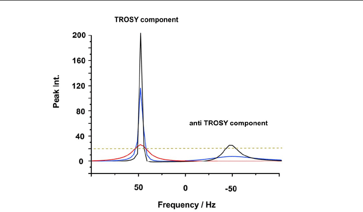
Simulation of the line broadening on each component of a double according to molecular
size is shown in Fig. 9. In this figure, the low-field component that shows a longer transverse
relaxation time and the other having a shorter transverse relaxation time are named as
TROSY and anti-TROSY, respectively. The slower transverse relaxation associated with the
low-field component is due to the mechanism used in TROSY (Transverse Relaxation
Optimized SpetroscopY). As demonstrated on the simulation, the anti-TROSY component
shows severe broadening even for the medium-size protein, 20 kDa.
The difference in line widths between the TROSY and anti-TROSY components will become
considerable for higher molecular weight proteins. As seen in the simulation, protein over
150 kDa gives severely broadened anti-TROSY signal, which already hard to observe.
Protein with 800 kDa never gives observable anti-TROSY signal. The size limitation in the
RDC based approach is clearly demonstrated in this simulation.
It should be noted, the TROSY component can retain observable signal intensity even for 800
kDa protein (Fig. 9). This motivated us to devise an approach to determine an alignment
tensor only using TROSY components.
4.5 Existing remedy for overcoming the size limitation in the RDC-based approach
Some remedies are proposed to overcome the size limitation the RDC application. They all
rely on the TROSY.
The difference in the transverse relaxation rates between the TROSY (low-field) and anti-
TROSY (high-field) components split along the
15
N axis are explained by the relaxation
interference. The same effect is active in the split signals along
1
H dimension. In observing
the
1
H-
15
N single bond correlation spectrum without decoupling during t
1
and also t
2
durations, each spin pair gives a quartet on a spectrum; split signals in both
1
H and
15
N
dimensions. The pure TROSY signal is the one having the longest transverse relaxation time
among the quartets. In using the protein labeled with
15
N and
2
H, where an unwanted
relaxation process is diminished by breaking the
1
H-
1
H dipolar interaction network in a
protein, TROSY effect is enhanced, and it allows
1
H-
15
N correlation spectrum for proteins
over 100 kDa (Pervushin et al. 1997).

Complementary Use of NMR to X-Ray Crystallography
for the Analysis of Protein Morphological Change in Solution
425
Fig. 9. Simulation of the molecular size dependency of the TROSY and anti-TROSY
components observed on a IPAP-HSQC spectrum. The data represent a slice peak along the
15
N axis. This simulation assumed a 750 MHz experiment. Black, blue, and red lines are the
simulated peaks for the sizes 20 kDa, 150 kDa, and 800 kDa, respectively. The dotted line is
assumed the expected noise level.
One proposed remedy is the combinatorial use of TROSY and HSQC. The difference
between TROSY and HSQC signals along the
15
N axis corresponds to a half of RDC. As
discussed above, the transverse relaxation rate of the HSQC signal is slower than that for the
anti-TROSY component in a IPAP-HSQC spectra. Therefore, the size limitation problem
should be alleviated by replacing anti-TROSY signal with HSQC counterpart. For a 81.4 kDa
protein, the transverse relaxation times for the TROSY, anti-TROSY, and HSQC signals are
reported to be 65 msec, 10 msec, and 30 msec, respectively (Tugarinov and Kay 2003). In
considering the difference between the transverse relaxation times between TROSY and
HSQC signals, the combinatorial use does not fully solve the problem, but just alleviates it.
Another remedy is the use of J-scaled TROSY, which is also referred to as J-enhanced (JE)
TROSY (Kontaxis, Clore and Bax 2000, Bhattacharya, Revington and Zuiderweg 2010). In
this experiment, short J-evolution step is added in the standard TROSY, which induces J-
dependent shift change from the standard TROSY shift. In the standard TROSY experiment,
the shift difference between the signals along the
15
N axis on the same
1
H chemical shift
corresponds to
1
NH
J
, whilst in the J-scaled TROSY, this shift difference is changed according
to the additional duration for J-evolution. From the magnitude of the shift change induced
by the applied J-evolution period, the apparent
1
NH
J
coupling value is estimated. If the J-
evolution period is applied to allow full recovery of
1
NH
J
coupling, the observed signal
position should be coincident with that of the HSQC signal. Usually, to gain the signal
intensity for the observed signal on J-scaled TROSY, rather limited evolution time is set. In
this J-evolution step, the coherences for TROSY and anti-TROSY are mixed; the equivalent

Current Trends in X-Ray Crystallography
426
mixing of the two gives the coherence for observed as HSQC signal. The more increased the
contribution of the anti-TROSY coherence to the observed signal leads to more broadened
signals observed. Therefore, in the J-scaled TROSY experiment, partial recovery of the J-
modulation is used to maintain the signal intensities on the J-scaled TROSY spectrum in the
observable level.
Signals observed on a J-scaled TROSY spectrum have longer transverse relaxation times
than those of the signals on a HSQC spectrum. Their transverse relaxation times, however,
are still shorter than those for real TROSY counterparts. The combined use of TROSY and J-
scaled TROSY is indeed advantageous over the TROSY/HSQC combination. In determining
more accurate RDCs, J-scaled TROSY requires more extent of the mixing of the anti-TROSY
coherences, which will result in the lesser sensitive J-scaled TROSY signals. The use of J-
scaled TROSY is not the complete remedy for the problem we concern.
In spite of the limitations in the existing approaches, they expanded the RDC application up
to 50 kDa protein (Jain, Noble and Prestegard 2003). However, it is also reported that the
rapid transverse relaxation of the non-TROSY component is already an obstacle in
measuring the RDCs for 81.4 kDa protein . The further expansion of the application limit is
expected, and our DIORITE is one of the possible methods used for this purpose.
5. Alignment tensor determination using only TROSY
As discussed above, the molecular size limitation problem in the RDC based domain
orientation analysis is not completely overcome, although the existing approaches have
given some successful results. Most of the biologically interesting multi domain proteins
tend to be over 100 kDa. The existing approaches are not thought to be applied to such
higher molecular weight protein. This is because they do not take fully advantages of
TROSY spectroscopy, which allows the longest transverse relaxation time for the observed
signals. In contrast to the existing approaches, our approach, DIOIRTE, uses only TROSY
spectra, where the signals having the longest transverse relaxation times are used. This may
give considerable advantages over the existing methods in respect to the size limitation
problem. In this section, we will describe the theoretical aspects of the TROSY based
alignment tensor determination, which will allow the domain orientation analysis for higher
molecular weight proteins ever.
5.1 Alignment induced TROSY shift changes
TROSY shift is changed when protein is transferred from isotropic to anisotropic states. This
TROSY shift change along the
15
N axis contains the effects of two anisotropic spin
interaction observed on a peptide plane;
1
H-
15
N dipolar interaction and
15
N CSA. As
depicted in Fig. 10, this alignment induced TROSY shift change,
TROSY
, contains a half of
RDC and the full RCSA effects. In a Cartesian representation using the Saupe order matrix,
the following relation should fold:
0
23
4
1
24 5
2
,,
2
3
,, ,,
0
12
23
cos( ) cos( )
cos( ) cos( )
{cos()cos()}
ij
ij
h
TROSY kl k l
r
kl x y z
kl kj lj jj
kl xyzj xyz
lk NH k l kl
kl
S
S
SD
(9)
