Chandrasekaran A. (ed.) Current Trends in X-Ray Crystallography
Подождите немного. Документ загружается.


Protein-Noble Gas Interactions Investigated by Crystallography
on Three Enzymes - Implication on Anesthesia and Neuroprotection Mechanisms
287
(David et al., 2010) while nitrous oxide reduces ischemic brain damage but increases tPA-
induced brain hemorrages (Haelewyn et al., 2011).
Xenon is thus a very promising neuroprotective drug with few or no adverse side effects in
models of acute ischemic stroke or perinatal hypoxia-ischemia (Homi et al., 2003; Ma et al.,
2003; Abraini et al., 2005; David et al., 2008; Luo et al., 2008). Despite this, the widespread
clinical use of xenon is limited by its scarceness and excessive cost of production, even if
close xenon delivery systems are now being developed.
Using a mixture of xenon and another anesthetic gas like nitrous oxide (Marassio et al.,
2011), argon (David et al., submitted), or helium (David et al., 2009) could combine the
efficiency of xenon and the low cost and availability of the second gas and is thought to be a
cost-efficient strategy.
Argon is an inert gas which is easily available and has no narcotic nor anesthetic action at
ambient pressure. It presents some mild to moderate neuroprotective properties (David et
al., submitted). Argon, contrary to xenon and nitrous oxide, may act directly by potentiating
GABA neurotransmission at the GABA
A
receptor (Abraini et al., 2003).
Krypton is significantly less potent as an anesthetic agent than xenon, consistently with the
Meyer-Overton rule which shows that krypton anesthetic potency is four fold less than
xenon potency (Cullen et al., 1951; Kennedy et al., 1992).
Xenon, which has the highest solubility in lipids, also has the highest anesthetic potency
(i.e. the lowest MAC-immobility) compared to krypton and argon (Table 1). Xenon also
has the highest polarizability due to its high number of electron, compared to krypton and
argon, so is predicted to be the gas which interact the most with proteins (Quillin et al.,
2000).
Gas Number
of
electrons
Polarizability
(Å
3
)
van der
Waals
radius (Å)
Solubility
in lipids
MAC-
immobility
(bar)
Ar 18 1.64 1.91 0.14 27
Kr 36 2.48 2.03 0.43 7.31
Xe 54 4.04 2.21 1.17 1.61
Table 1. Physical and anesthetic properties of argon, krypton and xenon (from (Koblin et al.,
1998; Quillin et al., 2000; Ruzicka et al., 2007)).
2. Determination of crystallographic structures of proteins under inert gases
pressure
To investigate the mechanism of interaction of gases with proteins, a structural approach
using protein crystallography under gas pressure was developed. Xenon binds reversibly to
proteins through non-covalent, weak energy van der Waals forces (Ewing et al., 1970). The
first structures of protein – xenon complexes were solved in 1965 with myoglobin and
haemoglobin under a xenon pressure of 2.5 bar, evidencing a xenon binding site in these
two globins (Schoenborn, 1965; Schoenborn et al., 1965). At a pressure of 7 bar, four xenon
binding sites were found in myoglobin indicating that the number of xenon binding sites
rises with pressure (Tilton et al., 1984).
Since then, many structures of protein-xenon complexes were solved, with xenon used as a
heavy atom in isomorphous replacement phasing method (MIR), because xenon has a high
Current Trends in X-Ray Crystallography
288
number of electrons (54 e
-
) and binds with very little perturbation of the protein structure
(Vitali et al., 1991; Schiltz et al., 1994; Bourguet et al., 1995; Colloc'h et al., 1997). On the other
hand, krypton, though lighter than xenon, was popularized as an internal reference in
anomalous phasing techniques MAD or SAD (Schiltz et al., 1997; Cohen et al., 2001) thanks
to its absoption K edge at a convenient and useful wavelength easy to tune at all
synchrotron places; for a review, see (Schiltz et al., 2003).
Xenon and other noble gas binds primarily in pre-existing hydrophobic cavities or pockets,
very often empty in the native gas-less structures (Prangé et al., 1998). Xenon diffusing
through protein atoms reaches easily its completely buried binding sites. Xenon was also
used as an oxygen probe, based on the hypothesis that xenon and dioxygen would have
equivalent binding sites (Duff et al., 2004). The comparison of the binding mode of xenon,
krypton and argon was done on the phage T4 lysozyme, showing that gas occupancy rises
with gas size and polarizability (Xe > Kr > Ar) (Quillin et al., 2000).
X-ray diffraction data of a protein under xenon pressure are collected either at liquid
nitrogen temperature (100 K) or at room temperature. In the first case, the crystal inserted in
a cryo-loop is placed in a xenon pressure chamber for a given time, then immediately after
frozen in liquid nitrogen, to minimize the amount of xenon which could escape the protein
crystal. The determination of the gas pressure within the crystal is thus quite imprecise. For
the present study which needs the determination of protein structures under a large range
of gas pressures, we have used a pressurisation cell in capillary, designed and developed for
the preparation of isomorphous xenon derivatives (Schiltz et al., 1994; Schiltz et al., 2003).
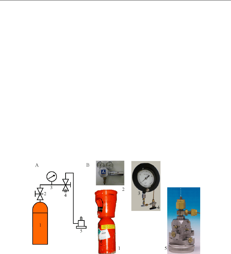
Fig. 1. The pressurisation cell setting. A. Connection between the five elements shown in B.
1- Xenon bottle, 2- Gas regulator, 3- High precision gauge, 4- Bleeding valve, 5-
Pressurisation cell.
Typically, a crystal of protein is placed inside a quartz capillary mounted on the
pressurisation cell. The pressurisation cell is fixed on a standard goniometer head, and
connected to a gas bottle. The pressure within the cell is determined precisely with a
calibrated Ashcroft precision gauge (Figure 1). The pressure is maintained constant during
all the data collection.
For the present study, we have investigated three different enzymes, urate oxidase, elastase
and lysozyme in complex with three gases, xenon, krypton and argon. In urate oxidase,

Protein-Noble Gas Interactions Investigated by Crystallography
on Three Enzymes - Implication on Anesthesia and Neuroprotection Mechanisms
289
xenon binds primarily in a large buried hydrophobic cavity close to the active site (Colloc'h
et al., 2007; Marassio et al., 2011). Xenon was used as an isomorphous derivative during the
determination of urate oxidase structure (Colloc'h et al., 1997). In elastase, like in most of the
serine proteases, xenon binds within the specificity pocket S1 of the active site (Schiltz et al.,
1995). In lysozyme, xenon binds weakly in an internal cavity and mainly in a pocket located
at a crystallographic interface (Schiltz et al., 1997; Prangé et al., 1998).
One of the drawbacks of using X-ray crystallography is the requirement to have a high gas
pressure to be able to observe it in the electron density map. A gas pressure about 5 to 10
fold the physiological concentration is estimated to correspond to physiological conditions
(Miller, 2002). In the present study, gas pressure ranges from 1 to 40 bar in order to reach a
maximum occupancy at saturation, however, only the data between 5 and 10 bar can be
compared to physiological conditions.
In the present study, diffraction data were collected at room temperature at the BM14, BM16
and BM30A beamlines at the European Synchrotron Radiation Facility (Grenoble, France).
Detectors used were a MAR CCD detector for BM14, an ADSC Q210r CCD detector for
BM16 and an ADSC Q315r CCD detector for BM30A. Data were indexed and integrated by
DENZO and scaled independently and reduced using SCALEPACK, both programs from the
HKL package (Otwinowski et al., 1997) or indexed and integrated by MOSFLM (Leslie, 2006)
or XDS (Kabsch, 2010) and scaled by SCALA; intensities were converted in structure factor
amplitudes and put on absolute scale using TRUNCATE and structure refinements were
carried out by REFMAC (Murshudov et al., 1997), all programs from the CCP4 package
(Collaborative Computational Project, 1994). The graphics program COOT (Emsley et al.,
2004) was used to visualize |2Fobs – Fcalc| and |Fobs – Fcalc| electron density maps and for
manual rebuilding. Cavity volume were calculated with the program CastP (Dundas et al.,
2006) with a probe radius of 1.3 Å. Structural figures were prepared using PyMol (deLano
W.L., DeLano Scientific, Palo Alto, CA, USA).
3. Structure of urate oxidase under inert gas pressure
3.1 Structure of urate oxidase under pressure of xenon and nitrous oxide and
comparison with in-vivo pharmacology effects
Aspergillus flavus urate oxidase (EC 1.7.3.3) is a homotetrameric enzyme of 301 residues
per subunit which is involved in the oxidation of uric acid in presence of molecular
oxygen. It crystallizes in the orthorhombic space group I222 with one monomer per
asymmetric unit (cell: a = 79.8 Å, b = 96.2 Å, c = 105.4 Å, = = = 90°). X-ray structures
of urate oxidase under various pressures of xenon and nitrous oxide have been
determined. Both gases were bound mainly in an internal cavity close to the active site of
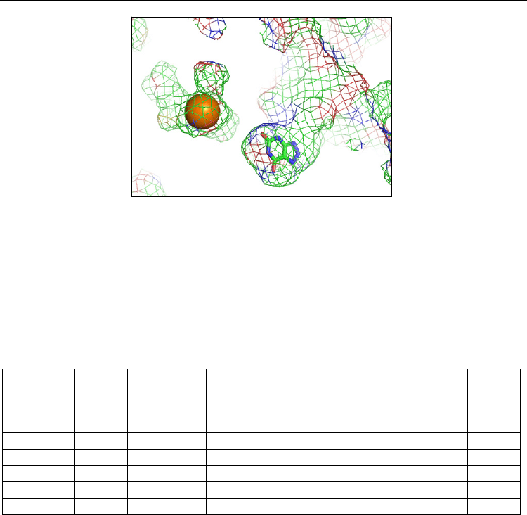
the enzyme, this cavity being empty in the native gas-less structure (Figure 2). This cavity,
completely buried within the monomer, is highly hydrophobic, with 86 % of the atoms
lining the cavity being carbons. Both gases were bound also very weakly to a second
location, a small extension of a solvent-accessible pocket quite hydrophobic (lined by 75 %
carbons). The gas occupancy in this second binding site remained very low (less than 30 %
at 30 bar of pressure). Gas occupancies in the main binding site were high, reaching
saturation at 100 % for xenon and 60 % for nitrous oxide (Table 2). The main effect of the
gas was to expand the volume of the cavity where it binds. This expansion increased with
gas occupancy and hence with gas pressure.
Current Trends in X-Ray Crystallography
290
Fig. 2. Hydrophobic cavity in urate oxidase where xenon is bound (shown as an orange
sphere). This cavity is close to the active site, where the competitive inhibitor 8-azaxanthine
is located (colored in stick by atomic type). The solvent-accessible surface is shown in mesh
representation colored by atomic type.
The ratio of gas-induced expansion of the hydrophobic cavity volume for xenon and nitrous
oxide in urate oxidase, which could be considered as a model of globular proteins where
inert gases bind and whose activity is disrupted by their presence, ranged between 1.1 and
1.5, depending on the applied pressure (Table 2). For the pressures estimated to correspond
to physiological conditions (i.e. 5-10 bar), this ratio ranged between 1.3 and 1.5.
Xenon
pressure
(bar)
Xe
occ.
(%)
Main
Xe-
induced
expansion
N
2
O
occ.
(%)
main
N
2
O-
induced
expansion
(%)
Ratio
Xe/N
2
O-
induced
expansion
Xe
occ.
(%)
2nd
N
2
O
occ.
(%)
2nd
5 18 10.8 0 8.5 1.3 0 0
10 60 18.8 40 12.4 1.5 10 0
15 100 19.5 50 17.8 1.1 20 0
20 100 23.1 60 18.4 1.3 22 0
30 100 23.2 60 20.1 1.2 27 25
Table 2. Gas pressure, xenon and nitrous oxide occupancies in the main binding site, gas-
induced expansion of the main gas binding site, ratio of expansion, and xenon and nitrous
oxide occupancies in the secondary binding site.
If we compared these data with in-vivo pharmacology studies, we noticed that this ratio
corresponded to the ratio of the narcotic potency of xenon compared to nitrous oxide (about
1.38) as estimated by the concentration of gas necessary to induce loss of righting reflex in
rodents (Koblin et al., 1998; David et al., 2003), considered to be a behavioural endpoint
closely related to MAC-awake (Campagna et al., 2003).
In comparison, the ratio of gas-induced volume expansion for xenon and nitrous oxide in
annexin V, a protein which could be considered as a prototype of NMDA receptor for its
properties of ion selectivity and voltage gating (Demange et al., 1994), did not correspond to
the ratio of anesthetic potency of xenon and nitrous oxide. However, when considering
urate oxidase and annexin V together as a model of simultaneous occupancy of globular
proteins and ion-channel receptors, the ratio of gas-induced expansion for xenon and
Xenon
8-azaxanthine

Protein-Noble Gas Interactions Investigated by Crystallography
on Three Enzymes - Implication on Anesthesia and Neuroprotection Mechanisms
291
nitrous oxide was close to 1, a value that corresponded to the ratio of anesthetic potency of
xenon compared to nitrous oxide, as assessed by their MAC-immobility (Russell et al., 1992;
Koblin et al., 1998).
These relationships between gas-induced structural effect and gas-induced narcotic effect
allowed proposing a step-by-step mechanism of anesthesia. Gas would first bind to globular
cytosolic or extracellular proteins which possess suitable gas binding site easily accessible.
Gas-induced disruption of their function would lead to the early stages of anesthesia, i.e.
amnesia and hypnosis. When all easily accessible gas binding sites are occupied, gas would
then bind to neuronal channel which possess smaller gas binding sites, the disruption of
their function would lead to surgical anesthesia, i.e. deep sedation and lack of responses to
noxious stimuli (Colloc'h et al., 2007).
3.2 Structure of urate oxidase under pressure of xenon and nitrous oxide and
comparison with in-vitro activity assays
As mentioned above, the main gas binding site in urate oxidase is very close to the active
site (Figure 2). To investigate if gas occupancy and gas-induced volume expansion may have
some functional relevance, we performed activity assays on urate oxidase in presence either
of air, and either of a mixture of 75 vol % xenon or nitrous oxide and 25 vol % oxygen. To
evaluate the alternative therapeutic strategy of using a mixture of xenon and nitrous oxide
to combine the efficiency of xenon and the low cost and availability of nitrous oxide, we
have also determined the structure of urate oxidase with various pressure of an equimolar
mixture of xenon and nitrous oxide, and we have performed an activity assay in presence of
a mixture of 37.5 vol% xenon, 37.5 vol% N
2
O and 25 vol% oxygen. We found that Xe:N
2
O
induced a higher expansion of the cavity volume than pure xenon, which in turn induced a
higher expansion than N
2
O as seen above. In-vitro activity assays revealed that Xe:N
2
O-
induced inhibition was higher than Xe-induced inhibition, itself higher than N
2
O-induced
inhibition. The relationship between structural effect of the gas, i.e. gas-induced volume
expansion, and the functional effect of the gas, i.e. gas-induced inhibition of the enzymatic
reaction, highlighted the way by which gases might disrupt protein function through an
indirect mechanism (Marassio et al., 2011).
The role of the void hydrophobic cavity in the catalytic mechanism was thus demonstrated
by the activity assays in presence of gas. This functional role was also suggested by the high-
pressure structural and functional study of urate oxidase. Under high hydrostatic pressure
(150 Ma; 1500 bar) the volume of the cavity was reduced as expected, while the volume of
the active site was expanded. High pressure also inhibited the catalytic mechanism of urate
oxidase, this loss of activity being a loss of substrate affinity (Girard et al., 2010).
In both cases (gas or pressure), there was a loss of flexibility of the cavity, either by the gas
presence which induced an expansion and inhibited its contraction, either by high pressure
which induced a cavity contraction but inhibited its expansion. The role of the cavity in the
functional mechanism of urate oxidase seems then to give some flexibility to the active site
to allow a structural fit for the ligand in the active site.
3.3 Structure of urate oxidase under pressure of krypton and comparison with gas
solubility in lipids
Structures of urate oxidase under krypton pressure of 2 to 30 bar were determined in the
present study. Krypton was bound to the exact same location than xenon, in the

Current Trends in X-Ray Crystallography
292
hydrophobic cavity close to the active site. Krypton occupancy increased with the applied
pressure up to 45 % at 30 bar (Table 3). Like xenon, krypton was also weakly bound to a
secondary binding site at the bottom of a solvent-accessible pocket, but only at pressure
above 20 bar.
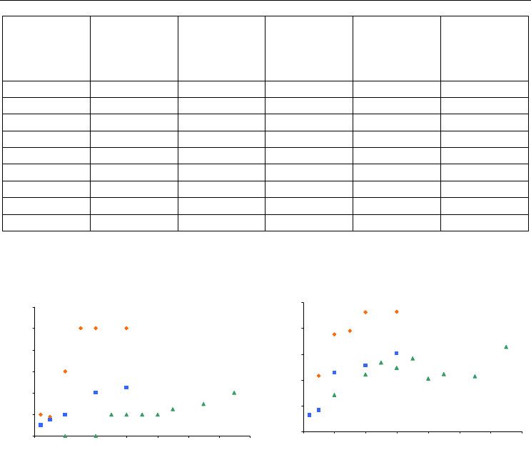
For an identical pressure, krypton occupancy was always lower than xenon occupancy
(Figure 3A). Xenon which has a higher number of electrons than krypton has a higher
polarizability (Table 1) and binds thus with a higher occupancy, as already observed in the
case of phage T4 lysozyme (Quillin et al., 2000).
Krypton
pressure
(bar)
Resolution
(Å)
Occ. in
main
binding
site (%)
Kr-
induced
expansion
(%)
Xe-
induced
expansion
(%)
Ratio
Xe / Kr
induced
expansion
Occ. in
2nd
binding
site (%)
2 1.60 10 3.2 3.3 1.0 0
5 1.60 15 4.1 10.8 2.6 0
10 1.55 20 11.4 18.8 1.6 0
20 1.65 40 12.8 23.1 1.8 10
30 1.65 45 15.2 23.2 1.5 15
Table 3. Krypton pressure, resolution of the crystallographic structure, krypton occupancy
in the main binding site, krypton-induced and xenon-induced volume expansion of the
main binding site, ratio of the Xe and Kr-induced volume expansion and krypton occupancy
in the secondary binding site.
If one refers to the Meyer-Overton rule, the narcotic potency of a gas would be related to its
solubility in lipids. The ratio of solubility in lipids of xenon compared to krypton is 1.17 /
0.14 = 2.7 (Table 1), a value which correspond to the ratio of Xe and Kr-induced volume
expansion at the pressure of 5 bar, well within the range of pressure estimated to
correspond to physiological condition. This result confirmed what was shown previously
when comparing the structural-induced effect of xenon and nitrous oxide on urate oxidase
to their in-vivo effect as evaluated by their MAC-awake (Colloc'h et al., 2007). However, the
MAC-immobility which prevents response to noxious stimuli for xenon in man is about 4.5
higher than the MAC-immobility of krypton (Table 1), which does not correspond to the
structural Xe- and Kr-induced structural effect in urate oxidase, considered as a model for
globular protein whose function is disrupted by the presence of gas.
3.4 Structure of urate oxidase under pressure of argon and comparison with in-vivo
pharmacology study
Structures of urate oxidase under argon pressure of 10 to 65 bar were determined in the
present study. Argon was bound to the exact same location than xenon and krypton, in the
large hydrophobic cavity close to the active site. Argon became visible in the electron
density map at a pressure of 30 bar and above, with an occupancy factor of 40 % at a
pressure of 65 bar (Table 4). 65 bar is the maximum pressure which could be reach in the
quartz capillary; above that pressure, the risk of breakage of the capillary became very high.
At the same pressure, argon occupancy was always lower than krypton and xenon
occupancies (Figure 3A). In the secondary binding site where xenon and krypton bind very
weakly, no argon is detectable in the electron density map, even at a pressure of 65 bar.
Protein-Noble Gas Interactions Investigated by Crystallography
on Three Enzymes - Implication on Anesthesia and Neuroprotection Mechanisms
293
Argon
pressure
(bar)
Resolution
(Å)
Occ. in main
binding site
(%)
Ar-induced
expansion
(%)
Xe-
induced
expansion
(%)
Ratio
Xe / Ar
Indiced
expansion
10 1.65 0 7.1 20.7 2.9
20 1.90 0 11.0 24.6 2.2
25 1.60 20 13.4
30 1.60 20 12.3 24.6 2
35 1.60 20 14.1
40 1.75 20 10.3
45 1.60 25 11.1
55 1.60 30 10.7
65 1.60 40 16.4
Table 4. Argon pressure, resolution of the crystallographic structure, argon occupancy,
argon-induced and xenon-induced cavity volume expansion, and ratio of the Xe and Ar-
induced volume expansion.
Fig. 3. A. Gas occupancy as a function of pressure. B. Gas-induced expansion of the main
binding site as a function of pressure.
As for the two other nobles gas, the main effect of argon was to expand the volume of the
cavity where it was bound. However, due to its quite low occupancy factor and its small
size, the argon-induced expansion remained low, around 10 % of expansion except for the
pressure of 65 bar where expansion reached 16 % (Table 4). For pressure of 10 and 20 bar
where argon was not detectable in the electron density map, there was already a volume
expansion indicating with little doubt the presence of argon within the cavity (Table 4,
Figure 3B).
The ratio of Xe- and Ar-induced expansions of the cavity volume was 2.9 for a pressure of 10
bar, which did not correspond to their inverse ratio of MAC-immobility (27/1.61 = 16.8) nor
their ratio of solubility in lipids (1.17 / 0.14 = 8.4) (Table 1). However, argon is not narcotic
at ambient pressure and needs to be pressurised to have some narcotic action.
In order to allow comparison of the effects of argon and xenon in urate oxidase, we further
calculated the theoretical expansion of the gas binding cavity produced by argon at 100 %
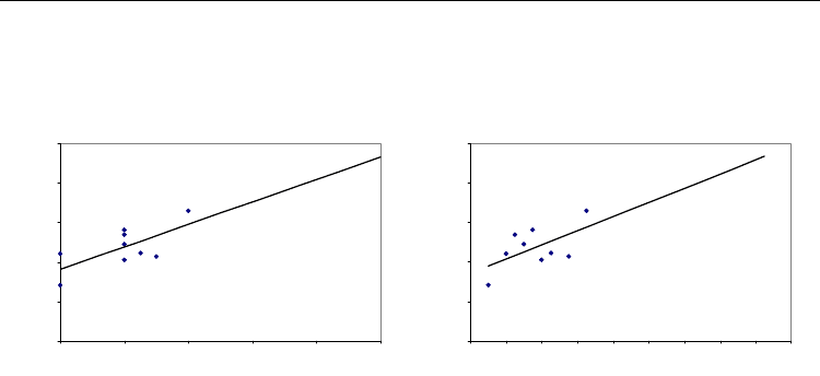
occupancy according to a linear regression model. For occupancy of 100% of argon, the
corresponding volume expansion would be of 23.3 % (Figure 4A). According to a linear
0
20
40
60
80
100
120
0 10203040506070
Pr e ssu re (b a r)
Occupancy (%)
0
5
10
15
20
25
0 10203040506070
Pr e ssu re (b a r)
Expansion (%)
Xe
Kr
Ar
Xe
Kr
Ar
Current Trends in X-Ray Crystallography
294
regression model, this expansion would be reach for a pressure of 164 bar (Figure 4B). This
pressure corresponds to about ten fold the pressure of 14 to 17 bar at which argon is known
to produces narcosis in rodents (Abraini et al., 1998; Koblin et al., 1998).
Fig. 4. Linear regression model for cavity volume expansion as a function of occupancy (A)
and as a function of pressure (B).
In addition, it should be mentioned that these estimated values for argon were also
consistent with crystallographic data that have demonstrated that xenon at full occupancy
(pressure of about 20 bar) produces a similar maximal expansion around 23-25 % of the gas
binding site (Table 2). This is consistent with the fact that the ratio between the efficient
estimated pressure of argon and the efficient experimental pressure of xenon at producing
full occupancy and maximal expansion of the gas binding site (164 / 20 = 8.2) is similar to
the ratio of their solubility in lipids (1.17 / 0.14 = 8.4) as predicted by the Meyer-Overton
rule (Abraini et al., 2003; Campagna et al., 2003).
Argon is narcotic only in hyperbaric condition. At ambient pressure, argon may thus have a
very limited influence on its target function. Since one of the major effect of hydrostatic
pressure is to contract the volume of internal cavities (Girard et al., 2010), it may explain
why argon needs hyperbaric condition to exert its influence.
3.5 Conclusion on urate oxidase structures under inert gas pressure
The three noble gases were bound to an identical location in urate oxidase, within an
internal hydrophobic cavity. The gas occupancies increased in the sequence argon < krypton
< xenon, as it was the case for T4 lysozyme (Quillin et al., 2000), who noticed that smaller
gases do not bind as well as larger ones as a result of their attenuated polarizability. Xenon
and krypton were bound also weakly in a secondary binding site, while argon was not
observed even at high pressure.
The main effect of the gas was to expand the cavity volume where it binds. The ratio of
expansion was related to the narcotic potency of the gas, as evaluated by their MAC-awake
or their solubility in lipids. The presence of xenon within the cavity induced an inhibition of
the catalytic mechanism, with a relationship between gas-induced expansion and gas-
induced inhibition, as shown by the comparison between xenon and nitrous oxide structural
and functional effects. No activity assays were performed in presence of krypton or argon,
but we can predict, based on the present structural results, that krypton should induce an
inhibition of the catalytic mechanism of urate oxidase. However, krypton-induced inhibition
should be lower than xenon-induced inhibition, according to their relative induced
0,00
5,00
10,00
15,00
20,00
25,00
0 20406080100
Occupancy (%)
Expansion (%)
0,00
5,00
10,00
15,00
20,00
25,00
0 20 40 60 80 100 120 140 160 180
P re ssu re
(
bar
)
Expansion (%)
Protein-Noble Gas Interactions Investigated by Crystallography
on Three Enzymes - Implication on Anesthesia and Neuroprotection Mechanisms
295
expansion in the range 5-10 bar (Table 3). Argon-induced inhibition should be very low or
inexistent, according to the very low gas occupancy and argon-induced expansion at a
pressure of 10 bar (Table 4).
4. Structure of elastase under inert gas pressure
4.1 Introduction on elastase structures under inert gas pressure
Pancreatic porcine elastase (EC 3.4.21.36) is a serine protease of 266 residues which hydrolyzes
peptide bonds in proteins, its main substrate being elastine. The catalytic triad of elastase is
composed, as for all serine proteases, of an activated serine (Ser 195) assisted by a proton relay
(His 57), which acts as a general base, and stabilized through an hydrogen bond by an aspartic
acid (Asp 102). Pancreatic porcine elastase crystallizes in the orthorhombic space group P2
1
2
1
2
1
with one monomeric enzyme per asymmetric unit (cell : a = 51.4 Å, b = 58.0 Å, c = 75.3 Å, =
= = 90°). In the crystallographic structure, a sulphate or an acetate ion is bound in the
oxyanion hole, depending on the concentration of the precipitating agents. The primary
specificity pocket S1 is a hydrophobic pocket located below the oxyanion hole and the Ser 195
which is specific for recognition of the peptidic substrate.
In the crystallographic structures of apo elastase, the S1 pocket was either empty, either
filled by a water molecule hydrogen-bonded to a water molecule outside of the S1 pocket
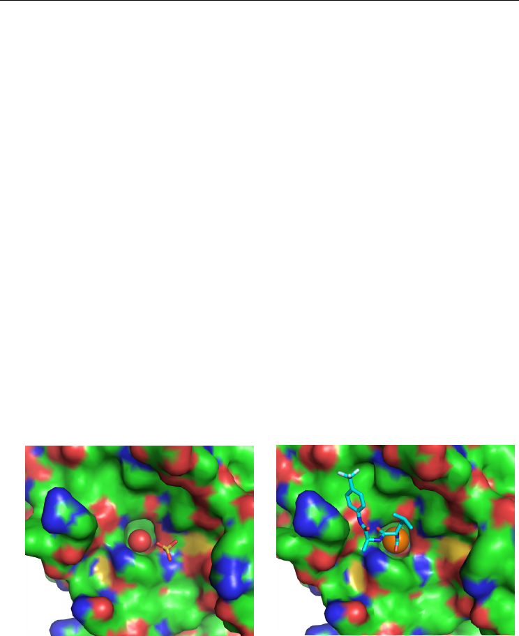
and to an oxygen atom of the sulphate ion (Figure 5A). When this water molecule (termed
W-S1) was present, its B-factor was quite elevated. In the different crystallographic
structures of elestase in complex with xenon deposited in the Protein Data Bank, 1C1M
(Schiltz et al., 1995), 1L1G and 1L0Z (Panjikar et al., 2002), 1UO6 and 2A7C (Mueller-
Dieckmann et al., 2004) and 2OQU (Kim et al., 2007), xenon was bound within the specificity
pocket S1 in the active site of elastase (Figure 5B). This xenon binding site is moderately
hydrophobic, lined with 60% carbons.
Fig. 5. Elastase shown with its solvent-accessible surface colored by atomic type. A. Native
gas-less elastase with the water molecule W-S1 in the S1 pocket. B. Elastase in complex with
xenon (shown as an orange sphere) or in complex with a peptidic inhibitor TFLA (shown in
cyan in stick representation).
In the present study, structures of native elastase (gas-less) were determined in the same
conditions than structures under inert gas pressure, i.e. at room temperature in a quartz
A
B
W
-
S1
Xe
TFLA

Current Trends in X-Ray Crystallography
296
capillary. Three structures have been solved, at 1.38 Å, 1.7 Å and 1.45 Å resolution. In two
native structures, the S1 pocket was empty while in one of them, there was the water
molecule W-S1 with a B-factor of 36.3 Å
2
.
4.2 Structure of elastase under pressure of xenon and comparison with in-vitro
activity assays
Structures of elastase under xenon pressure from 1 bar to 30 bar were determined in the
present study. Whatever the applied pressure, xenon was bound to a unique site, within the
specificity pocket S1, with an occupancy which increased with the applied pressure.
Occupancy reached 100 % for a pressure of 30 bar (Table 5). The S1 pocket where xenon
binds is moderately hydrophobic, with 60 % of atoms lining it being carbons. This gas
binding site is less hydrophobic than the main gas binding site in urate oxidase, which was
lined by 86 % carbons. The atom the closest to xenon is the side chain atom O of the
catalytic Ser 195.
Xenon
pressure
(bar)
Resolution
(Å)
Occ. (%) Xe-induced
expansion
(%)
1 1.40 15 3.0
2 1.45 25 3.3
5 1.50 30 7.8
10 1.50 70 9.4
20 1.60 90 12.7
30 1.65 100 13.6
Table 5. Xenon pressure, resolution of the crystallographic structure, occupancy and xenon-
induced expansion of the S1 pocket in elastase.
Whatever the pressure, there was no water molecules in the S1 pocket, so if the water
molecule W-S1 was present in the native gas-less structure, it was not displaced but
replaced by xenon. However, xenon did not take the exact location of the W-S1 and was
closer to the O of the catalytic Ser 195 (3.4 Å instead of 3.8 Å).
The presence of xenon within the S1 pocket expanded its volume, its expansion rising with
the applied pressure. However, since the gas was bound directly within the active site, the
gas-induced inhibition is likely to be a direct inhibition and the expansion by itself has
probably no functional relevance. Xenon took indeed the place of peptidic inhibitors, like
the trifluroacetyl-leu-ala (TFLA) known to be an excellent inhibitor of elastase (Li de la
Sierra et al., 1990) (Figure 5B).
To investigate the direct inhibition by xenon, we performed activity assays on elastase in
presence either of air, either of 100 vol % xenon. Initial velocity in presence of xenon when
compared to air (taken as 100 %) was 81.5 + 2.1 % revealing an inhibition of the catalytic
activity of elastase of around 20 % by xenon. However, this inhibition was lower than xenon
occupation in the range 5-10 bar (30 – 70 % occupation).
Tissue-type plasminogen activator (tPA), the only approved treatment for thrombolysis
after an ischemic stroke, is also a serine protease. As in the case of elastase, xenon inhibited
tPA enzymatic activity (David et al., 2010). This inhibition is likely to be a direct inhibition
with xenon binding directly in the S1 pocket in the active site of tPA. Serine proteases have
