Brigatti M.F., Galan E. and Theng B.K.G. Structures and mineralogy of clay minerals
Подождите немного. Документ загружается.


Preisinger (1963) reported an orthorhombic model for palygor skite similar to the
orthorhombic sepiolite of Brauner and Preisinger (1956) except for the ribbon width.
Christ et al. (1969) studied five palygorskite samples by XRD and found three
orthorhombic (Pn) and two different monoclinic cells. Although there are not suffi-
cient data to define exactly the difference between them, it is clear that at least two
symmetries are possible for palygorskite, one orthorhombic and another monoclinic.
Monoclinic structures have an n-glide plane parallel to (1 0 0) (Table 2.2). One of the
monoclinic symmetries is similar to the one proposed by Zvyagin et al. (1963) (P2/a,
b ¼ 95.831). The other, with the Z-axis as the monoclinic axis and g ¼ 92.231 for the
monoclinic angle, has no structural interpretation up to the present time.
More recently, Chisholm (1992) analysed the structural models given by Christ et
al. (1969) and other authors, and found two palygorskite structures, one ortho-
rhombic (PbmnPnmb) and another monoclinic (C2/mA/2 m). Most palygorskite
samples appear to contain both forms. There are samples of pure or nearly pure
monoclinic palygorskite but there is no pure orthorhombic palygorskite. The co-
existence of both structures lies behind the confusion that arises from indexing XRD
patterns in terms of a single phase, and using different unit cell s and space groups for
palygorskite. The two structures determined by Chisholm (1992) agree with those
Table 2.3. Octahedral and tetrahedral occupancy ranges for bulk and EDX analyses of se-
piolite and palygorskite (in bracket mean value) (Gala
´
n and Carretero, 1999).
Bulk
analyses
EDX analyses
by (Paquet et
al., 1987)
Other EDX
analyses from
literature
EDX analyses
by (Gala
´
n and
Carretero, 1999)
Sepiolite
VI
R 6.95–8.11 6.93–8.5 7.61–7.87 7.93–7.98
(7.72) (7.74) (7.95)
VI
Mg 4.96–8.1 5.6–8.5 6.05–7.73 7.93–7.98
(7.36) (7.11) (7.95)
VI
(R
2
+R
3
) 0–2.28 0–1.8 0–1.8 0
(0.32) (0.62)
IV
(Al+Fe
3+
) 0–0.72 — 0–0.2 (0.10) 0
(0.19)
Palygorskite
VI
R 3.45–4.33 2.63–4.63 3.36–4.17 3.95–4.09
(3.96) (3.88) (4.00)
VI
Mg 1.12–2.82 0.83–3.08 1.32–2.60 1.71–2.10
(2.00) (1.97) (1.96)
VI
(R
2
+R
3
) 1.12–2.50 1.5–2.66 1.46–2.41 1.87–2.24
(1.96) (1.91) (2.04)
IV
(Al+Fe
3+
) 0–0.67 — 0.07–0.49 0–0.29
(0.29) (0.30) (0.14)
VI
R ¼ all octahedral cations.
VI
(R
2
+R
3
) ¼ octahedral cations other than Mg. They are mainly Al and Fe
3+
.
2.9. Palygorskite and Sepiolite 59
proposed by Drits and Sokolova (1971) for the monoclinic form (b about 105.21,is
not far from Drits and Sokolova’s value of 1071), and that by Preisinger (1963) for
the orthorhombic form.
Each form has some reflections (with l6¼0) that are not shown by the other; these
can be used for discrimination (Figs. 2.22 and 2.23). For example, the lines at
0.425 nm (121), 0.309 nm (123) , and 0.2536 nm (161) indicate the presence of ortho-
rhombic palygorskite, whi le those at 0.436 nm (120) and 0.251 nm (162, overlapping
with 200) are indicative of monoclinic palygorskite. Two lines near 0.320 nm also
indicate the presence of the monoclinic form, while a single line at 0.319 nm is
expected for the orthorhombic forms. The d-values for monoclinic palygorskite de-
pend on b, even in the narrow range of 106–1081 (Figs. 2.22 and 2.23).
One palygorskite structure must be considered as orthorhombic: Pnmb (Prei-
singer, 1963) and another as monoclinic: A2/m (Bradley, 1940; Drits and Sokolova,
1971). Monoclinic cells proposed by Zvyagin et al. (1963) and Christ et al. (1969)
with smaller values of b may represent alternative choices of axes in the monoclinic
system, as noted by Bailey (1980) who also gave the following unit-cell parameters:
a ¼ 0.52 nm, b ¼ 1.79 nm, c sin b ¼ 1.27 nm, b ¼ 90, 96 or 1071. Channel s in the
structure are 0.37 0.64 nm in dimens ion and run parallel to the fibre length. Powder
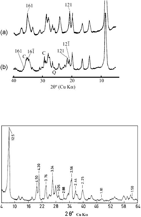
XRD patterns of palygorskite and sepiolite are shown in Figs. 2.24 and 2.25.
Zoltai (1981) has described the palygorskite and sepiolite structures as biopyri-
boles (bio ¼ biotite, pyr ¼ pyroxenes, iboles ¼ amphiboles) built of tri-di-octahedral
modules, the tri-module being M
3
A
2
Si
4
O
10
, and di-module M
2
A
2
Si
4
O
10
. M is the
octahedral cation and A is the anion not bonded to Si within to module; it can be
oxygen when bonded to Si and is (OH) when bonded to more that one M cations.
One half of each A anion is H
2
O molecule when the anion bonded to only one M
cation. The width of these modules is one tetrahedral chain, and their height (t)is
four times the height of an ideal polyhedral layer. Com binations of these modules
can give rise to complet e crystal structures with a vertical displacement between the
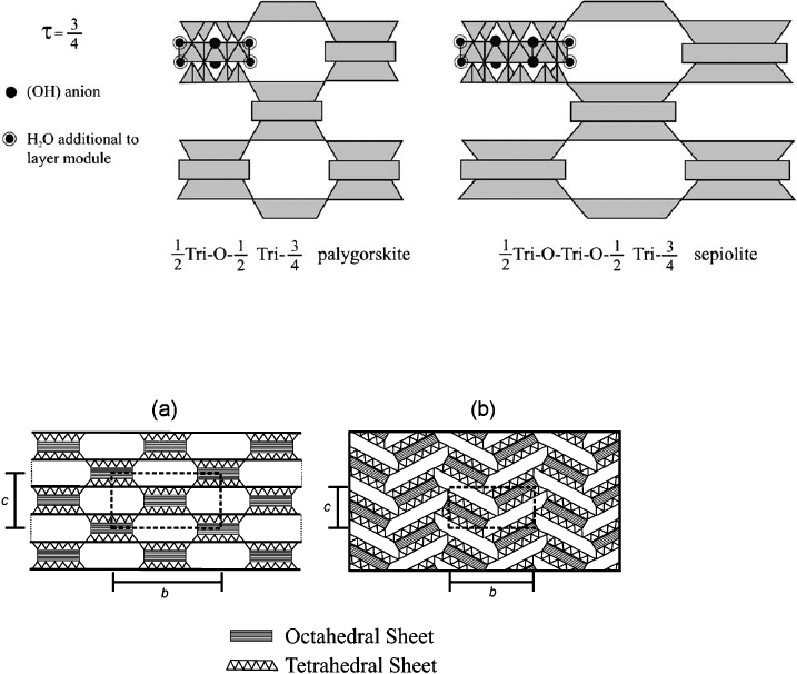
modules equal to n t (with n ¼ 0, 1/2, 3/4). If n ¼ 0, the major layer silicates are
produced. A sequence of n ¼ 0 and 3/4 between modules produces palygorskite, and
the sequence 0, 0, and 3/4 gives the sepiolite structure (Fig. 2.26). Symbol 0 is relative
to the orientation of tetrahedral chains, indicating that the faces of adjacent tet-
rahedra point in opposite directions (‘0’ chains).
Although the crystallographic description by Zoltai (1981) is attractive, palygors-
kite and sepiolite should be considered as phyllosilicates (see above) with special
features rather than as biopyriboles. This is because the physicochemical propert ies
and genetic environments of palygorskite and sepiolite are akin to those of clay
minerals. In common with many other phyllosilicates, a detailed single-crystal
structure of sepiolite and palygorskite is still wanting. IR studi es combined with
powdered diffraction EM and TA have provided insight into the nature of the water
in sepiolite and palygorskite, and the structural changes that occur after heating/
dehydration (Hayashi et al., 1969 ; Serna et al., 1975, 1977; Mifsud et al., 1978; Van
Scoyoc et al., 1979; Blanco et al., 1988).
Chapter 2: Structures and Mineralogy of Clay Minerals60
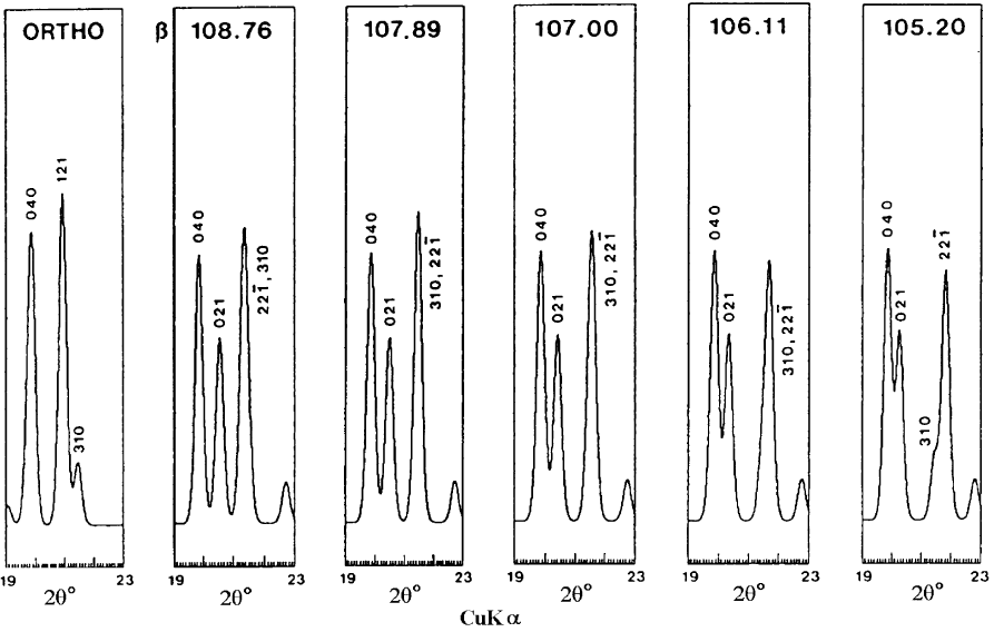
Fig. 2.22. XRD powder patterns in the 19–231 range of 2y (Cu Ka radiation), showing the 0.40–0.45 nm diagnostic region calculated
for idealized orthorhombic and monoclinic palygorskites. The 121 reflection is characteristic of the orthorhombic form, while the 021
and strong 2 2
1 reflections are characteristic of the monoclinic form. After Chisholm (1992).
2.9. Palygorskite and Sepiolite 61
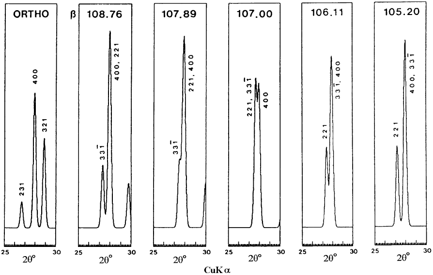
Fig. 2.23. XRD powder patterns in the 25–301 range of 2y (Cu Ka radiation), showing the 0.305–0.33 nm diagnostic region calculated
for idealized orthorhombic and monoclinic palygorskites. The 321 line is characteristic of the orthorhombic form. The appearance of
two lines close together near 400 indicate the presence of the monoclinic form; the exact position of these lines is sensitive to the value of
b in the range 106–1081. After Chisholm (1992).
Chapter 2: Structures and Mineralogy of Clay Minerals62
A more recent study by McKeown et al. (2002), using polarized Raman and FTIR
spectroscopy, indicates that the Si–O stretching and O–Si–O bending force constants
for palygorskite are similar to the corresponding values previously calculated for
other phyllosilicates. However, the values for Mg–O stretching are about half of
those obtained for the Al–O and Mg–O stretching force constants in other phyllo-
silicates (i.e., the octahedral sheets in micas). This finding suggests that the respective
interatomic bonds within the octahedral ribbo ns of palygorskite and sepiol ite are
weaker than those in a continuous octahedral sheet.
Fig. 2.24. XRD pattern of the orthorhombic form (a) and monoclinic form (b) of palygors-
kite (C ¼ calcite, Q ¼ quartz). After Christ et al. (1969). Indices for the monoclinic form are
taken from Chisholm (1992).
Fig. 2.25. XRD diagram of sepiolite from Vallecas. The numbers at the top of each peak refer
to A
˚
ngstrom units (1 A
˚
¼ 0.1 nm). After Pe
´
rez-Rodrı
´
guez and Gala
´
n (1994).
2.9. Palygorskite and Sepiolite 63
Four water molecules (zeolitic water) are present in the channels, and four others
are bound to the octahedral edge inside the channels. Accordingly, the DTA curves
can be divided into three parts: (i) the low-temperature region (o300 1C) where the
minerals lose water adsorbed on outer surfaces and zeolitic water (peak at
120–150 1C); (ii) the central region (300–600 1C), where two endothermic peaks oc-
cur at about 350 1C and 500–550 1C for sepiolite, but only one (about 450–500 1C) for
palygorskite; and (iii) the high-temperature region (>600 1C) where an endothermic
effect (at about 800 1C) is immediately followed by an exothermic maiximum.
In sepiolite, the first endotherm in the central region is narrower and more intense
than the second one (Fig. 2.27). The first endothermic peak is ascribed to the loss of
the first two water molecules coordinated to the inner octahedral edge, causing
rotation of alternate ribbons and particle folding (Nagata et al., 1974; Serna et al.,
1975; Van Scoyoc et al., 1979). The second central endotherm in sepiolite is due to
the loss of the other two edge-coordinated water molecules that are ‘trapped’ inside
Fig. 2.26. Palygorskite and sepiolite structures. After Zoltai (1981).(t ¼ displacement be-
tween a pair of 1/2 Tri modules; 1/2 indicates that only half of the extra trioctahedral sites are
occupied, that is, those sites between the two linked modules.)
Fig. 2.27. General scheme for an unfolded (a) and folded (b) fibrous clay mineral. After Jones
and Gala
´
n, 1988).
Chapter 2: Structures and Mineralogy of Clay Minerals64
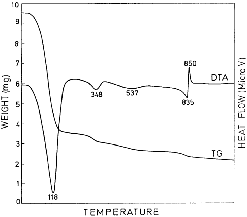
the collapsed channels (Pe
´
rez-Rodrı
´
guez and Gala
´
n, 1994). In palygorskite, coor-
dination water is gradually lost through the whole interval, starting when zeolitic
water is lost and ending when dehydroxylation begins (Mifsud et al., 1978). The
high-temperature endotherm repres ents dehydroxylation of the structure, and the
exothermic peak that follows is due to the formation of clinoenstatite. Typical
DTA–TG curves for sepiolite are shown in Fig. 2.28.
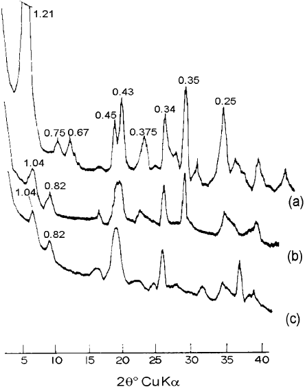
The structural changes that occur on heating also lead to a decrease in the in-
tensity of the principal XRD peaks. For instance, in sepiolite (Fig. 2.29) the reflec-
tions at 1.2, 0.45, 0.38, and 0.34 nm decrease when the mineral is heated at 250 1C for
1 h, while new reflections appear at 1.04 and 0.82 nm. Further heating to 450 1C
increases the intensity of these new reflections, which persist up to 700 1C(Hayashi
et al., 1969; Ferna
´
ndez a
´
lvarez, 1970; Nagata et al., 1974). In palygorskite, the
intensity of the reflections at 1.05, 0.45, and 0.323 nm decreases on heating, and new
peaks appear at 0.92 and 0.47 nm. On heating to 325 1 C, these changes become more
marked. Heating to 600 1C completely eliminates the 1.05 nm reflect ion. At the same
time, the 0.92 nm peak becomes less intense (Hayashi et al., 1969) and shifts to
0.87 nm. At 700 1 C, palygorskite is practically X-ray amorphous.
The decrease in intensity of the principal reflection occurs because structural
disorder produced by heating is more prominent along the principal cleavage face
(011) and less along the (040) plane (Lokanatha and Bhattacherjee, 1984). In
the 200–300 1C range, the particle size of palygorskite slightly increases as water
Fig. 2.28. DTA and TG curves of sepiolite from Vallecas. (temperature in 1C, weight sample ¼
42 mg, heating rate ¼ 121 min
1
, ambient conditions). After Pe
´
rez-Rodrı
´
guez and Gala
´
n (1994).
2.9. Palygorskite and Sepiolite 65
molecules are expelled from the channels, but decreases markedly at 600 1C when the
anhydrous stage is reached. As particle size decreases, the parameter a along the fibre
axis increases until the structure collapses. Both palygorskite and sepiolite can
rehydrate following particle folding. However, rehydration is difficult once the an -
hydrous state is reached when new interparticle bonds are formed.
From an historical point of view, the chemical analysis of sepiolite (in the form of
a ‘meerschaum’ pipe from Turkey) was first attempted in the second half of the 18th
century by Johann Christian Wiegleb . In 1794, Martin Heinrich Klaproth made a
chemical analys is of a sepiolite from Eskis
-
ehir, Turkey. Since then most papers on
sepiolite and palygor skite have information abo ut their respective chemical compo-
sitions.
However, published analytical data mostly refer to bulk samples. As such, they
are affected by both crystallochemical variations and admix ed contaminants (other
clay minerals and associated minerals). The most frequent admixtures in sepiolite
and palygorskite are smectite, illite, chlorite, quartz, felds pars, carbonates, zeolites,
iron, and silica gels.
Fig. 2.29. XRD diffraction pattern of sepiolite recorded under vacuum at 25 1C, 4 h (a);
200 1C, 4 h (b); 530 1C, 6 h (c). After Serna et al. (1975).
Chapter 2: Structures and Mineralogy of Clay Minerals66
Gala
´
nandCarretero(1999)have reviewed the literature on chemical analyses, includ-
ing bulk chemical analyses, and EDX analyses of selected individual particles and pure
samples. Their assessment indicates that sepiolite is a true trioctahedral mineral with
eight octahedral positions filled by Mg
2+
(Table 2.3) and negligible structural substi-
tutions. A very pure (near end-member) specimen has close to the theoretical formula of
Mg
8
Si
12
O
30
ðOHÞ
4
ðOH
2
Þ
4
ðH
2
OÞ
8
Palygorskite is intermediate between di- and tri-octahedral. The octahedral sheet con-
tains mainly Mg
2+
,Al
3+
,andFe
3+
,withanR
2+
/R
3+
ratio close to 1, and has four of
the five structural positions occupied (Table 2.3). The theoretical formula is
ðMg
2þ
2
R
3þ
2
&
1
ÞðSi
4þ
8x
Al
3þ
x
ÞO
20
ðOHÞ
2
ðOH
2
Þ
4
R
2þ
x=2
ðH
2
OÞ
4
; x ¼ 0 0:5
The proposed formula is very close to that given by Smith and Norem (1986) for very
pure palygorskite samples obtained by electron-microprobe analysis.
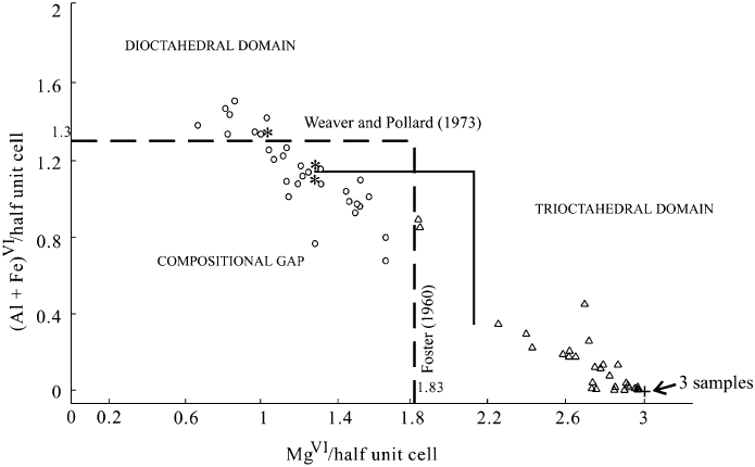
Figs. 2.30 and 2.31 indica te that the compositions of sepiolite and palygorskite are
more limited than previously reported. The two minerals show no compositional gap
if bulk analytical data are plotted on the Martin-Vivaldi an d Cano (1956) diagram
Fig. 2.30. Selected bulk analysis data for sepiolite and palygorskite, plotted as the content of
6-coordinate Mg atoms per half unit cell against the content of 6-coordinate (Al+Fe) atoms
per half unit cell. (
J
): data for palygorskite from the literature; n: data for sepiolite from the
literature. The EDX results for palygorskite (
) and sepiolite (+) from Gala
´
n and Carretero
(1999) are also included. Broken line represents the conventional plot; solid line denotes the
relationship proposed by Paquet et al. (1987).
2.9. Palygorskite and Sepiolite 67
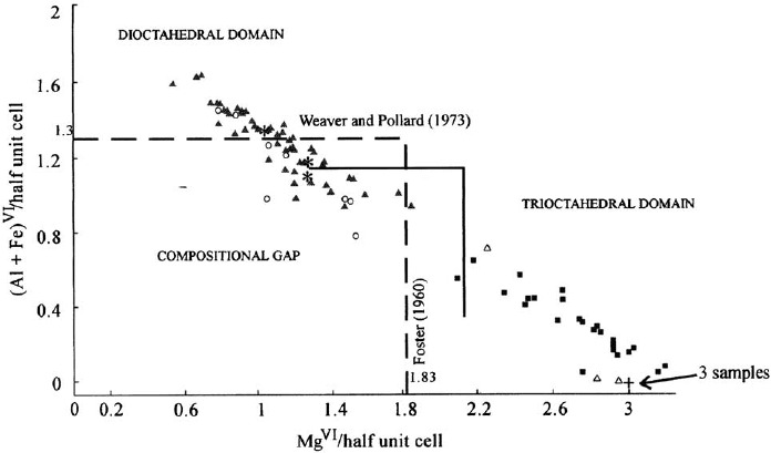
(Fig. 2.32). On the contrary, a gap can be recognized if the EDX results alone are
considered.
On the basis of trace elements content, sepiolite is a more ‘restricted’ mineral than
palygorskite. According to Torres-Ruiz et al. (1994), sepiolite contains three or four
times more trace elements than palygorskite. Both minerals are rich in F
–
and Li
+
.
Sepiolite ca n contain up to 1.5% F
–
, and 800 ppm Li
+
(Leguey et al., 1995; Torres-
Ruiz et al., 1994). Inherited minerals have higher values of REE and transition-metal
elements, and lower values of F
–
and Li
+
, than those formed by chemical precip-
itation in depositional basins (Torres-Ruiz et al., 1994; Lo
´
pez-Galindo et al., 1996).
The CEC of both minerals is quite low, ranging from 4 to 40 cmol/kg; higher
values are probably related to impurities (Gala
´
n, 1996).
In some sepiolites cations other than Mg
2+
may occur in octahedral positions. If
Ni
2+
>Mg
2+
the species is known as falcon doite (Springer, 1976). Iron- and alu-
minium-rich varieties are named Fe
3+
-sepiolite and Al
3+
-sepiolite, respectively.
Loughlinite is Na
+
-sepiolite where two Na
+
substitute for two Mg
2+
, and two Na
+
are in the channels (Fahey and Axelrod, 1948; Echle, 1978). Since it is very difficult
to conceive of a stable sepiolite structure containing octahedral Na
+
, Jones and
Gala
´
n (1988) have suggested that the occurrence of loughlinite as a different mineral
from sepiolite is still to be conclusively demonstrated. Other varieties of sepiolite are
Fig. 2.31. EDX analysis data for sepiolite and palygorskite.
J
: data for palygorskite from the
literature; n: data for sepiolite from the literature. The results for palygorskite (
) and se-
piolite (+) from Gala
´
n and Carretero (1999) are also included. Broken line represents the
conventional plot; solid line denotes the relationship proposed by Paquet et al. (1987) from
their sepiolite (’) and palygorskite (m) data.
Chapter 2: Structures and Mineralogy of Clay Minerals68
