BD Diagnostic Systems (publ.). Difco Manual (Manual of Microbiological Culture)
Подождите немного. Документ загружается.


226 The Difco Manual
Inositol Assay Medium Section II
Bacto
®
Inositol Assay Medium
Intended Use
Bacto Inositol Assay Medium is used for determining inositol concen-
tration by the microbiological assay technique.
Summary and Explanation
Vitamin Assay Media are prepared for use in the microbiological assay
of vitamins. Three types of media are used for this purpose:
1. Maintenance Media: For carrying the stock culture to preserve the
viability and sensitivity of the test organism for its intended purpose;
2. Inoculum Media: To condition the test culture for immediate use;
3. Assay Media: To permit quantitation of the vitamin under test.
Inositol Assay Medium, a modification of the formula described by
Atkin et al.,
1
is used in the microbiological assay of inositol using
Saccharomyces cerevisiae ATCC
®
9080 (Saccharomyces uvarum) as
the test organism.
Principles of the Procedure
Inositol Assay Medium is an inositol-free dehydrated medium
containing all other nutrients and vitamins essential for the cultivation
of S. cerevisiae ATCC
®
9080. The addition of inositol in specified
increasing concentrations gives a growth response that can be measured
turbidimetrically.
Formula
Inositol Assay Medium
Formula Per Liter
Bacto Dextrose . . . . . . . . . . . . . . . . . . . . . . . . . . . . . . . . . 100 g
Potassium Citrate . . . . . . . . . . . . . . . . . . . . . . . . . . . . . . . . 10 g
Citric Acid . . . . . . . . . . . . . . . . . . . . . . . . . . . . . . . . . . . . . . . 2 g
2. ISP Medium 4
IRRITANT. IRRITATING TO EYES, RESPIRATORY SYSTEM
AND SKIN. Avoid contact with skin and eyes. Do not breath dust.
Wear suitable protective clothing. Keep container tightly closed.
TARGET ORGAN(S): Lungs, Intestines.
FIRST AID: In case of contact with eyes, rinse immediately with
plenty of water and seek medical advice. After contact with skin,
wash immediately with plenty of water. If inhaled, remove to fresh
air. If not breathing, give artificial respiration. If breathing is
difficult, give oxygen. Seek medical advice. If swallowed seek
medical advice immediately and show this container or label.
3. Follow proper established laboratory procedures in handling and
disposing of infectious materials.
Storage
Store the dehydrated medium below 30°C. The dehydrated medium is
very hygroscopic. Keep container tightly closed.
Expiration Date
The expiration date applies to the product in its intact container when
stored as directed. Do not use a product if it fails to meet specifications
for identity and performance.
Procedure
Materials Provided
ISP Medium 1
ISP Medium 2
ISP Medium 4
Materials Required But Not Provided
Glassware
Distilled or deionized water
Autoclave
Incubator (30°C)
Sterile tubes
Sterile Petri dishes
Method of Preparation
1. Suspend the appropriate amount of medium in 1 liter distilled or
deionized water:
ISP Medium 1 8 g/l
ISP Medium 2 38 g/l
ISP Medium 4 37 g/l
2. Heat to boiling to dissolve completely.
3. Autoclave at 121°C for 15 minutes.
4. Mix thoroughly while dispensing.
Specimen Collection and Preparation
Obtain and process specimens according to the techniques and
procedures established by laboratory policy.
Test Procedure
For a complete discussion on the isolation and maintenance of
Streptomyces species refer to appropriate references.
2,3
Results
Refer to appropriate references and procedures for results.
Limitations of the Procedure
1. Since the nutritional requirements of organisms vary, some strains
may be encountered that fail to grow or grow poorly on the medium.
References
1. Shirling, E. B., and D. Gottlieb. 1966. Methods for characteriza-
tion of Streptomyces species. Int. J. Syst. Bacteriol. 16:313-340.
2. Isenberg, H. D. (ed.). 1992. Clinical microbiology procedures
handbook. American Society for Microbiology, Washington, D.C.
3. Murray, P. R., E. J. Baron, M. A. Pfaller, F. C. Tenover, and
R. H. Yolken (ed.). 1995. Manual of clinical microbiology, 6th ed.
American Society for Microbiology, Washington, D.C.
Packaging
ISP Medium 1 500 g 0769-17
ISP Medium 2 500 g 0770-17
ISP Medium 4 500 g 0772-17

The Difco Manual 227
Section II Inositol Assay Medium
User Quality Control
Identity Specifications
Dehydrated Appearance: White to off-white, free-flowing,
homogeneous.
Solution: 6.1% (single strength) or 12.2%
(double strength) solution, soluble
in distilled or deionized water on
boiling. Light amber, clear, may have
a slight precipitate.
Prepared Medium: Light amber, clear, may have a slight
precipitate.
Reaction of 6.1%
Solution at 25°C: pH 5.2 ± 0.2
Cultural Response
Prepare Inositol Assay Medium per label directions. Dispense
medium into 50 ml flasks with a titration from 0 to 10 µg of
Inositol. Inoculate flasks with one drop of S. cerevisiae ATCC
®
9080 inoculum suspension (washed three times and diluted
1:1000). Incubate flasks at 25-30°C for 20-24 hours. The curve
obtained from turbidimetric readings should be typical.
Monopotassium Phosphate. . . . . . . . . . . . . . . . . . . . . . . . . 1.1 g
Potassium Chloride. . . . . . . . . . . . . . . . . . . . . . . . . . . . . . 0.85 g
Magnesium Sulfate . . . . . . . . . . . . . . . . . . . . . . . . . . . . . . 0.25 g
Calcium Chloride . . . . . . . . . . . . . . . . . . . . . . . . . . . . . . . 0.25 g
Manganese Sulfate . . . . . . . . . . . . . . . . . . . . . . . . . . . . . . . 50 mg
Ferric Chloride . . . . . . . . . . . . . . . . . . . . . . . . . . . . . . . . . . 50 mg
DL-Tryptophane . . . . . . . . . . . . . . . . . . . . . . . . . . . . . . . . . 80 mg
L-Cystine. . . . . . . . . . . . . . . . . . . . . . . . . . . . . . . . . . . . . . . 0.1 g
L-Isoleucine . . . . . . . . . . . . . . . . . . . . . . . . . . . . . . . . . . . . 0.5 g
L-Leucine . . . . . . . . . . . . . . . . . . . . . . . . . . . . . . . . . . . . . . 0.5 g
L-Lysine . . . . . . . . . . . . . . . . . . . . . . . . . . . . . . . . . . . . . . . 0.5 g
L-Methionine . . . . . . . . . . . . . . . . . . . . . . . . . . . . . . . . . . . 0.2 g
DL-Phenylalanine . . . . . . . . . . . . . . . . . . . . . . . . . . . . . . . . 0.2 g
L-Tyrosine . . . . . . . . . . . . . . . . . . . . . . . . . . . . . . . . . . . . . . 0.2 g
L-Asparagine . . . . . . . . . . . . . . . . . . . . . . . . . . . . . . . . . . . 0.8 g
DL-Aspartic Acid . . . . . . . . . . . . . . . . . . . . . . . . . . . . . . . . 0.2 g
DL-Serine . . . . . . . . . . . . . . . . . . . . . . . . . . . . . . . . . . . . . . 0.1 g
Glycine . . . . . . . . . . . . . . . . . . . . . . . . . . . . . . . . . . . . . . . . 0.2 g
DL-Threonine . . . . . . . . . . . . . . . . . . . . . . . . . . . . . . . . . . . 0.4 g
L-Valine. . . . . . . . . . . . . . . . . . . . . . . . . . . . . . . . . . . . . . . . 0.5 g
L-Histidine . . . . . . . . . . . . . . . . . . . . . . . . . . . . . . . . . . . 0.124 g
L-Proline . . . . . . . . . . . . . . . . . . . . . . . . . . . . . . . . . . . . . . . 0.2 g
DL-Alanine . . . . . . . . . . . . . . . . . . . . . . . . . . . . . . . . . . . . . 0.4 g
L-Glutamic Acid . . . . . . . . . . . . . . . . . . . . . . . . . . . . . . . . . 0.6 g
L-Arginine. . . . . . . . . . . . . . . . . . . . . . . . . . . . . . . . . . . . . 0.48 g
Thiamine Hydrochloride . . . . . . . . . . . . . . . . . . . . . . . . . 500 µg
Biotin. . . . . . . . . . . . . . . . . . . . . . . . . . . . . . . . . . . . . . . . . . 16 µg
Calcium Pantothenate . . . . . . . . . . . . . . . . . . . . . . . . . . . . . . 5 mg
Pyridoxine Hydrochloride . . . . . . . . . . . . . . . . . . . . . . . . . . 1 mg
Final pH 5.2 ± 0.2 at 25°C
Precautions
1. For Laboratory Use.
2. Great care must be taken to avoid contamination of media or glass-
ware in microbiological assay procedures. Extremely small
amounts of foreign material may be sufficient to give erroneous
results. Scrupulously clean glassware free from detergents and
other chemicals must be used. Glassware must be heated to 250°C
for at least 1 hour to burn off any organic residues that might be
present.
3. Take precautions to keep sterilization and cooling conditions
uniform throughout the assay.
4. Follow proper established laboratory procedures in handling and
disposing of infectious materials.
Storage
Store the dehydrated medium at 2-8°C. The dehydrated medium is very
hygroscopic. Keep container tightly closed.
Expiration Date
The expiration date applies to the product in its intact container when
stored as directed. Do not use a product if it fails to meet specifications
for identity and performance.
Procedure
Materials Provided
Inositol Assay Medium
Materials Required But Not Provided
Glassware
Autoclave
Stock culture of Saccharomyces cerevisiae ATCC® 9080
Inositol
Sterile tubes
Sterile 0.85% saline
Distilled or deionized water
Lactobacilli Agar AOAC
Centrifuge
Spectrophotometer
Method of Preparation
1. Suspend 12.2 grams in 100 ml distilled or deionized water.
2. Boil to dissolve.
3. Dispense 5 ml amounts into flasks.
4. Add standard or test samples.
5. Adjust flask volume to 10 ml with distilled or deionized water.
6. Autoclave at 121°C for 5 minutes.
Specimen Collection and Preparation
Assay samples are prepared according to references given in the specific
assay procedures. The samples should be diluted to approximately the
same concentration as the standard solution.
Test Procedure
Remove a loopful of culture from a stock culture slant of S. cerevisiae
ATCC
®
9080 and suspend it in 10 ml sterile 0.85% saline. Centrifuge
cells at moderate speed for 10 minutes. Decant the supernatant and
resuspend cells in 10 ml 0.85% sterile saline. Wash the cells three times
with 10 ml sterile 0.85% saline. After the third wash, resuspend the
228 The Difco Manual
Bacto
®
KF Streptococcus Agar
User Quality Control
Identity Specifications
Dehydrated Appearance: Light greenish-beige, free-flowing,
homogeneous.
Solution: 7.64% solution, soluble in distilled or
deionized water on boiling. Solution
is light purple, very slightly to
slightly opalescent.
Prepared Medium: Light purple, very slightly to
slightly opalescent.
Reaction of 7.64%
Solution at 25°C: 7.2 ± 0.2
Cultural Response
Prepare KF Streptococcus Agar per label directions. Inoculate
using the pour plate technique and incubate at 35 ± 2°C for 46-48 hours.
INOCULUM
ORGANISM ATCC
®
CFU GROWTH APPEARANCE
Enterobacter aerogenes 13048* 1,000-2,000 marked to –
complete inhibition
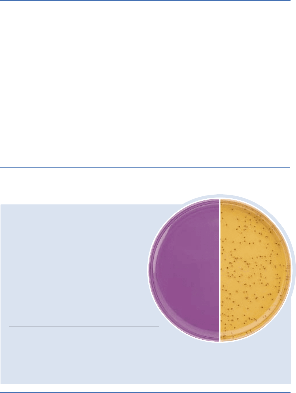
Enterococcus faecalis 19433* 30-300 good red centers
Enterococcus faecalis 29212* 30-300 good red centers
Escherichia coli 25922* 1,000-2,000 marked to –
complete inhibition
Enterococcus faecalis
ATCC
®
19433
Uninoculated
plate
The cultures listed are the minimum that should be used for performance testing.
*These cultures are available as Bactrol
™
Disks and are to be used as directed per Bactrol Disk Technical Information.
cells in 10 ml 0.85% saline. Dilute 1 ml of the cell suspension in 1000 ml
of sterile 0.85% saline. This diluted suspension is the inoculum. Use
1 drop of inoculum suspension to inoculate each assay flask.
The concentrations of inositol required for the preparation of the standard
curve may be prepared by dissolving 200 mg inositol in 100 ml
distilled water. Mix thoroughly. Dilute 1 ml of this solution with 999 ml
distilled water to make a final solution containing 2 µg inositol per ml.
Use 0.0, 0.5, 1, 2, 3, 4 and 5 ml per flask. Prepare this stock solution
fresh daily.
It is essential that a standard curve be constructed each time an assay is
run. Autoclave and incubation conditions can impact the standard curve
readings and cannot always be duplicated. The standard curve is
obtained by using inositol at levels of 0.0, 1, 2, 4, 6, 8 and 10 µg per
assay flask (10 ml).
Following inoculation, flasks are incubated at 25-30°C for 20-24 hours.
Place flasks in the refrigerator for 15-30 minutes to stop growth. Growth
is measured turbidimetrically using any suitable spectrophotometer.
Results
1. Prepare a standard concentration response curve by plotting the
response readings against the amount of standard in each tube, disk
or cup.
KF Streptococcus Agar Section II
2. Determine the amount of vitamin at each level of assay solution by
interpolation from the standard curve.
3. Calculate the concentration of vitamin in the sample from the
average of these volumes. Use only those values that do not vary
more than ± 10% from the average. Use the results only if two
thirds of the values do not vary more than ± 10%.
Limitations of the Procedure
1. The test organism used for inoculating an assay medium must be
grown and maintained on media recommended for this purpose.
2. Aseptic technique should be used throughout the assay procedure.
3. The use of altered or deficient media may cause mutants having
different nutritional requirements that will not give a satisfactory
response.
4. For successful results of these procedures, all conditions of the
assay must be followed precisely.
References
1. Atkin, Schultz, Williams, and Frey. 1943. End. & Eng. Chem.,
Ann. Ed. 15:141.
Packaging
Inositol Assay Medium 100 g 0995-15

The Difco Manual 229
Section II KF Streptococcus Agar
Intended Use
Bacto KF Streptococcus Agar is used with Bacto TTC Solution 1% in
isolating and enumerating fecal streptococci according to APHA.
Also Known As
Kenner Fecal Streptococcus Agar
Summary and Explanation
Kenner et al. developed KF Streptococcal Agar for use in detecting
streptococci in surface waters by direct plating or by the membrane
filtration method.
1
These investigators compared the performance of
their formulation to other media used for enumerating fecal streptococci
and achieved greater recoveries with KF Streptococcal Agar.
This medium is currently recommended for use in determining counts
of fecal streptococci in foods and water.
2,3
Principles of the Procedure
Peptone provides a source of nitrogen, amino acids and carbon. Yeast
Extract is a source of trace elements, vitamins and amino acids. Maltose
and Lactose are fermentable carbohydrates and carbon sources. Sodium
Azide is a selective agent. Brom Cresol Purple is an indicator dye.
The addition of 1% triphenyltetrazolium chloride (TTC) causes
enterococci to develop a deep red color following reduction of
tetrazolium to an acid azo dye.
Formula
KF Streptococcus Agar
Formula Per Liter
Bacto Proteose Peptone No. 3 . . . . . . . . . . . . . . . . . . . . . . 10 g
Bacto Yeast Extract . . . . . . . . . . . . . . . . . . . . . . . . . . . . . . . 10 g
Sodium Chloride . . . . . . . . . . . . . . . . . . . . . . . . . . . . . . . . . . 5 g
Sodium Glycerophosphate . . . . . . . . . . . . . . . . . . . . . . . . . 10 g
Bacto Maltose . . . . . . . . . . . . . . . . . . . . . . . . . . . . . . . . . . . 20 g
Bacto Lactose . . . . . . . . . . . . . . . . . . . . . . . . . . . . . . . . . . . . 1 g
Sodium Azide . . . . . . . . . . . . . . . . . . . . . . . . . . . . . . . . . . . 0.4 g
Bacto Brom Cresol Purple . . . . . . . . . . . . . . . . . . . . . . . 0.015 g
Bacto Agar . . . . . . . . . . . . . . . . . . . . . . . . . . . . . . . . . . . . . 20 g
Final pH 7.2 ± 0.2 at 25°C
Precautions
1. For Laboratory Use
2. HARMFUL. HARMFUL BY INHALATION AND IF SWAL-
LOWED. IRRITATING TO EYES, RESPIRATORY SYSTEM
AND SKIN. Avoid contact with skin and eyes. Do not breathe dust.
Wear suitable protective clothing. Keep container tightly closed.
TARGET ORGAN(S): Cardiovascular, Lungs, Nerves.
FIRST AID: In case of contact with eyes, rinse immediately with
plenty of water and seek medical advice. After contact with skin,
wash immediately with plenty of water. If inhaled, remove to
fresh air. If not breathing, give artificial respiration. If breathing is
difficult, give oxygen. Seek medical advice. If swallowed seek
medical advice immediately and show this container or label.
3. Follow proper established laboratory procedure in handling and
disposing of infectious materials.
Storage
Store the dehydrated medium below 30°C. The dehydrated medium is
very hygroscopic. Keep container tightly closed.
Expiration Date
The expiration date applies to the product in its intact container when
stored as directed. Do not use a product if it fails to meet specifications
for identity and performance.
Procedure
Materials Provided
KF Streptococcus Agar
TTC Solution 1%
Materials Required But Not Provided
Glassware
Incubator (35°C)
Pipettes
Sterile Petri dishes, 50 x 9 mm
Membrane filter equipment
Sterile 47 mm, 0.45 µm, gridded membrane filters
Dilution blanks
Stereoscopic microscope
Method of Preparation
1. Suspend 76.4 grams in 1 liter distilled or deionized water.
2. Heat to boiling to dissolve completely.
2. Heat an additional 5 minutes. Avoid overheating which could
decrease the productivity of the medium. DO NOT AUTOCLAVE.
3. Add 10 ml TTC Solution 1% to the medium at 50°C and mix well.
4. Pour medium into sterile Petri dishes if using the Membrane Filter
procedure. If using the Pour Plate technique, hold the liquid
medium at 45°C.
Specimen Collection and Preparation
Consult appropriate references for specific procedures using KF
Streptococcus Agar in the examination of waters and food.
2,3
The pour
plate technique or the membrane filter procedure can be used for
detection and enumeration of enterococci.
Test Procedure
Pour Plate Technique
1. Prepare appropriate dilutions of the test material.
2. Place the selected volume of sample in a Petri dish.
3. Pour 15 ml of the prepared medium at 45°C into each plate.
4. Thoroughly mix the medium and sample to uniformly disperse the
organisms.
5. Allow the agar to solidify.
6. Incubate plates in the inverted position at 35 ± 2°C for 46-48 hours.
Membrane Filter Procedure
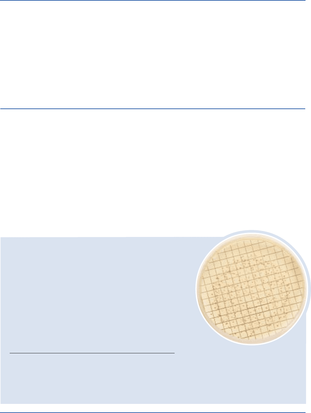
1. Filter a suitable volume of sample through a sterile membrane, as
directed.
2. Place the inoculated membrane filter on the solidified agar in the
Petri dish, inoculum side up.
3. Incubate the plates, inverted, at 35 ± 2°C for 46-48 hours.
230 The Difco Manual
KF Streptococcus Broth Section II
Results
Enterococci will appear as red or pink colonies. The use of a stereoscopic
microscope with 15X magnification can aid in counting colonies.
Limitations of the Procedure
1. Many strains of S. bovis and S. equinus are inhibited by azide.
2. Overheating may lower the pH, causing a decrease in the produc-
tivity of the medium.
References
1. Kenner, B. A., H. F. Clark, and P. W. Kabler. 1961. Fecal
streptococci. I. Cultivation and enumeration of streptococci in
surface waters. Appl. Microbiol. 9:15.
Bacto
®
KF Streptococcus Broth
Intended Use
Bacto KF Streptococcus Broth is used for isolating fecal streptococci.
Also Known As
Kenner Fecal Streptococcus Broth
Summary and Explanation
Kenner et al. developed KF Streptococcal Broth for the detection and
enumeration of enterococci in waters.
1,2
They found that this formulation
was superior to other liquid media in the recovery of enterococci in
Most Probable Number (MPN) test systems. The medium is not
specific for presumptive identification of group D streptococci. Other
tests are required.
2-4
This medium currently is recommended for use in enumerating
enterococci in foods.
5
Principles of the Procedure
Proteose Peptone No. 3 provides a source of nitrogen, amino acids and
carbon. Yeast Extract is a source of trace elements, vitamins and amino
acids. Maltose and Lactose are the fermentable carbohydrates and
carbon source. Sodium Azide is the selective agent. Brom Cresol Purple
is the indicator dye.
The addition of 1% triphenyltetrazolium chloride, in the membrane
filter procedure, causes the enterococci to have a deep red color as a
result of tetrazolium reduction to an acid azo dye.
User Quality Control
Identity Specifications
Dehydrated Appearance: Light greenish-beige, free-flowing, homogeneous.
Solution: 5.64% solution, soluble in distilled or deionized water with
frequent agitation on boiling. Solution is reddish to light
purple, clear to very slightly opalescent.
Prepared Tubes: Purple, clear to very slightly opalescent.
Reaction of 5.64%
Solution at 25°C: 7.2 ± 0.2.
Cultural Response
Prepare KF Streptococcus Broth per label directions. Supplement with TTC
Solution 1%. Using the membrane filter technique, inoculate and incubate at 35 ± 1°C
in an atmosphere saturated with water vapor for 46-48 hours.
INOCULUM
ORGANISM ATCC
®
CFU GROWTH APPEARANCE
Enterobacter aerogenes 13048* 300-1,000 inhibited –
Enterococcus faecalis 19433* 30-200 good red
Enterococcus faecalis 29212* 30-200 good red
Escherichia coli 25922* 300-1,000 inhibited –
The cultures listed are the minimum that should be used for performance testing.
*These cultures are available as Bactrol
™
Disks and should be used as directed in Bactrol Disks Technical Information.
2. Donnelly, C. W., R. E. Bracket, D. Doores, W. H. Lee, and
J. Lovett. 1992. Compendium of methods for the microbiological
examination of foods, 3rd ed. American Public Health Association,
Washington, D.C.
3. Bordner, R., and J. Winter. 1978. Microbiological methods for
monitoring the environment, water and wastes. EPA, Cincinnati, OH.
4. MacFadden, J. F. 1985. Media for isolation-cultivation-
identification-maintenance of medical bacteria, vol. I. Williams
& Wilkens, Baltimore, MD.
Packaging
KF Streptococcus Agar 500 g 0496-17
Enterococcus faecalis
ATCC
®
29212

The Difco Manual 231
Section II KF Streptococcus Broth
Formula
KF Streptococcus Broth
Formula Per Liter
Bacto Proteose Peptone No. 3 . . . . . . . . . . . . . . . . . . . . . . 10 g
Bacto Yeast Extract . . . . . . . . . . . . . . . . . . . . . . . . . . . . . . . 10 g
Sodium Chloride . . . . . . . . . . . . . . . . . . . . . . . . . . . . . . . . . . 5 g
Sodium Glycerophosphate . . . . . . . . . . . . . . . . . . . . . . . . . 10 g
Bacto Maltose . . . . . . . . . . . . . . . . . . . . . . . . . . . . . . . . . . . 20 g
Bacto Lactose . . . . . . . . . . . . . . . . . . . . . . . . . . . . . . . . . . . . 1 g
Sodium Azide . . . . . . . . . . . . . . . . . . . . . . . . . . . . . . . . . . . 0.4 g
Bacto Brom Cresol Purple . . . . . . . . . . . . . . . . . . . . . . . 0.015 g
Final pH 7.2 ± 0.2 at 25°C
Precautions
1. For Laboratory Use.
2. HARMFUL. HARMFUL BY INHALATION AND IF SWAL-
LOWED. IRRITATING TO EYES, RESPIRATORY SYSTEM
AND SKIN. Avoid contact with skin and eyes. Do not breathe dust.
Wear suitable protective clothing. Keep container tightly closed.
TARGET ORGAN(S): Cardiovascular, Lungs, Nerves.
FIRST AID: In case of contact with eyes, rinse immediately with
plenty of water and seek medical advice. After contact with skin,
wash immediately with plenty of water. If inhaled, remove to fresh
air. If not breathing, give artificial respiration. If breathing is diffi-
cult, give oxygen. Seek medical advice. If swallowed seek medical
advice immediately and show this container or label.
3. Follow proper established laboratory procedures in handling and
disposing of infectious materials.
Storage
Store the dehydrated medium below 30°C. The dehydrated medium is
very hygroscopic. Keep container tightly closed.
Expiration Date
The expiration date applies to the product in its intact container when
stored as directed. Do not use a product if it fails to meet specifications
for identity and performance.
Procedure
Materials Provided
KF Streptococcus Broth
Materials Required But Not Provided
Glassware
Autoclave
Incubator (35°C)
Pipettes
Culture tubes
Membrane filter equipment
TTC Solution 1%
Incubator (35°C), saturated with water vapor
Sterile Petri dishes, 50 x 9 mm
Sterile 47 mm, 0.45 µm, gridded membrane filters
Sterile absorbent pads
Stereoscopic microscope
Method of Preparation
MPN Procedure
1. For an inoculum of 1 ml or less, suspend 56.4 g in 1 liter distilled
or deionized water.
For an inoculum of 10 ml, suspend 84.6 g in 1 liter distilled or
deionized water.
2. Heat to boiling to dissolve completely.
3. For an inoculum of 1 ml or less, dispense 10 ml amounts into
culture tubes.
For an inoculum of 10 ml, dispense 20 ml amounts into culture tubes.
4. Autoclave at 121°C for 10 minutes.
Membrane Filter Procedure
1. Suspend 56.4 g in 1 liter distilled or deionized water.
2. Heat to boiling to dissolve completely.
3. Dispense 100 ml amounts into flasks and autoclave at 121°C for
10 minutes.
4. Cool to 60°C.
5. Add 1 ml TTC Solution 1% per 100 ml of medium.
Specimen Collection and Preparation
Water or food samples should be collected and prepared according to
appropriate references.
Test Procedure
MPN Procedure
1. Inoculate tubes of the KF Streptococcus Broth with the appropriate
amount of inoculum.
2. Incubate tubes at 35 ± 1°C, with loosened caps, for 46-48 hours.
Membrane Filter Procedure
1. Place a sterile absorbent pad in each sterile Petri dish.
2. Saturate the pads with the sterile medium containing TTC.
3. Place an inoculated membrane filter, inoculated side up, on the
saturated pad.
4. Incubate at 35 ± 1°C in an atmosphere saturated with water vapor
for 46-48 hours.
Results
MPN Procedure
MPN tubes positive for enterococci are turbid with growth that ap-
pears yellow in color and does not produce foaming. When foaming
occurs, confirmation for enterococci should be made by Gram staining.
Membrane Filter Procedure
All red or pink colonies visible with 15x magnification are counted as
enterococci colonies.
Limitations of the Procedure
1. Many strains of S. bovis and S. equinus are inhibited by azide.
2. The pH of KF Streptococcus Broth should be between 7.2 and 7.3.
If below 7.0, it should not be used.
3. Overheating may lower the pH, resulting in a decrease in productivity
of the medium.

232 The Difco Manual
References
1. Kenner, B. A., H. F. Clark, and P. W. Kabler. 1960. Fecal
streptococci. II. Quantification of streptococci in feces. Am. J. Public
Health 50:1553.
2. Kenner, B. A., H. F. Clark, and P. W. Kabler. 1961. Fecal
streptococci. I. Cultivation and enumeration of streptococci in surface
waters. Appl. Microbiol. 9:15.
3. MacFadden, J. F. 1985. Media for isolation-cultivation-
identification-maintenance of medical bacteria, vol. 1. Williams
& Wilkens, Baltimore, MD.
KL Virulence Agar
Bacto
®
KL Virulence Agar
.
KL Virulence Enrichment
KL Antitoxin Strips
KL Virulence Agar Section II
Intended Use
Bacto KL Virulence Agar is used with Bacto KL Virulence
Enrichment, Bacto Chapman Tellurite Solution 1% and Bacto KL
Antitoxin Strips in differentiating virulent (toxigenic) from nonvirulent
strains of Corynebacterium diphtheriae.
Also Known As
KL Virulence Agar conforms with Klebs-Loeffler Virulence Agar.
Summary and Explanation
Elek
2
was the first to describe the agar plate diffusion technique for
demonstrating the in vitro toxigenicity (virulence) of Corynebacterium
diphtheriae. King, Frobisher, and Parsons
3
expanded on Elek’s
technique and, by using a carefully standardized medium, obtained
results in agreement with animal inoculation tests. These authors
demonstrated that Difco Proteose Peptone possessed properties
essential for toxin production. Incorporating Difco Proteose Peptone
into the test medium assured consistent results. The authors used
rabbit, sheep and horse serum as enrichments, finding human serum to
be unsatisfactory. To overcome irregularities encountered in previous
formulations, Hermann, Moore, and Parsons
1
refined the medium used
for the in vitro KL Virulence Test, simplifying the basal medium and
developing a nonserous enrichment. The medium and enrichment
described by these authors have been standardized for use in the
KL Virulence Test.
KL Virulence Agar and KL Virulence Enrichment are prepared
according to the formulation of Hermann, Moore and Parsons.
1
Principles of the Procedure
Proteose Peptone provides the carbon and nitrogen sources required
for good growth of a wide variety of organisms and for toxin
production. Sodium Chloride maintains the osmotic balance of the
medium. Bacto Agar is incorporated as a solidifying agent.
KL Virulence Enrichment, composed of Casamino Acids, Glycerol and
Tween
®
80, provides a source of nonserous enrichment. Casamino
Acids is derived from acid-hydrolyzed casein that has low sodium
chloride and iron concentrations. The low iron concentration is
beneficial because iron is known to prevent the production of diph
theria toxin when present in more than minute amounts. Glycerol
(glycerine) contains no heavy metals and is used by bacteria as a source
of carbon. Tween
®
80 improves growth of certain strains of
Corynebacterium diphtheriae. Toxin produced by bacteria and diffused
into the medium is detected by precipitation with the antitoxin present
on the KL Antitoxin Strip. Chapman Tellurite Solution 1%
(1% potassium tellurite solution) inhibits gram-negative and most
gram-positive bacteria except Corynebacterium spp., Streptococcus
mitis, S. salivarius, enterococci, and possibly Staphylococcus
epidermidis. This permits direct testing of mixed primary cultures.
Formula
KL Virulence Agar
Formula Per Liter
Bacto Proteose Peptone . . . . . . . . . . . . . . . . . . . . . . . . . . . 20 g
Sodium Chloride . . . . . . . . . . . . . . . . . . . . . . . . . . . . . . . . . 2.5 g
Bacto Agar . . . . . . . . . . . . . . . . . . . . . . . . . . . . . . . . . . . . . 15 g
Final pH 7.8 ± 0.2 at 25°C
KL Virulence Enrichment
Formula Per 100 ml
Bacto Casamino Acids . . . . . . . . . . . . . . . . . . . . . . . . . . . . . 1 g
Glycerol. . . . . . . . . . . . . . . . . . . . . . . . . . . . . . . . . . . . . . . . . 1 ml
Tween
®
80 . . . . . . . . . . . . . . . . . . . . . . . . . . . . . . . . . . . . . . . 1 ml
KL Antitoxin Strips
KL Antitoxin Strips are 1 x 7 cm filter paper strips containing antitoxin
to diphtheria toxin.
Precautions
1. For Laboratory Use.
2. Follow proper established laboratory procedure in handling and
disposing of infectious materials.
4. Facklam, R. R., and M. D. Moody. 1970. Presumptive identification
of group D streptococci: the bile-esculin test. Appl. Microbiol.
20:245-250.
5. Donnelly, C. W., R. E. Bracket, D. Doores, W. H. Lee, and J.
Lovett. 1992. Compendium of Methods for the Microbiological
Examination of Foods, 3rd ed. American Public Health Association,
Washington, D.C.
Packaging
KF Streptococcus Broth 500 g 0997-17
The Difco Manual 233
User Quality Control
Identity Specifications
KL Virulence Agar
Dehydrated Appearance: Light beige with some small dark specks,
free- flowing, homogeneous.
Solution: 3.75% solution, soluble in distilled or
deionized water on boiling. Solution
is light to medium amber, slightly
opalescent, with a slight precipitate.
Reaction of 3.75%
Solution at 25°C: pH 7.8 ± 0.2
Prepared Medium: Light medium amber, slightly opalescent,
may have a slight precipitate.
KL Virulence Enrichment
Appearance: Colorless to very light amber, clear liquid.
KL Antitoxin Strips
Appearance: White, filter paper strips, 1 x 7 cm.
Cultural Response
Prepare KL Virulence Agar per label directions, including KL
Virulence Enrichment, Chapman Tellurite Solution 1% and one
KL Antitoxin Strip per plate. Inoculate and incubate at 35 ± 2°C under
CO
2
for up to 72 hours.
ORGANISM ATCC
®
GROWTH
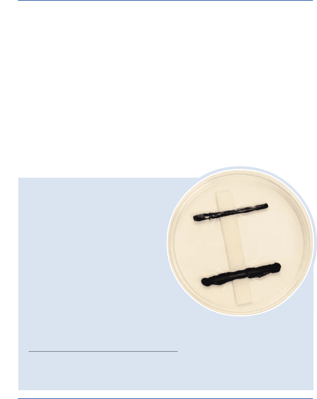
Corynebacterium diphtheriae Type gravis 8028 +
Corynebacterium diphtheriae Type intermedius 8032 +
Staphylococcus aureus 25923* –
+ = positive, line of precipitation at 45% angle to the strip
– = negative, no line of precipitation
Corynebacterium diphtheriae
ATCC
®
8028
Precipitate lines are graphically enhanced for
demonstration purposes (see Results).
Section II KL Virulence Agar
The cultures listed are the minimum that should be used for
performance testing.
*This organism is available as a Bactrol
™
culture and should
be used as directed.
Storage
Store KL Virulence Agar below 30°C. The dehydrated medium is very
hygroscopic. Keep container tightly closed.
Store KL Virulence Enrichment and KL Antitoxin Strips at 2-8°C.
Store prepared plates at 2-8°C.
Expiration Date
The expiration date applies to the product in its intact container when
stored as directed. Do not use a product if it fails to meet specifications
for identity and performance.
Procedure
Materials Provided
KL Virulence Agar
KL Virulence Enrichment
KL Antitoxin Strips
Materials Required But Not Provided
Glassware
Autoclave
Water bath (55-60°C)
Incubator (35°C)
Chapman Tellurite Solution 1%
Method of Preparation
1. Suspend 37.5 grams of KL Virulence Agar in 1 liter distilled or
deionized water and heat to boiling to dissolve completely.
2. Autoclave at 121°C for 15 minutes.
3. Cool in a water bath to 55-60°C.
4. Aseptically dispense 10 ml of KL Virulence Agar into a Petri dish
containing 2 ml KL Virulence Enrichment and 0.5 ml Chapman
Tellurite Solution 1%; mix thoroughly.
5. Using aseptic technique, submerge a KL Antitoxin Strip or
equivalent beneath the agar prior to solidification.
Specimen Collection and Preparation
For cases of suspected diphtheria, material for culture is obtained on
a swab from the inflamed membranes of the throat and nasopharynx,
or from wounds.
4
Care must be taken not to contaminate the swab
with normal skin flora. The specimen should be immediately
transported to a laboratory and inoculated onto the proper media.
If the specimen is to be shipped to a laboratory, it should be placed
in a sterile tube or a special packet containing a desiccant such
as silica gel.
5

234 The Difco Manual
Test Procedure
Inoculate the medium by streaking a loopful of a 24-hour culture in
a single line across the plate perpendicular to (right angle to)
the antitoxin strip. (Do not touch the actual strip itself). As many as
eight cultures may be tested on a single plate.
6
Place test isolates about
1 cm apart. Also inoculate a toxigenic (positive control) and a
nontoxigenic (negative control) C. diphtheriae strain approximately
1 cm on either side of the test isolates.
6
Incubate the inverted plates
at 37°C for 72 hours. Examine at 24-, 48- and 72-hour intervals.
Results
Toxigenic (virulent) cultures of C. diphtheriae will show fine lines
of precipitation at approximately 45° angles from the culture streak.
This line forms where toxin (from the bacteria) combines with
antitoxin from the strip. Primary precipitin lines form an arc of
identity with the precipitin line produced by an adjacent positive
control strain.
7
Nontoxigenic strains of C. diphtheriae will show no
lines of precipitation.
Limitations of the Procedure
1. Each test should include positive and negative controls.
5
2. False-positive reactions may be seen after 24 hours as weak bands
near the antitoxin strip. These can be recognized when compared
with the positive control.
8
3. Corynebacterium ulcerans and C. pseudotuberculosis may also
produce lines of toxin-antitoxin.
9
References
1. Hermann, G. J., M. S. Moore, and E. I. Parsons. 1958. A
substitute for serum in the diphtheria in vitro test. Am. J. Clin.
Pathol. 29:181-183.
2. Elek, S. D. 1948. The recognition of toxicogenic bacterial strains
in vitro. Brit. Med. J. 1:493.
3. King, E. O., M. Frobisher, Jr., and E. I. Parsons. 1949. The
in vitro test for virulence of Corynebacterium diphtheriae.
Am. J. Public Health 39:1314.
4. Clarridge, J. E., and C. A. Spiegel. 1995. Corynebacterium and
miscellaneous irregular gram-positive rods, Erysipelothrix and
Gardnerella, p. 357-378. In P. R. Murray, E. J. Baron, M. A. Pfaller,
F. C. Tenover, and R. H. Yolken (eds.), Manual of clinical
microbiology, 6th ed. American Society for Microbiology,
Washington, D.C.
5. Krech, T., and D. G. Hollis. 1991. Corynebacterium and related
organisms, p. 277-286. In A. Ballows, W. J. Hausler, Jr.,
K. Herrmann, H. D. Isenberg, and H. J. Shadomy (eds.), Manual of
clinical microbiology, 5th ed. American Society for Microbiology,
Washington, D.C.
6. MacFaddin, J. F. 1985. Media for isolation-cultivation-
identification-maintenance of medical bacteria, vol. 1, p. 410-414.
Williams & Wilkins, Baltimore, MD.
7. Washington, J. A., Jr. 1981. Laboratory procedures in clinical
microbiology. Springer-Verlag, New York, NY.
8. Lennette, E. H., A. Balows, W. J. Hausler, Jr., and J. P. Truant
(eds.). 1980. Manual of clinical microbiology, 3rd ed. American
Society for Microbiology, Washington, D.C.
9. Branson, D. 1972. Methods in clinical bacteriology. Charles
C. Thomas, Springfield, IL.
Packaging
KL Virulence Agar 500 g 0985-17
KL Virulence Enrichment 12 x 20 ml 0986-64
KL Antitoxin Strips 12 strips 3101-30
Chapman Tellurite Solution 1% 6 x 1 ml 0299-51
6 x 25 ml 0299-66
Kligler Iron Agar Section II
Bacto
®
Kligler Iron Agar
Intended Use
Bacto Kligler Iron Agar is used for differentiating pure cultures of
gram-negative bacilli based on the fermentation of dextrose and
lactose and production of hydrogen sulfide.
Also Known As
Kligler Iron Agar is also known as KIA.
Summary and Explanation
Kligler Iron Agar is a modification of Kligler’s
1
original formula. It
is recommended to identify pure cultures of colonies picked from
primary plating media, such as MacConkey Agar. Kligler’s
1
original
medium was a soft nutrient agar containing dextrose, Andrade
indicator and lead acetate. Russell
2
devised a medium containing
glucose, lactose, and an indicator for the differentiation of
lactose-fermenting and nonlactose-fermenting gram negative bacilli.
Kligler
3
found that lead acetate for the detection of hydrogen sulfide
could be successfully combined with Russell double sugar medium for
the differentiation of the typhoid, paratyphoid and dysentery groups.
Bailey and Lacy
4
simplified the formula by using phenol red as
the pH indicator instead of Andrade indicator. A similar medium
containing saccharose, Tryptone, ferrous sulfate and thiosulfate was
developed by Sulkin and Willett.
5
Kligler Iron Agar is recommended for differentiation of enteric gram-
negative bacilli from clinical specimens
6-8
and food samples.
9,10
Principles of the Procedure
Kligler Iron Agar combines the principles of Russell double sugar agar
and lead acetate agar into one medium. This combination permits
the differentiation of the gram-negative bacilli both by their ability to
ferment dextrose or lactose and to produce hydrogen sulfide. Beef
Extract, Yeast Extract, Bacto Peptone, and Proteose Peptone provide
nitrogen, vitamins and minerals. Ferrous sulfate and sodium thiosulfate
are the indicators of hydrogen sulfide production. Phenol red is the
pH indicator. Sodium chloride maintains the osmotic balance of the
medium. Bacto
®
Agar is the solidifying agent.
The Difco Manual 235
Section II Kligler Iron Agar
User Quality Control
Identity Specifications
Dehydrated Appearance: Pinkish beige, free flowing,
homogeneous.
Solution: 5.5% solution; soluble in distilled or
deionized water on boiling. Orange-red,
slightly opalescent with precipitate.
Prepared Tubes: Slightly orange-red, slightly
opalescent, slight precipitate.
Reaction of 5.5%
Solution at 25°C: pH 7.4 ± 0.2
Cultural Response
Prepare Kligler Iron Agar per label directions. Inoculate and
incubate tubes at 35°C for 18-48 hours.
SLANT/
ORGANISM ATCC
®
CFU GROWTH BUTT GAS H
2
S
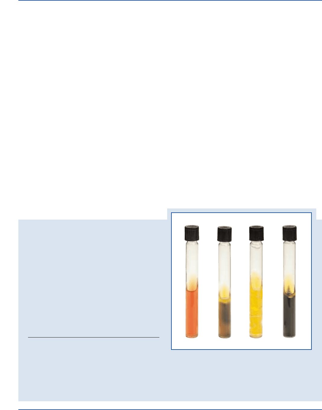
Citrobacter freundii 8090* undiluted good A/A + +
Escherichia coli 25922* undiluted good A/A + –
Proteus vulgaris 6380 undiluted good K/A – +
A = acid reaction (yellow) K = alkaline reaction (no color change)
+gas = cracks, splits or bubbles in medium –gas = no cracks, splits, or bubbles in medium
+H
2
S = black precipitate in butt –H
2
S = no black precipitate in butt
The cultures listed are the minimum that should be used for performance testing.
*These cultures are available as Bactrol
™
Disks and should be used as directed in Bactrol Disks Technical Information.
Proteus vulgatus
ATCC
®
6380
Citrobacter freundii
ATCC
®
8090
Escherichia coli
ATCC
®
25922
Uninoculated
tube
Formula
Kligler Iron Agar
Formula Per Liter
Bacto Beef Extract . . . . . . . . . . . . . . . . . . . . . . . . . . . . . . . . 3 g
Bacto Yeast Extract . . . . . . . . . . . . . . . . . . . . . . . . . . . . . . . . 3 g
Bacto Peptone . . . . . . . . . . . . . . . . . . . . . . . . . . . . . . . . . . . 15 g
Bacto Proteose Peptone . . . . . . . . . . . . . . . . . . . . . . . . . . . . 5 g
Bacto Lactose . . . . . . . . . . . . . . . . . . . . . . . . . . . . . . . . . . . 10 g
Bacto Dextrose . . . . . . . . . . . . . . . . . . . . . . . . . . . . . . . . . . . 1 g
Ferrous Sulfate . . . . . . . . . . . . . . . . . . . . . . . . . . . . . . . . . . 0.2 g
Sodium Chloride . . . . . . . . . . . . . . . . . . . . . . . . . . . . . . . . . . 5 g
Sodium Thiosulfate. . . . . . . . . . . . . . . . . . . . . . . . . . . . . . . 0.3 g
Bacto Agar . . . . . . . . . . . . . . . . . . . . . . . . . . . . . . . . . . . . . 12 g
Bacto Phenol Red . . . . . . . . . . . . . . . . . . . . . . . . . . . . . . 0.024 g
Final pH 7.4 ± 0.2 at 25°C
Precautions
1. For Laboratory Use.
2. Follow proper established laboratory procedure in handling and
disposing of infectious materials.
Storage
Store the dehydrated medium below 30°C. The dehydrated medium
is very hygroscopic. Keep container tightly closed. Store prepared
tubes at 2-8°C.
Expiration Date
The expiration date applies to the medium in its intact container when
stored as directed. Do not use a product if it fails to meet specifications
for identity and performance.
Procedure
Materials Provided
Kligler Iron Agar
Materials Required But Not Provided
Flasks with closures
Distilled or deionized water
Bunsen burner or magnetic hot plate
Tubes with closures
Autoclave
Incubator (35°C)
Method of Preparation
1. Suspend 55 grams in 1 liter distilled or deionized water.
2. Heat to boiling to dissolve completely.
3. Dispense into tubes with closures.
4. Autoclave at 121°C for 15 minutes. Cool in slanted position
with deep butts.
Specimen Collection and Preparation
1. Collect specimens or food samples in sterile containers or with
sterile swabs and transport immediately to the laboratory
following recommended guidelines.
6-10
2. Process each specimen, using procedures appropriate for that
specimen or sample.
6-10
