BD Diagnostic Systems (publ.). Difco Manual (Manual of Microbiological Culture)
Подождите немного. Документ загружается.


106 The Difco Manual
or spreading may be observed on moist surfaces. Growth of normal
enteric bacteria is markedly to completely inhibited. Growth of fungi
is markedly to completely inhibited on Campylobacter Agar Blaser.
Colonies are selected for further biochemical characterization.
Identification is based on a positive oxidase reaction and characteristic
darting motility in a wet mount.
1
For further differentiation into
species and biotypes, test for catalase activity, urease, hydrogen
sulfide production, nitrate reduction, hippurate, indoxyl acetate, DNA
hydrolysis and susceptibility to cephalothin and nalidixic acid.
1
Limitations of the Procedure
1. Campylobacter Agar prepared with either Campylobacter Antimi-
crobic Supplement S or Campylobacter Antimicrobic Supplement
B is selective primarily for Campylobacter species. Biochemical
testing using a pure culture is necessary for complete identification.
Consult appropriate references for further information.
1,3,7
2. Growth of Campylobacter fetus subsp. intestinalis may be
dramatically inhibited on Campylobacter Agar Blaser due to the
presence of cephalothin. The use of Campylobacter Agar Skirrow
and incubation at 35°C is suggested when isolating this organism
from mixed populations.
3. Some strains of C. fetus subsp. jejuni may be encountered that fail
to grow or grow poorly on prepared Campylobacter Agar.
4. Some strains of normal enteric organisms may be encountered that
are not inhibited or only partially inhibited on Campylobacter Agar.
References
1. Murray, P. R., E. J. Baron, M. A. Pfaller, F. C. Tenover, and
R. H. Yolken (ed.). Manual of clinical microbiology, 6th ed.
American Society for Microbiology, Washington, D.C.
2. Skirrow, M. D. 1977. Campylobacter enteritis: A “new” disease.
Br. Med. J. 2:9-11.
3. Vanderzant, C., and D. F. Splittstoesser (ed). 1992. Compen-
dium of methods for the microbiological examination of food,
3rd ed. American Public Health Association, Washington, D.C.
4. Association of Official Analytical Chemists. 1995. Bacteriological
analytical manual, 8th ed. AOAC International, Gaithersburg, MD.
5. Blaser, M. J., V. Berkowitz, F. M. LaForce, J. Cravens, L. B.
Reller, and W. L. Wang. 1979. Campylobacter enteritis: clinical
and epidemiologic features. Ann. Intern. Med. 91:179-185.
6. Blaser, M. J., J. Cravens, B. W. Powers, and W. L. Wang. 1978.
Campylobacter enteritis associated with canine infection. Lancet
(ii):979-980.
7. Koneman E. W., S. D. Allen, W. M. Janda, P. C.
Schreckenberger, W. C. Winn. 1983. Color atlas and textbook
of diagnostic microbiology, 5th ed. J. B. Lippencott-Raven
Publishers. Washington, D.C.
Packaging
Campylobacter Agar Base 2 kg 1820-07
Campylobacter Agar Kit Blaser 3279-32
To prepare: 6 x 1 liter
Campylobacter Agar Base 6 x 39.5 grams
Campylobacter
Antimicrobic Supplement B 6 x 10 ml
Campylobacter Agar Kit Blaser 3279-40
To prepare: 6 x 500 ml
Campylobacter Agar Base 6 x 19.75 grams
Campylobacter
Antimicrobic Supplement B 6 x 5 ml
Campylobacter Agar Kit Skirrow 3280-32
To prepare: 6 x 1 liter
Campylobacter Agar Base 6 x 39.5 grams
Campylobacter
Antimicrobic Supplement S 6 x 10 ml
Campylobacter Agar Kit Skirrow 3280-40
To prepare: 6 x 500 ml
Campylobacter Agar Base 6 x 19.75 grams
Campylobacter
Antimicrobic Supplement S 6 x 5 ml
Bacto
®
Candida BCG Agar Base
Candida BCG Agar Base Section II
Intended Use
Bacto Candida BCG Agar Base is used with added neomycin in isolating
and differentiating Candida from primary specimens.
Also Known As
Candida BCG Agar Base is an abbreviation for Candida Brom Cresol
Green Agar Base.
Summary and Explanation
Candida BCG Agar Base is prepared according to the formulation of
Harold and Snyder.
1
Candida BCG Agar Base was developed after a
study demonstrated triphenyltetrazolium chloride (TTC) employed in
Pagano Levin medium retarded the growth of some Candida species.
Harold and Snyder
1
used brom cresol green as the indicator, which is
nontoxic to Candida species. This medium is primarily used for
demonstrating morphological and biochemical reactions characterizing
the different Candida species for clinical diagnosis.
Candidiasis is the most frequently encountered opportunistic fungal
infection.
2
It is caused by a variety of species of Candida, with
Candida albicans being the most frequent etiological agent, followed
by Candida tropicalis and Candida (Torulopsis) glabrata.
2
Candida
species can be present in clinical specimens as a result of environmental
contamination, colonization, or actual disease process.
3
Principle of the Procedure
Bacto Peptone provides the nitrogen and amino acids in Candida BCG
Agar Base. Yeast Extract is the vitamin source. The high concentration
of Dextrose provides carbon as an energy source in this formula. Bacto
Agar is the solidifying agent. Brom cresol green is the pH indicator,
and acid production changes the medium from blue-green to yellow.
Due to pH changes, specific color patterns appear in the base and
surface of colonies for differentiation of Candida species.

The Difco Manual 107
User Quality Control
Identity Specifications
Dehydrated Appearance: Beige to blue-green, free-flowing,
homogeneous.
Solution: 6.6% solution, soluble in distilled or
deionized water on boiling, blue-green,
slightly opalescent to opalescent,
may have a precipitate.
Prepared Medium: Blue-green to greenish blue,
slightly opalescent to opalescent;
may have a precipitate.
Reaction of 6.6%
Solution at 25°C: pH 6.1 ± 0.1
Cultural Response
Prepare Candida BCG Agar Base per label directions. Inoculate medium
using the streak plate technique, and incubate at 30 ± 2°C for 24-48 hours.
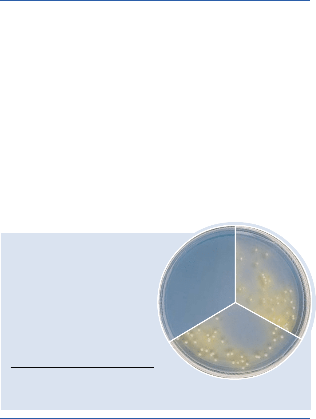
INOCULUM COLOR OF
ORGANISM ATCC
®
CFU GROWTH MEDIUM
Candida albicans 10231 100-1,000 good yellow
Candida tropicalis 3869 100-1,000 good yellow
Escherichia coli 25922* 1,000-2,000 inhibited green
Candida albicans
ATCC
®
10231
Uninoculated
plate
Candida tropicalis
ATCC
®
3869
The cultures listed are the minimum that should be used for performance testing.
*These cultures are available as Bactrol
™
Disks and should be used as directed in Bactrol Disks Technical Information.
Neomycin is added to the medium in a concentration of 500 µg/ml.
Neomycin and brom cresol green act as selective agents to inhibit
bacteria in Candida BCG Agar Base.
4
Formula
Candida BCG Agar Base
Formula Per Liter
Bacto Peptone . . . . . . . . . . . . . . . . . . . . . . . . . . . . . . . . . . . 10 g
Bacto Yeast Extract . . . . . . . . . . . . . . . . . . . . . . . . . . . . . . . . 1 g
Bacto Dextrose . . . . . . . . . . . . . . . . . . . . . . . . . . . . . . . . . . 40 g
Bacto Agar . . . . . . . . . . . . . . . . . . . . . . . . . . . . . . . . . . . . . 15 g
Brom Cresol Green . . . . . . . . . . . . . . . . . . . . . . . . . . . . . . 0.02 g
Final pH 6.1 ± 0.1 at 25°C
Precautions
1. For Laboratory Use.
2. Follow proper established laboratory procedures in handling and
disposing of infectious materials.
Storage
Store the dehydrated medium below 30°C. The dehydrated medium is
very hygroscopic. Keep container tightly closed.
Expiration Date
The expiration date applies to the product in its intact container when
stored as directed. Do not use a product if it fails to meet specifications
for identity and performance.
Procedure
Materials Provided
Candida BCG Agar Base
Materials Required But Not Provided
Glassware
Autoclave
Incubator (30°C)
Waterbath (45-55°C)
Neomycin (500 µg/ml)
Sterile Petri dishes
Method of Preparation
1. Suspend 66 grams in 1 liter distilled or deionized water.
2. Heat to boiling to dissolve completely.
3. Autoclave at 121°C for 15 minutes.
4. Cool the medium to 50-55°C. Add sterile neomycin (500 µg/ml).
Mix well.
Specimen Collection and Preparation
Obtain and process specimens according to the techniques and procedures
established by laboratory policy.
Test Procedure
Refer to the scheme for yeast identification.
3
For a complete discussion
on the isolation and identification of Candida species refer to the
procedures described in the appropriate references.
2,3,5
Results
Identification of Candida species on the basis of colony morphology
on Candida BCG Agar follows:
C. albicans: Colonies appear as blunt cones 4.5-5.5 mm diameter with
smooth edges and surfaces; coarse feathery growths may arise from
the center of the colony base to penetrate the medium. The color of
Section II Candida BCG Agar Base

108 The Difco Manual
the base and surface of the colonies is yellowish to bluish green
with the intensity diminishing from a gray green center spot to
paleness at the edge, although some strains may show a distinct
green outer ring.
C. stellatoidea: Colonies appear convex 4.0-5.0 mm in diameter, with
smooth edges and smooth to irregular surfaces; there is a fine central
basal feathery growth penetrating the medium. The color of both
base and surface of colonies is yellow to green, the intensity of which
may or may not diminish from center to border but is usually light.
C. tropicalis: Colonies appear convex or as low cones 4.5-5.0 mm in
diameter with smooth to undulate edges, and smooth to granular or
ridged surfaces; deeply stained feathery growth arises from several
points in the base of the colony to form an effusive cloud. The
color of the submerged growth is normally an intense blue green
compared with that of the base which is much lighter; the surface
is uniformly pale and may be yellowish green to green, reflecting a
lower pH than observed of the base.
C. pseudotropicalis: Colonies appear convex, 4.5-5.5 mm in diameter
with undulate to smooth edges, and smooth surfaces; occasionally
the surface is membranous but all colonies are shiny in appearance,
and there is feathering growth emerging from several points in the
base of the colony. The color of a large central area in the base of the
colony is a medium green, which diminishes in intensity toward the
edge; a similar distribution of color occurs on the surface, but this
green is bright in hue and is never grayed as it is with C. tropicalis.
C. krusei: Colonies appear as low cones 4.5-5.0 m in diameter with
pseudohyphal edges, which may be weakly contractile or spreading,
and have dull surfaces. There is abundant lightly colored growth
penetrating the medium from the base of the colony. The base of
the colony is a medium blue green in the center diminishing in
intensity to paleness at the edge; the surface is usually a light green
to yellow green without much concentration in any part.
C. parapsilosis: Colonies appear as convex to low cones 3.5-4.5 mm
in diameter with smooth or slightly spreading edges, but vary from
smooth to granular or rough surfaces; there is no submerged
growth. The color for both base and surface of the colony is blue
green over much of the colony, being more intense in the base than
the surface which is modified by a thin grayish film of cells; the
intensity in color fades abruptly leaving a broad pale edge.
C. guilliermondii: Colonies appear as low cones 4.0-5.0 mm in
diameter with very smooth edges and highly glossy surfaces; there
maybe a weak, fine feathered submerged growth. Both base and
surface of the colony tend to have blue centers of medium intensity
fading into a pale edge; however the surface may be blue green
with the central third lightened with gray.
C. glabrata: Colonies are smooth and convex, 4.6-5.0 mm diameter;
the surface color pattern is pale green in the center which becomes
medium green at the edge, and the base has the same color pattern
but of less intensity.
Limitations of the Procedure
1. Since the nutritional requirements of yeast vary, some strains may
be encountered that fail to grow or grow poorly on this medium.
References
1. Harold, W., and M. Snyder. 1968. Personal Communication.
2. Baron, E. J., L. R. Peterson, and S. M. Finegold. 1994. Bailey &
Scott’s diagnostic microbiology, 9th ed. Mosby-Year Book, Inc.,
St. Louis, MO.
3. Warren, N. G., and K. C. Hazen. 1995. Candida, Cryptococcus,
and other yeasts of medical importance, p. 723-737. In P. R.
Murray, E. J. Baron, M. A. Pfaller, F. C. Tenover, and R. H. Yolken
(ed.), Manual of clinical microbiology, 6th ed. American Society
for Microbiology, Washington, D.C.
4. MacFaddin, J. D. 1985. Media for isolation-cultivation-
identification-maintenance of medical bacteria, vol. 1, p. 136-137.
Williams & Wilkins, Baltimore, MD.
5. Isenberg, H. D. (ed.). 1992. Clinical microbiology procedures
handbook. American Society for Microbiology, Washington, D.C.
Packaging
Candida BCG Agar Base 500 g 0835-17
Candida Isolation Agar Section II
Bacto
®
Candida Isolation Agar
Intended Use
Bacto Candida Isolation Agar is used for isolating and differentiating
Candida albicans.
Summary and Explanation
Candida Isolation Agar is a nutritionally rich medium that supports
growth of many yeasts and molds and is differential for Candida
albicans. Candida Isolation Agar was developed using a modification
of YM Agar as described by Fung and Liang.
1
Goldschmidt demonstrated
that YM Agar with Aniline Blue WS could be used to identify
C. albicans in clinical samples with high accuracy and predictability.
2
Aniline Blue is metabolized by C. albicans to produce a fluorescent
moiety that can be detected under long wave UV light.
2
Principles of the Procedure
Yeast Extract provides nitrogen, carbon, vitamins and cofactors. Malt
Extract provides carbon, protein and nutrients. Bacto Peptone provides
additional carbon and nitrogen. Dextrose is an energy source. Aniline
Blue is a fluorescent indicator. Bacto Agar is a solidifying agent.
Formula
Candida Isolation Agar
Formula Per Liter
Bacto Yeast Extract . . . . . . . . . . . . . . . . . . . . . . . . . . . . . . . . 3 g
Bacto Malt Extract . . . . . . . . . . . . . . . . . . . . . . . . . . . . . . . . 3 g
Bacto Peptone . . . . . . . . . . . . . . . . . . . . . . . . . . . . . . . . . . . . 5 g
Bacto Dextrose . . . . . . . . . . . . . . . . . . . . . . . . . . . . . . . . . . 10 g
Bacto Agar . . . . . . . . . . . . . . . . . . . . . . . . . . . . . . . . . . . . . 20 g
Aniline Blue . . . . . . . . . . . . . . . . . . . . . . . . . . . . . . . . . . . . 0.1 g
Final pH 6.2 ± 0.2 at 25°C
Precautions
1. For Laboratory Use.
2. Follow proper established laboratory procedure in handling and
disposing of infectious materials.

The Difco Manual 109
Section II Candida Isolation Agar
Storage
Store dehydrated medium below 30°C. The dehydrated medium is very
hygroscopic. Keep container tightly closed.
Expiration Date
The expiration date applies to the product in its intact container when
stored as directed. Do not use a product if it fails to meet specifications
for identity and performance.
Procedure
Materials Provided
Candida Isolation Agar
Materials Required but not Provided
Glassware
Autoclave
Method of Preparation
1. Suspend 41.1 grams in 1 liter distilled or deionized water.
2. Heat to boiling to dissolve completely.
3. Autoclave at 121°C for 15 minutes.
Specimen Collection and Preparation
1. Specimens should be collected in sterile containers or with sterile
swabs and transported immediately to the laboratory according to
recommended guidelines.
3,4
Test Procedure
1. Process each specimen as appropriate for that specimen and inocu-
late directly onto the surface of the medium. Streak for isolation.
2. Incubate plates aerobically at 30°C for 18-72 hours.
3. Examine plates for growth after 18-72 hours of incubation.
Results
Colonies of C. albicans fluoresce yellow-green under long wave UV
light following incubation at 30°C for 18-24 hours. Non-C. albicans
isolates do not fluoresce.
Limitations of the Procedure
1. Strains of Candida albicans have been reported that are false
negative for fluorescence on this medium.
2
2. Strains of C. parapsilosis, C. krusei, and C. pulcherrima that fluoresce
on this medium may be encountered.
2
These strains may be distin-
guished from C. albicans based on germ tube formation in serum.
2,5
3. Since the nutritional requirements of organisms vary, some strains
may be encountered that fail to grow or grow poorly on this medium.
References
1. Fung, D. Y. C., and C. Liang. 1988. A new fluorescent agar for
the isolation of Candida albicans. Bull. Inf. Lab. Serv. Vet. (France)
29/30:1-2.
2. Goldschmidt, M. C., D. Y. C. Fung, R. Grant, J. White, and T.
Brown. 1991. New aniline blue dye medium for rapid identification
and isolation of Candida albicans. J. Clin. Micro. 29:1095-1099.
3. Miller, J. M., and H. T. Holmes. 1995. Specimen collection and
handling, p. 19- 32. In P. R. Murray, E. J. Baron, M. A. Pfaller, F. C.
Tenover, and R. H. Yolken, (ed.), Manual of clinical microbiology,
6th ed. American Society for Microbiology, Washington, D.C.
4. Splittstoesser, D. F., and C. Vanderzant (ed.). 1992. Compen-
dium of methods for the microbiological examination of foods,
3rd ed. American Public Health Association, Washington, D.C.
5. Murray, P. R, E. J. Baron, M. A. Pfaller, F. C. Tenover, and
R. H. Yolken (ed.). 1995. Manual of clinical microbiology, 6th ed.
American Society for microbiology, Washington, D.C.
Packaging
Candida Isolation Agar 500 g 0507-17
User Quality Control
Identity Specifications
Dehydrated
Medium Appearance: Beige, free-flowing, homogeneous.
Solution: 4.1% solution, soluble in distilled or deionized
water on boiling. Solution is medium blue,
very slightly opalescent.
Prepared Plates: Medium blue, slightly opalescent.
Reaction of 4.1%
Solution at 25°C: pH 6.2 ± 0.2
Cultural Response
Prepare Candida Isolation Agar per label instructions. Inoculate
and incubate plates aerobically at 30 ± 2°C for 18-72 hours.
INOCULUM
ORGANISM ATCC
®
CFU GROWTH FLUORESCENCE
Bacillus subtilis 6633 100-1,000 good negative
Candida albicans 10231 100-1,000 good positive
Escherichia coli 25922* 100-1,000 good negative
The cultures listed are the minimum that should be used for performance testing.
*This culture is available as Bactrol™ Disks and should be used as directed in Bactrol Disks Technical Information.
Candida albicans
ATCC
®
10231

110 The Difco Manual
Casamino Acids, Casamino Acids, Technical & Vitamin Assay Casamino Acids Section II
Bacto
®
Casamino Acids
.
Bacto Casamino Acids, Technical
Bacto Vitamin Assay Casamino Acids
User Quality Control
Identity Specifications
Casamino Acids
Dehydrated Appearance: Very light beige, free-flowing,
homogeneous.
Solution: 1% solution-very light amber, clear
solution.
2% solution-Light amber, clear,
soluble in distilled or deionized water
upon slight heating.
Reaction of a 2%
Solution at 25°C: pH 5.8-6.65
Casamino Acids, Technical
Dehydrated Appearance: Very light beige, free-flowing,
homogeneous.
Solution: 1% solution, soluble in distilled or
deionized water. Solution is colorless
to very light amber and clear.
Reaction of 1%
Solution at 25°C: pH 5.0-7.5
Vitamin Assay Casamino Acids
Dehydrated Appearance: Light beige, free-flowing, homogeneous.
Solution: 3% solution, soluble in distilled or
deionized water on boiling. Very
light to light amber, clear, may have
a slight precipitate.
Reaction of 3%
Solution at 25°C: pH 6.5-8.5
Cultural Response
Casamino Acids and Casamino Acids, Technical
Prepare a 1% solution and adjust the pH to 7.2 ± 0.2. Inoculate
tubes with the test organisms, and incubate at 35 ± 2°C for
18-48 hours.
INOCULUM
ORGANISM ATCC
®
CFU GROWTH
Escherichia coli 25922* 100-1,000 good
Salmonella typhi 19430 100-1,000 good
*This culture is available as Bactrol
™
Disks and should be used as
directed in Bactrol Disks Technical Information.
Vitamin Assay Casamino Acids
Vitamin Assay Casamino Acids is prepared in various vitamin
assay media to determine the vitamin content. It should not
contain a vitamin content higher than 20% above the
following values:
Vitamin B
12
0.2 nanograms/gram
Biotin 0.3 nanograms/gram
Folic Acid 3.3 nanograms/gram
Niacin 0.17 micrograms/gram
Pantothenate 0.04 micrograms/gram
Riboflavin 0.1 micrograms/gram
Thiamine 0.1 micrograms/gram
The cultures listed are the minimum that should be used for
performance testing.
Intended Use
Bacto Casamino Acids is used in preparing microbiological
culture media.
Bacto Casamino Acids, Technical is used in the preparation of
microbiological culture media.
Bacto Vitamin Assay Casamino Acids is used in vitamin assay
procedures.
Also Known As
Casamino Acids are also referred to as Casein Hydrolysate (Acid) or
Casein Peptone, Acid Hydrolysate.
Summary and Explanation
Casamino Acids is acid hydrolyzed casein having low sodium chloride
and iron concentrations. Casamino Acids is recommended for use in
microbiological culture media that require a completely hydrolyzed
protein as a nitrogen source. Casamino Acids is prepared according to
the method described by Mueller and Miller
1
and Mueller and Johnson.
2
Mueller
3
prepared diphtheria toxin in a medium containing a casein
hydrolysate as the source of nitrogen. It was shown that the high
sodium chloride content was the limiting factor in the amount of toxin
that could be produced in this medium. Mueller and Miller
1
described
a method to reduce the sodium chloride and iron content of the
hydrolyzed casein. This hydrolyzed casein, supplemented with
inorganic salts, growth factors, cystine, maltose and an optimum
amount of iron, was used to prepare diphtheria toxin.
1,3
Casamino
Acids duplicates this specially treated hydrolyzed casein.
In Casamino Acids, hydrolysis is carried out until all the nitrogen in
the casein is converted to amino acids or other compounds of relative
chemical simplicity. Casamino Acids is particularly well suited for
nutritional studies, microbiological assays, and in the semi-synthetic
medium for testing disinfectants.
4
Casamino Acids is also used in the
preparation of tetanus toxins, and pertussis vaccines, and for sulfonamide
inhibitor studies.
5
Casamino Acids, Technical is acid hydrolyzed casein. The hydrolysis
is carried out as in the preparation of Casamino Acids, but the sodium
chloride and iron content of this product have not been decreased to
the same extent. Casamino Acids, Technical is recommended for use
in culture media where amino acid mixtures are required for a nitrogen
source, and the sodium chloride content is slightly increased. It is
particularly valuable in studying the growth requirements of bacteria.
Casamino Acids, Technical is prepared according to the method
suggested by Mueller
1
for use in the preparation of diphtheria toxin.
Mueller and Hinton
6
used Casamino Acids, Technical in a medium for
primary isolation of gonococcus and meningococcus. Casamino Acids,
Technical was used in agar-free media for the isolation of Neisseria,
and in a tellurite medium for the isolation of Corynebacterium,
described by Levin.
7
Wolf
8
used Casamino Acids, Technical in the
preparation of a medium for the testing of disinfectants.
Vitamin Assay Casamino Acids is an acid digest of casein specially
treated to markedly reduce or eliminate certain vitamins. It is

The Difco Manual 111
Section II Casamino Acids, Casamino Acids, Technical & Vitamin Assay Casamino Acids
recommended for use in microbiological assay media and in studies of
the growth requirements of microorganisms. Vitamin Assay Casamino
Acids is commonly used as the amino acid source in early phases of
nutrition work.
9
Sarett
10
used Vitamin Assay Casamino Acids as the
acid hydrolyzed casein in his studies on p-aminobenzoic acid and
p-teroylglutamic acid as growth factors for Lactobacillus species.
Several media containing Casamino Acids are specified in standard
methods for multiple applications.
11,12,13
Principles of the Procedure
Casamino Acids, Casamino Acids, Technical and Vitamin Assay
Casamino Acids are acid hydrolyzed casein. Casein is milk protein,
and a rich source of amino acid nitrogen. Casamino Acids, Casamino
Acids, Technical and Vitamin Assay Casamino Acids provide nitrogen,
vitamins, carbon and amino acids in microbiological culture media.
Although Casamino Acids, Casamino Acids, Technical, and Vitamin
Assay Casamino Acids are added to media primarily because of their
organic nitrogen and growth factor components, their inorganic
components also play a vital role.
14
Formula
Casamino Acids is a dehydrated acid hydrolyzed casein in which
Sodium Chloride and Iron are present in low concentrations permitting
toxin production.
Casamino Acids, Technical is a dehydrated acid hydrolyzed casein.
The Sodium Chloride and Iron content have not been reduced to same
extent as Casamino Acids.
Vitamin Assay Casamino Acids is an acid hydrolyzed casein used to
prepare media for microbiological assay of vitamins.
Typical Analysis
Physical Characteristics
Ash (%) 24.4 Loss on Drying (%) 4.5
Clarity, 1% Soln (NTU) 0.5 pH, 1% Soln 6.4
Filterability (g/cm
2
) 2.9
Nitrogen Content (%)
Total Nitrogen 10.5 AN/TN 83.8
Amino Nitrogen 8.8
Amino Acids (%)
Alanine 3.26 Lysine 5.71
Arginine 2.20 Methionine 1.28
Aspartic Acid 4.76 Phenylalanine 2.11
Cystine 0.16 Proline 6.17
Glutamic Acid 15.30 Serine 2.19
Glycine 1.31 Threonine 2.41
Histidine 1.66 Tryptophan <0.01
Isoleucine 3.34 Tyrosine 0.47
Leucine 5.47 Valine 4.30
Inorganics (%)
Calcium <0.001 Phosphate 3.325
Chloride 7.400 Potassium 0.410
Cobalt <0.001 Sodium 8.710
Copper <0.001 Sulfate 0.045
Iron <0.001 Sulfur 0.420
Lead <0.001 Tin <0.001
Magnesium 0.002 Zinc <0.001
Manganese <0.001
Vitamins (µg/g)
Biotin <0.1 PABA <5.0
Choline
(as Choline Chloride)
160.0 Pantothenic Acid <0.1
Cyanocobalamin <0.1 Pyridoxine <0.1
Folic Acid <0.1 Riboflavin 1.8
Inositol <100.0 Thiamine 1.2
Nicotinic Acid <20.0 Thymidine <30.0
Biological Testing (CFU/g)
Coliform negative Standard Plate Count 950
Salmonella negative Thermophile Count 25
Spore Count 390
Precautions
1. For Laboratory Use.
2. Follow proper established laboratory procedures in handling and
disposing of infectious materials.
Storage
Store the dehydrated product below 30°C. The dehydrated product is
very hygroscopic. Keep container tightly closed.
Expiration Date
The expiration date applies to the product in its intact container when
stored as directed. Do not use a product if it fails to meet specifications
for identity and performance.
Procedure
Materials Provided
Casamino Acids
Casamino Acids, Technical
Vitamin Assay Casamino Acids
Materials Required But Not Provided
Materials vary depending on the medium being prepared.
Method of Preparation
Refer to the final concentration of Casamino Acids, Casamino Acids,
Technical or Vitamin Assay Casamino Acids in the formula of the
medium being prepared. Add Casamino Acids, Casamino Acids,
Technical or Vitamin Assay Casamino Acids as required.
Specimen Collection and Preparation
Obtain and process specimens according to the techniques and procedures
established by laboratory policy.
Test Procedure
See appropriate references for specific procedures using Casamino
Acids, Casamino Acids, Technical or Vitamin Assay Casamino Acids.
Results
Refer to appropriate references and procedures for results.
References
1. Mueller and Miller. 1941. J. Immunol. 40:21.
2. Mueller and Johnson. 1941. J. Immunol. 40:33.
3. Mueller. 1939. J. Immunol. 37:103.

112 The Difco Manual
Casein Digest Section II
Bacto
®
Casein Digest
User Quality Control
Identity Specifications
Dehydrated Appearance: Tan, free-flowing, homogeneous.
Solution: 1%, 2%, and 10% solutions, soluble
in distilled or deionized water:
1%-Light amber, clear;
2%-Medium amber, clear;
10%-Dark amber, clear, no significant
precipitate.
Reaction of 1%
Solution at 25°C: pH 7.2 ± 0.2
Cultural Response
Prepare NZM Broth per formula. Inoculate and incubate at
35 ± 2°C for 18-24 hours.
INOCULUM
ORGANISM ATCC
®
CFU GROWTH
Bacillus subtilis
†
6633 100-1,000 good
Escherichia coli (HB101) 33694 100-1,000 good
Escherichia coli (JM107) 47014 100-1,000 good
Escherichia coli (DH5) 53868 100-1,000 good
Saccharomyces cerevisiae 9763 100-1,000 good
Streptomyces avermitilis 31267 100-1,000 good
The cultures listed are the minimum that should be used for
performance testing.
†
Bacillus subtilis is available as Subtilis Spore Suspension.
Intended Use
Bacto Casein Digest is used in preparing microbiological culture media.
Also Known As
Casein Digest is similar to N-Z-Amine A.
Summary and Explanation
Casein Digest, an enzymatic digest of casein, was developed for use in
molecular genetics media. This product is digested under conditions
different from other enzymatic digests of casein, including Tryptone
and Casitone.
Casein Digest is contained in the formulas of NZ media (NZCYM
Broth, NZYM Broth and NZM Broth), which are used for cultivating
recombinant strains of Escherichia coli. E. coli grows rapidly in these
rich media because they provide amino acids, nucleotide precursors,
vitamins and other metabolites that the cells would otherwise have to
synthesize.
1
Consult appropriate references for recommended test
procedures using NZ media.
1,2
Principles of the Procedure
Casein Digest is a nitrogen and amino acid source for microbiological
culture media. Casein is raw milk protein, a rich source of amino acid
nitrogen.
Precautions
1. For Laboratory Use.
2. Follow proper established laboratory procedures in handling and
disposing of infectious materials.
Storage
Store Casein Digest below 30°C. The product is very hygroscopic.
Keep container tightly closed.
Expiration Date
The expiration date applies to the product in its intact container when
stored as directed. Do not use a product if it fails to meet specifications
for identity and performance.
Procedure
Materials Provided
Casein Digest
Materials Required But Not Provided
Materials vary depending on the medium being prepared.
4. Klarman and Wright. 1945. Soap and San. Chem. 21:113.
5. Straus, Dingle and Finland. 1941. J. Immunol. 42:331.
6. Mueller and Hinton. 1941. Proc. Soc. Exp. Biol. Med. 48:330.
7. Levin. 1943. J. Bacteriol. 46:233.
8. Wolf. 1945. J. Bacteriol. 49:463.
9. Nolan, R. A. 1971. Amino acids and growth factors in vitamin-free
casamino acids. Mycol. 63:1231-1234.
10. Sarett. 1947. J. Biol. Chem. 171:265.
11. Vanderzant, C., and D. F. Splittstoesser (ed.). 1992. Compendium
of methods for the microbiological examination of food, 3rd. ed.
American Public Health Association, Washington, D.C.
12. Association of Official Analytical Chemists. 1995. Bacteriological
analytical manual, 8th ed. AOAC International, Gaithersburg, MD.
13. Eaton, A. D., L. S. Clesceri, and A. E. Greenberg (ed.). 1995.
Standard methods for the examination of water and wastewater,
19th ed. American Public Health Association, Washington, D.C.
14. Nolan, R. A., and W. G. Nolan. 1972. Elemental analysis of
vitamin-free casamino acids. Appl. Microbiol. 24:290-291.
Packaging
Casamino Acids 100 g 0230-15
500 g 0230-17
2 kg 0230-07
10 kg 0230-08
Casamino Acids, Technical 500 g 0231-17
10 kg 0231-08
Vitamin Assay Casamino Acids 100 g 0288-15
500 g 0288-17

The Difco Manual 113
Section II Casitone
Method of Preparation
Refer to the final concentration of Casein Digest in the formula of the
medium being prepared. Add Casein Digest as required.
Specimen Collection and Preparation
Obtain and process specimens according to the techniques and proce-
dures established by laboratory policy.
Test Procedure
See appropriate references for specific procedures using Casein Digest.
Results
Refer to appropriate references and procedures for results.
References
1. Ausubel, F. M., R. Brent, R. E. Kingston, D. D. Moore, J. G.
Seidman, J. A. Smith, and K. Struhl (ed.). 1994. Current
protocols in molecular biology, vol.1. Current Protocols,
New York, NY.
2. Sambrook, J., E. F. Fritsch, and T. Maniatis. 1989. Molecular
cloning: a laboratory manual, 2nd ed. Cold Spring Harbor
Laboratory, Cold Spring Harbor, NY.
Packaging
Casein Digest 500 g 0116-17
Bacto
®
Casitone
User Quality Control
Identity Specifications
Dehydrated Appearance: Tan, free-flowing granules.
Solution: 1%, 2% and 10% solutions are
soluble in distilled or deionized water.
1%-Light amber, clear, no precipitate.
2%-Light to medium amber, clear,
may have a slight precipitate.
10%-Medium to dark amber, clear
to very slightly opalescent, may
have a precipitate.
Reaction of 1%
Solution at 25°C: pH 6.8 - 7.4
Cultural Response
All solutions are prepared with the pH adjusted to 7.2 - 7.4.
TEST SOLUTION ORGANISM ATCC
®
RESULT INOCULUM
Fermentable 2% Escherichia 25922* negative –
Carbohydrates coli
Indole 0.1% Escherichia 25922* positive –
Production coli
Acetylmethylcarbinol 0.1% Enterobacter 13048* positive –
Production aerogenes
Hydrogen Sulfide 1% Salmonella 6539 positive –
Production typhi
Toxicity 2%w/0.5% Escherichia 25922* good 100-1,000
NaCl & coli growth
1.5% Agar
Toxicity 2%w/0.5% Staphylococcus 25923* good 100-1,000
NaCl & aureus growth
1.5% Agar
The cultures listed are the minimum that should be used for
performance testing.
*These cultures are available as Bactrol
™
Disks and should be used
as directed in Bactrol Disks Technical Information.
Intended Use
Bacto Casitone is used in preparing microbiological culture media.
Summary and Explanation
Casitone, is recommended for preparing media where an enzymatic
hydrolyzed casein is desired. Casitone is used to support the growth of
fastidious microorganisms. The high tryptophan content of Casitone
makes it valuable for use in detecting indole production.
Dubos Broth and Dubos Oleic Agar media that support the growth of
Mycobacterium tuberculosis contain Casitone. Media used for the
enumeration of coliforms in water, m Endo Agar and m Endo Broth
MF
®
, use Casitone as a nitrogen source. Several Thioglycollate media
used for detecting microorganisms in normally sterile materials,
include Casitone as a nitrogen and amino acid source.
Casitone is recommended for preparing media for sterility testing
according to US Pharmacopeia XXIII (USP).
1
Several media containing
Casitone are specified in standard methods
2,3,4,5
for multiple applications.
Principles of the Procedure
Casitone is a pancreatic digest of casein. Casein is the main protein of
milk, and a rich source of amino acid nitrogen.
Precautions
1. For Laboratory Use.
2. Follow proper established laboratory procedures in handling and
disposing of infectious materials.
Storage
Store the dehydrated product below 30°C. The dehydrated product is
very hygroscopic. Keep container tightly closed.
Expiration Date
The expiration date applies to the product in its intact container when
stored as directed. Do not use a product if it fails to meet specifications
for identity and performance.
Typical Analysis
Physical Characteristics
Ash (%) 7.0 Loss on Drying (%) 3.7
Clarity, 1% Soln (NTU) 0.6 pH, 1% Soln 7.2
Filterability (g/cm
2
)1.7
Carbohydrate (%)
Total 0.2

114 The Difco Manual
Nitrogen Content (%)
Total Nitrogen 13.3 AN/TN 35.3
Amino Nitrogen 4.7
Amino Acids (%)
Alanine 3.01 Lysine 13.62
Arginine 3.76 Methionine 1.71
Aspartic Acid 6.61 Phenylalanine 4.02
Cystine 0.02 Proline 8.57
Glutamic Acid 20.03 Serine 4.82
Glycine 1.97 Threonine 3.74
Histidine 2.17 Tryptophan 0.14
Isoleucine 4.16 Tyrosine 2.09
Leucine 8.74 Valine 4.06
Inorganics (%)
Calcium 0.010 Phosphate 2.604
Chloride 0.110 Potassium 0.162
Cobalt <0.001 Sodium 3.073
Copper <0.001 Sulfate 0.339
Iron 0.003 Sulfur 0.676
Lead <0.001 Tin <0.001
Magnesium 0.019 Zinc 0.004
Manganese <0.001
Vitamins (µg/g)
Biotin 0.2 PABA 15.9
Choline
(as Choline Chloride)
550.0 Pantothenic Acid 7.7
Cyanocobalamin <0.1 Pyridoxine 1.3
Folic Acid 0.8 Riboflavin 0.4
Inositol 980.0 Thiamine <0.1
Nicotinic Acid 20.3 Thymidine 342.9
Biological Testing (CFU/g)
Coliform negative Standard Plate Count 1850
Salmonella negative Thermophile Count 100
Spore Count 300
Procedure
Materials Provided
Casitone
Materials Required But Not Provided
Materials vary depending on the medium being prepared.
Method of Preparation
Refer to the final concentration of Casitone in the formula of the
medium being prepared. Add Casitone as required.
Specimen Collection and Preparation
Obtain and process specimens according to the techniques and
procedures established by laboratory policy.
Test Procedure
See appropriate references for specific procedures using Casitone.
Results
Refer to appropriate references and procedures for results.
References
1. The United States Pharmacopeial Convention. 1995. The United
States Pharmacopeia, 23rd ed. Sterility test, p. 1686-1690. The
United States Pharmacopeial Convention Inc., Rockville, MD.
2. Vanderzant, C., and D. F. Splittstoesser (ed.). 1992.
Compendium of methods for the microbiological examination of
food, 3rd ed. American Public Health Association, Washington, D.C.
3. Association of Official Analytical Chemists. 1995. Bacteriological
analytical manual, 8th ed. AOAC International, Gaithersburg, MD.
4. Eaton, A. D., L. S. Clesceri, and A. E. Greenberg (ed.). 1995.
Standard methods for the examination of water and wastewater,
19th ed. American Public Health Association, Washington, D.C.
5. Marshall, R. T. (ed.). 1993. Standard methods for the examination
of dairy products, 16th ed., American Public Health Association,
Washington, D.C.
Packaging
Casitone 100 g 0259-15
500 g 0259-17
10 kg 0259-08
Casman Medium Base Section II
Bacto
®
Casman Medium Base
Intended Use
Bacto Casman Medium Base is used with blood in isolating fastidious
microorganisms under reduced oxygen tension.
Summary and Explanation
In 1947, Casman
1,2,3
described an infusion-free medium enriched with
5% blood for fastidious microorganisms incubated anaerobically. This
medium replaced labor intensive formulas containing fresh meat
infusion and unheated and heated blood.
1
Casman adjusted the
medium after experiments revealed that nicotinamide disrupted the
action of a blood enzyme that inactivates V factor (NAD).
2
Using
unheated human blood in the formula, Haemophilus influenzae grew
well and Neisseria was inhibited. The concentration of nicotinamide
was lowered to support growth of Neisseria species.
2,3
Casman Agar Base with rabbit blood can be used for the cultivation
and maintenance of Gardnerella vaginalis.
4
Principles of the Procedure
Proteose Peptone No.3, Tryptose and Beef Extract provide nitrogen,
vitamins and amino acids. Nicotinamide enhances growth of
N. gonorrhoeae and H. influenzae by impeding the removal of
coenzyme (V factor) by nucleotidase from the enriched blood. The
small amount of Dextrose is added to enhance growth of pathogenic
cocci. Sodium chloride maintains the osmotic balance of the medium.
Para-aminobenzoic acid is a preservative. Corn starch is added to
ensure that any toxic metabolites produced are absorbed, to neutralize
glucose inhibition of beta-hemolysis
4
and to enhance growth of
Neisseria species. Agar Noble is a solidifying agent.

The Difco Manual 115
Section II Casman Medium Base
User Quality Control
Identity Specifications
Dehydrated Appearance: Light tan, free-flowing, homogeneous.
Solution: 4.3% solution, soluble in distilled or
deionized water with frequent agitation
on boiling. Light to medium amber
with a ground glass appearance.
Prepared Medium: Without blood, light to medium
amber with a ground glass appearance.
With 5% blood, cherry red opaque.
Reaction of 4.3%
Solution at 25°C: pH 7.3 ± 0.2
Cultural Response
Prepare Casman Medium Base per label directions, enrich
with 5% sterile blood and 0.15% sterile water-lysed blood
solution. Inoculateprepared medium and incubate at 35 ± 2°C
under increased CO
2
for 18-48 hours.
INOCULUM GROWTH
ORGANISM ATCC
®
CFU w/BLOOD
Haemophilus influenzae 10211 100-1,000 good
Neisseria gonorrhoeae
CDC
116 100-1,000 good
Streptococcus pneumoniae 6305 100-1,000 good
Streptococcus pyogenes 19615* 100-1,000 good
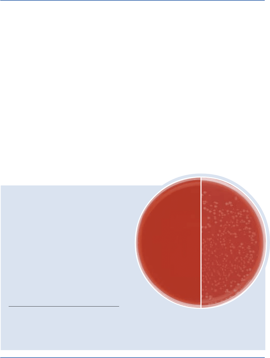
Neisseria gonorrhoeae
CDC 116
with enrichment
Uninoculated plate
with enrichment
The cultures listed are the minimum that should be used for performance testing.
*These cultures are available as Bactrol
™
Disks and should be used as directed in Bactrol Disks Technical Information.
Formula
Casman Medium Base
Formula Per Liter
Bacto Proteose Peptone No. 3 . . . . . . . . . . . . . . . . . . . . . . 10 g
Bacto Tryptose . . . . . . . . . . . . . . . . . . . . . . . . . . . . . . . . . . 10 g
Bacto Beef Extract . . . . . . . . . . . . . . . . . . . . . . . . . . . . . . . . 3 g
Nicotinamide. . . . . . . . . . . . . . . . . . . . . . . . . . . . . . . . . . . 0.05 g
p-Aminobenzoic Acid. . . . . . . . . . . . . . . . . . . . . . . . . . . . 0.05 g
Bacto Dextrose . . . . . . . . . . . . . . . . . . . . . . . . . . . . . . . . . . 0.5 g
Corn Starch . . . . . . . . . . . . . . . . . . . . . . . . . . . . . . . . . . . . . . 1 g
Sodium Chloride . . . . . . . . . . . . . . . . . . . . . . . . . . . . . . . . . . 5 g
Agar Noble . . . . . . . . . . . . . . . . . . . . . . . . . . . . . . . . . . . . . 14 g
Final pH 7.3 ± 0.2 at 25°C
Precautions
1. For Laboratory Use.
2. Follow proper established laboratory procedures in handling and
disposing of infectious materials.
Storage
Store the dehydrated medium below 30°C. The dehydrated medium is
very hygroscopic. Keep container tightly closed.
Expiration Date
The expiration date applies to the product in its intact container when
stored as directed. Do not use a product if it fails to meet specifications
for identity and performance.
Procedure
Materials Provided
Casman Medium Base
Materials Required But Not Provided
Glassware
Autoclave
Incubator (35°C)
Waterbath (45-50°C) (optional)
Sterile defibrinated blood
Sterile water-lysed blood
Method of Preparation
1. Suspend 43 grams in 1 liter distilled or deionized water.
2. Heat to boiling to dissolve completely.
3. Autoclave at 121°C for 15 minutes. Cool to 50°C.
4. Add 5% sterile blood and 0.15% sterile water-lysed blood solution
(one part blood to three parts water). Omit water-lysed blood if
sterile blood is partially lysed.
5. Dispense into sterile Petri dishes or as desired.
Specimen Collection and Preparation
Obtain and process specimens according to the techniques and
procedures established by laboratory policy.
