Whittemore S. The Circulatory System
Подождите немного. Документ загружается.


50
Anatomy of the
Circulatory System
5
As described in Chapter 2, the human circulatory system is divided
into two separate circuits, the systemic circuit and the pulmonary
circuit (Figure 5.1). Each of these circuits consists of a similar
sequence of blood vessels. Blood is pumped out of the heart into
arteries of decreasing size that merge into arterioles before reaching
the sites of exchange with the capillaries. Blood leaving the capillaries
is gathered into venules and then veins before returning to the heart.
The systemic circuit, the left side of the heart, provides the pressure
to propel blood to the entire body. The pulmonary circuit, the right
side of the heart, takes blood to the lungs for gas exchange with
the atmosphere.
In this chapter, you will examine the anatomy of the heart and
blood vessels. You will also learn about two common circulatory
diseases afflicting millions of Americans, atherosclerosis and
myocardial infarction (heart attack).
ANATOMY OF THE HEART
The heart beats steadily from early in embryonic development until
death. If a person lives until 75 years of age, and during that time his
or her heart beats an average of 75 times per minute, by the time the
person dies, the heart will have beat a total of 3 billion times and
pumped more than 200 million liters of blood.
The heart is located in the chest, or thoracic, cavity with the lungs.
It lies slightly left of the midline of the body. Because the heart takes
CH.YBW.Cir.C05.Final.q 12/20/06 3:19 PM Page 50

51
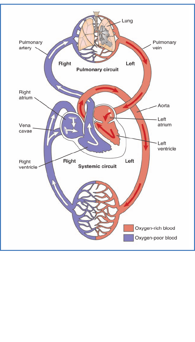
Figure 5.1 An overview of the pulmonary and systemic circuits of the
human circulatory system is illustrated here. The human heart has four
chambers. The right atrium and ventricle pump blood into the pulmonary
circuit, while the left atrium and ventricle move blood into the systemic
circuit. For both circuits, blood leaving the heart travels through arteries,
then the arterioles, and the capillaries. In the pulmonary circuit, gas
exchange occurs in the capillaries in the lungs. In the systemic circuit,
gas exchange occurs with the bodily tissues. Blood leaving the capillaries
is gathered into venules and then veins before returning to the heart.
In this diagram, blue blood represents blood of low oxygen content, and
the red blood represents fully oxygenated blood.
CH.YBW.Cir.C05.Final.q 12/20/06 3:19 PM Page 51

THE CIRCULATORY SYSTEM
up more space on the left side of the chest cavity, the left lung
has two lobes, compared to the three lobes of the right lung.
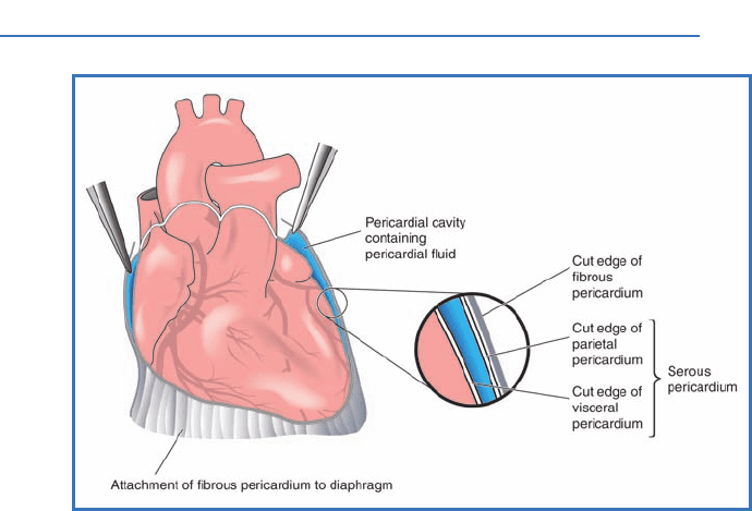
The heart is surrounded by a pericardium, a lining that separates
the heart from the lungs and the chest wall (Figure 5.2). The
pericardium consists of two membranes with fluid between
them—the fibrous portion and the serous portion. This peri-
cardial fluid lubricates the heart and reduces friction during
beating. The tough outer pericardial membrane (fibrous
pericardium) lines the outer surface of the heart and helps keep
the heart in position while beating.
The heart possesses four chambers, two
atria (plural for
atrium) and two ventricles (Figure 5.3). The right side of
the heart, consisting of the right atrium and right ventricle, is
separated from the left side of the heart by a wall, or septum.
The right and left side of the heart may beat as one unit, but
52
Figure 5.2 The heart is surrounded by the pericardium, two
layers of membrane separated by pericardial fluid. The fluid helps
lubricate the heart and reduce friction. The tough outer membrane
of the pericardium (fibrous pericardium) helps keep the heart in
place during its vigorous beating actions.
CH.YBW.Cir.C05.Final.q 12/20/06 3:19 PM Page 52

they are completely separate from each other with respect
to the blood they contain. Both the right and left atria are
separated from their respective ventricles by the
atrioventricular
(AV) valves, folds of tough tissue that open in one direction
only, from the atrium into the ventricle. Atrioventricular valves
are also known as cuspid valves.
The atria receive blood returning to the heart and then
pump that blood into the ventricles. The ventricles are the
more muscular pumps of the heart, because they must generate
enough force to propel the blood out into circulation against the
pressures existing in the two circuits. The muscular walls of the
atria are thinner than those of the ventricles, reflecting the fact
that they do not have to generate the high forces required of the
53
Anatomy of the Circulatory System
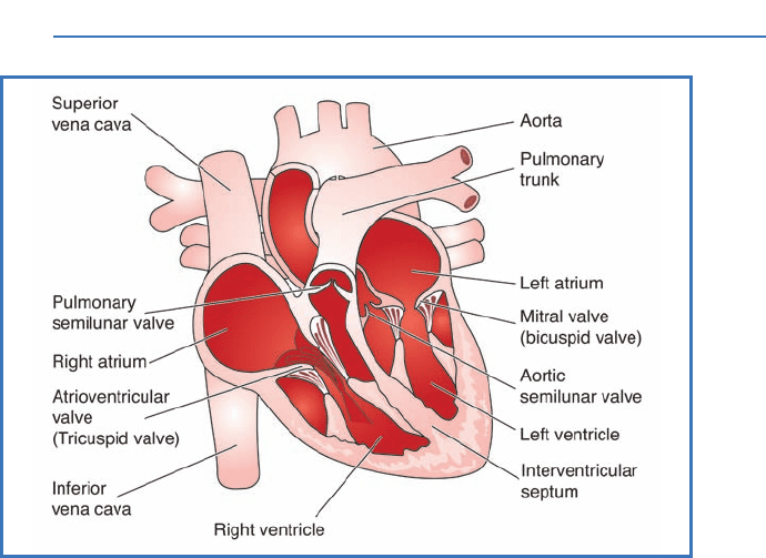
Figure 5.3 The basic anatomy of the human heart includes the
chambers, known as atria and ventricles, the attached major blood
vessels, and the two types of heart valves, the semilunar and
atrioventricular valves. Recall that the right side of the heart sends
blood to the lungs, and the left side supplies the entire body. Note
the thickness of the left ventricular wall.
CH.YBW.Cir.C05.Final.q 12/20/06 3:19 PM Page 53

THE CIRCULATORY SYSTEM
ventricles. Similarly, because the left ventricle must generate
enough force to overcome the higher pressure existing in the
systemic circuit and propel blood for longer distances, its
muscular walls are thicker than those of the right ventricle. The
right ventricle supplies the pulmonary circuit, where the distance
traveled by the blood is short and blood pressure is lower.
The primary component of both the atrial and ventricular
walls is
cardiac muscle. Although all muscle tissue is special-
ized for contraction, cardiac muscle has some characteristics
that differ from the skeletal muscle used to move the joints,
reflecting its unique function. For example, individual cardiac
muscle cells are smaller than skeletal muscle cells, and they
contain a single nucleus (Figure 5.4). Cardiac muscle cells
are well-connected to each other through regions known as
intercalated discs. A high density of adhesion molecules
known as
desmosomes keep the cells tightly attached to each
other in these regions, ensuring that the forces generated during
the beating actions of the heart do not rip apart the heart
muscle.
Gap junctions allow ions to move from one cardiac cell
to another, and, as you will learn in Chapter 6, these junctions
help the heart muscle to synchronize its actions.
The muscular walls of the atria are easily stretched and can
accommodate large volumes of blood returning to the heart.
The right atrium receives blood returning from the systemic
circuit via two large veins, the
superior vena cava, which
drains all regions above the heart, and the
inferior vena cava,
which collects blood returning from the lower body regions
(refer again to Figure 5.3). The AV valve separating the right
atrium and ventricle is sometimes called the
tricuspid valve
because it is composed of three flaps of tissue. This valve opens
only when blood pressure in the atria exceeds ventricular
pressure, thus preventing any backflow into the atria when
the pressure gradient is reversed. The left AV valve, or
bicuspid
valve, serves a similar function between the left atrium and
ventricle, but consists of two flaps instead of three.
54
CH.YBW.Cir.C05.Final.q 12/20/06 3:19 PM Page 54

The cone-shaped left and right ventricles are similar in
design. The right ventricle pushes blood out into the pulmonary
circuit through a
pulmonary semilunar valve, which separates the
ventricular chamber from the pulmonary trunk. This valve opens
when ventricular pressure exceeds pulmonary trunk pressure,
otherwise it remains closed. In a similar fashion, the
aortic
semilunar valve separates the left ventricle from the ascending
aorta. Both semilunar valves prevent blood from flowing back
into the heart once it has had been forced out into circulation.
55
Anatomy of the Circulatory System
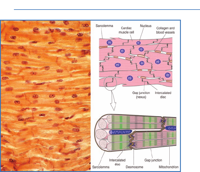
Figure 5.4 Cardiac muscle cells are smaller than skeletal
muscle cells and are connected through structures known as
intercalated discs. Desmosomes, or adhesion molecules, help to
hold the cardiac cells together during contractions. Gap junctions
allow for synchronization of heart contractions. A photograph of
actual cardiac muscle is shown on the left. The illustrations on
the right depict the components of cardiac muscle.
CH.YBW.Cir.C05.Final.q 12/20/06 3:19 PM Page 55

THE CIRCULATORY SYSTEM
The importance of the AV and semilunar valves is under-
scored by conditions that lead to their malfunction. Rheumatic
fever, a condition that may develop after an infection with
Streptococcus, can lead to valve dysfunction even decades
after the infection occurred. Some individuals are born with
malformations of their heart valves. Regardless of the cause,
malfunctioning valves can cause debilitating reductions in
cardiac function. Surgical repair and replacement, often with
valves obtained from the similar-sized pig heart, is a common
treatment for these valvular diseases.
56
CORONARY-ARTERY DISEASE
AND HEART ATTACK
Coronary arteries bring oxygen-rich blood to the hardworking
heart muscle. The blockage of these arteries, as well as others
in the body, most often arises from a condition known as
atherosclerosis. With this disease, calcified fatty deposits
build up to form so-called plaques in the inner lining of these
arteries. If these plaques grow large enough to reduce blood
flow, the heart’s access to oxygen and nutrients may be
affected and its ability to function impaired. If a plaque
ruptures, the blood clot that forms as a result may block
the artery completely or break free and lodge in another
smaller artery. In either of these cases, if the flow of blood
to a region of the heart is interrupted for more than a few
minutes, permanent damage to that region in the form of
a myocardial infarction (better known as a heart attack) is
very likely to occur. The extent and severity of the damage
determines whether the individual who suffered the attack
will live or die. Warning signs of an impending heart attack
may include
angina, or chest pain. People suffering from
angina often experience the pain when they exert them-
selves. As their level of activity increases, the heart
works harder to compensate and is more likely to become
oxygen-deprived.
CH.YBW.Cir.C05.Final.q 12/20/06 3:19 PM Page 56

THE CORONARY ARTERIES
The heart is a hardworking muscle and therefore needs
an ample supply of oxygen and fuel. The heart muscle does
not gain these essentials from the blood it is pumping;
instead, it requires its own extensive blood supply. The
coronary arteries serve this function. Recall that the left
ventricle pumps blood out into the systemic circuit,
through the left semilunar valve, and into the ascending
aorta. At the base of the ascending aorta, the right and left
coronary arteries branch off to bring oxygen-rich blood to
57
Anatomy of the Circulatory System
The risk factors associated with atherosclerosis include
high levels of “bad” cholesterol, or LDL, and low levels of
HDL, or “good” cholesterol. There is increased incidence
for older individuals, for people with high blood pressure
or diabetes, and for those who smoke, are obese, or are
inactive. Genetics also appears to play a role. Medical
practitioners use blood tests, ECGs (electrocardiograms),
stress tests, and techniques that visualize the blood flow
through the coronary or other arteries to diagnose the presence
of atherosclerosis.
Clogged coronary arteries can be opened using
angioplasty.
Plaques can be removed or pressed into the arterial wall using
an inflated balloon. If these procedures fail to increase blood
flow to the heart muscle adequately, then coronary bypass
surgery may be performed. For this treatment, small vessels,
like the great saphenous vein of the leg, are removed to
replace a diseased section of a coronary artery. A quadruple
bypass surgery means that four separate coronary arteries are
bypassed using this technique during one operation. Bypass
surgery has become safer and fairly routine and is successful at
improving heart function and reducing angina in most individuals
with coronary-artery disease.
CH.YBW.Cir.C05.Final.q 12/20/06 3:19 PM Page 57

THE CIRCULATORY SYSTEM
their respective sides of the heart. Blood flow through these
arteries can increase up to nine times the resting rate during
intensive exercise, when the heart is pumping maximally.
The coronary arteries of a large number of Americans are
diseased, reducing the ability of their hearts to function
properly (see box on page 56).
THE BLOOD VESSELS
The circulatory system consists of the heart, the blood vessels,
and blood. We have just examined the structure and function
of the heart, the muscular pump that provides the force
to circulate blood throughout the body. Previously, we
discussed the composition of blood, the fluid medium that
transports oxygen, nutrients, and water to our cells and
removes wastes. Now, we will examine the types of blood vessels
found in the human circulatory system.
Within each of the two circuits, there are five basic types of
blood vessels: arteries, arterioles, capillaries, venules, and veins.
Each type of blood vessel differs in form and function.
When blood is first ejected from the heart, it enters large
arteries that immediately begin to branch into medium-sized
and then smaller arteries. The arteries receive the pressurized
blood from the heart and distribute it to all of the body’s
tissues, including the heart itself. Arterial walls are thick
because they are very muscular. The larger arteries have elastic
walls that can withstand the enormous changes in blood
pressure that accompany the actions of the heart. These vessels
are designed for efficiently transporting blood away from
the heart.
The medium-sized arteries distribute blood to the skeletal
muscles and major organs. These arteries, in general, have a
thinner layer of muscle, although the difference in structure
from the larger arteries is subtle. Overall, the diameters of the
vessels decrease and proportion of the muscle of the arterial
wall decreases as the arteries become smaller.
58
CH.YBW.Cir.C05.Final.q 12/20/06 3:19 PM Page 58

Arterioles are small arteries that have an inner layer of
smooth muscle cells. These vessels play the more critical role
in determining blood pressure. If the arterioles receive a signal
to
vasodilate, or increase their diameter, blood pressure is
reduced. Conversely, when stimulated to decrease their diame-
ter, or
vasoconstrict, they can initiate a profound increase in an
individual’s blood pressure. For this reason, the arterioles are
called the
resistance vessels. When they are vasoconstricted,
they resist blood flow and increase blood pressure.
Arterioles connect to capillaries, the sites for exchange
between the blood and the tissues. Fick’s law of diffusion
dictates that capillary walls should be thin to minimize the
diffusion distance and maximize the exchange rate, and this is
indeed the case. The typical capillary wall consists only of a
single layer of endothelium surrounded by a thin basement
membrane. The diameter of these vessels is so small that red
blood cells can barely squeeze through single file. The rate of
blood flow through the capillaries is quite slow, permitting
ample time for exchange with the tissues.
Capillaries are organized into interconnected units called
capillary beds. Blood may be restricted from entering a capil-
lary bed if rings of smooth muscle, or
precapillary sphincters,
are constricted. When the sphincters are relaxed, blood flows
into the bed. In this way, blood flow to a specific region can be
adjusted based on the need for oxygen. How this change in flow
to a region is regulated will be discussed in Chapter 7. The
precapillary sphincters typically exhibit cycles of relaxation
(opening) and constriction (closing) such that blood flow
through capillary beds is pulsatile (on and off), exhibiting a
pattern known as
vasomotion.
Capillaries empty into
venules, small-diameter veins.
The venules merge into medium-sized veins, which then
merge into larger-diameter veins. Veins return blood to the
heart and differ in a number of ways from arteries. For
example, the walls of veins are thinner and the lumens
59
Anatomy of the Circulatory System
CH.YBW.Cir.C05.Final.q 12/20/06 3:19 PM Page 59
