Sullivan R.J. Digestion and Nutrition
Подождите немного. Документ загружается.


30
Digestion,
Absorption,
and Elimination
4
Let’s get back to Amy and her lunch mentioned in Chapter 1. She will
eat her hamburger, fries, and chocolate shake, but how do these
nutrients get to the tissues in the body that need them? Digestion
is the process of preparing foods to enter the body. This may
sound strange, but any foods inside the digestive system are not
yet actually in the body. The digestive system is a long tube (about
30 feet when relaxed) with openings at both ends (Figure 4.1).
This tube is contained within the body and anything that enters it
must pass into the cells lining the tube in order to get into the body’s
tissues. As food passes through the digestive tube, it is processed and
broken down gradually so that the nutrients (e.g., sugars, proteins,
and fats) can be absorbed by microscopic cells. This process occurs
through the steps of digestion (including ingestion and propulsion),
and absorption.
The hamburger bun, the fries, and the shake contain sugars.
Carbohydrates (types of sugars) must be broken down to individ-
ual units called monosaccharides. Some sugars, such as the starch
in the bread and potatoes, have hundreds of monosaccharides.
Other sugars, such as table sugar, the milk in the shake, or beer,
have only two sugar units and are called disaccharides. Anything
larger than a monosaccharide will not be absorbed through the
31
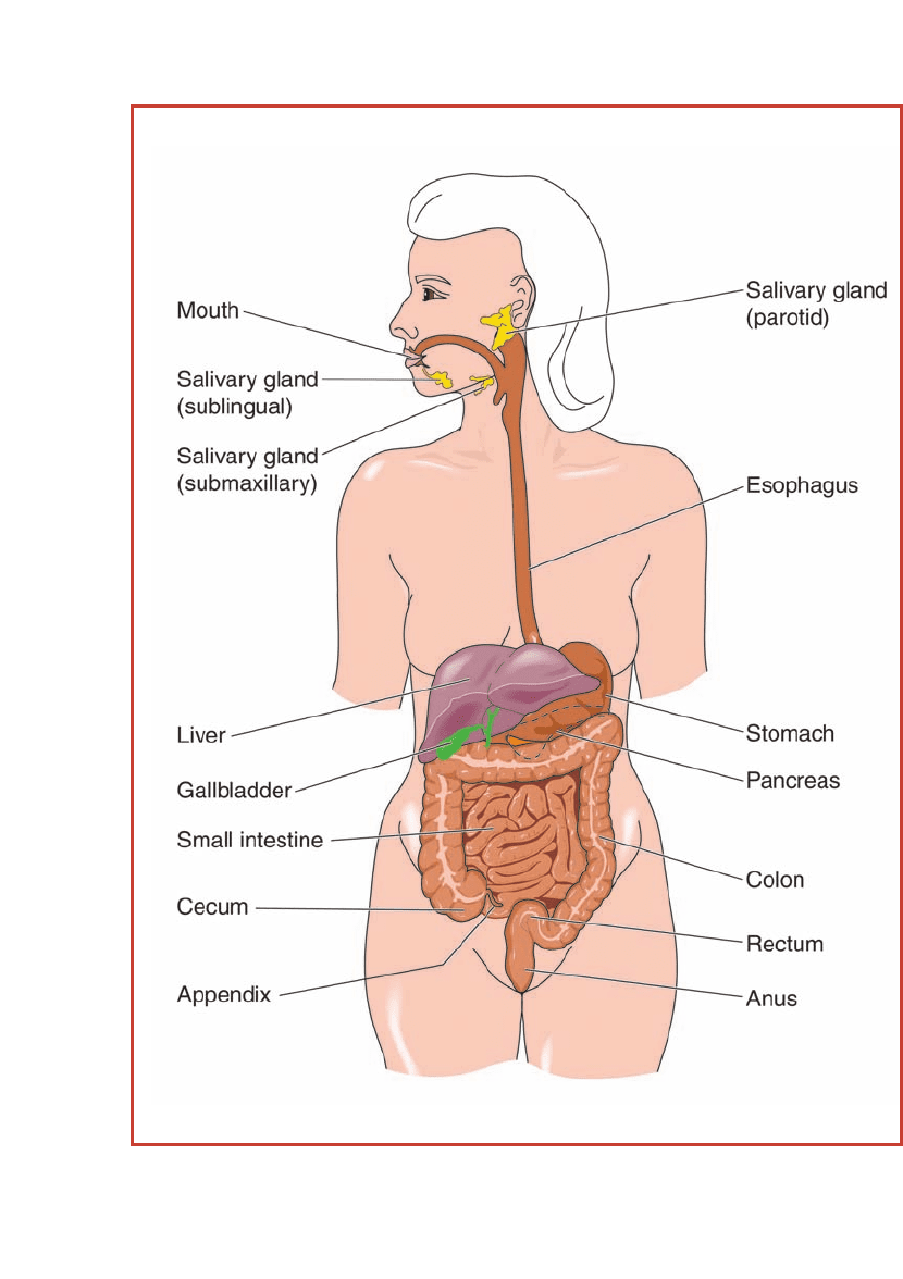
Figure 4.1 The digestive system is a tube within the body, with
an opening at the mouth (for intake) and an opening at the anus
(for excretion). The digestive system includes the mouth, esophagus,
stomach, small and large intestines, and the rectum.

DIGESTION AND NUTRITION
digestive tube and will be used by bacteria living in the
intestines. As a result of this bacterial metabolism, some
people experience abdominal cramping and diarrhea. This
occurs when a person is lactose intolerant, which is discussed
in Chapter 9.
The meat in the burger is a good source of protein.
Proteins are composed of hundreds of amino acids and must
be broken down into individual amino acids in order to be
absorbed into the cells lining the digestive tube. The body will
use these building blocks to make body proteins. Proteins must
be broken down in order to be used by the body.
The beef of the hamburger also contains fats, as does
the oil in which the fries are prepared. Fats, also called
lipids, may or may not be broken down to get them into
the lining cells of the digestive tube. Different types of
fats were described in Chapter 2. Cholesterol is absorbed
whole, while triglycerides are broken apart every time they
must enter or leave a cell. Triglycerides cannot pass through
any cell membrane intact, but cholesterol can. Triglyc-
erides are composed of a single glycerol and three fatty
acid chains. The fatty acid chains can be either saturated or
unsaturated. Saturated fatty acids contain the maximum
number of hydrogen atoms, or are saturated with them,
while unsaturated fats are missing two or more hydrogen
atoms. Because the fatty acid chains are absorbed through
the digestive tube “as is,” the body will build up a supply of
triglycerides that contains whichever type of dietary fatty
acids we ingest. If a person eats food high in saturated
fatty acids, the fatty acids will be transported to the tissue
of the body and stored there. Fats must be mixed with
proteins in order to travel in the bloodstream. Otherwise,
the combination of these fats and blood would look like
Italian salad dressing, with vinegar (blood) on the bottom
and oil (fats) on the top. Because these saturated fats
separate from the proteins carrying them in the circulatory
32

system more frequently than unsaturated fats, these fats tend
to float separately and get stuck in small blood vessels. This
may cause a blockage of blood in the heart or around the
brain. If this blockage is severe enough, it might cause a
heart attack or stroke. Cholesterol can also separate from
its protein carrier, adding to the potential blockage of
the blood vessels and increasing the risks of heart attack
and stroke.
STRUCTURE OF THE DIGESTIVE TUBE
Throughout the digestive tube, the walls of the organs are
made up of four layers:
mucosa, submucosa, muscularis, and
the
serosa or adventitia (Figure 4.2).
The innermost layer of the digestive tube is the mucosa.
This layer is composed of three parts: the
epithelium, the
lamina propria, and the muscularis mucosae. The innermost
part of the mucosa is the epithelium. Most of the epithelial
layer is made up of a single layer of cells called
columnar
epithelial cells. These cells are lined up like columns with one
end exposed to the material in the digestive tube and the other
end forming the connection between the epithelial layer
and the tissue beneath the lining. Everything absorbed into
the body must pass through these cells. In addition to the
columnar cells, mucus-secreting cells called
goblet cells
because of their unique shape (narrow bottom and wider top)
are found throughout the tube. The mucus becomes especially
important farther along in the tube, when the intestinal
contents are dehydrated into feces.
At the beginning of the digestive tube, the epithelium is
made up of
squamous epithelial cells, which are special-
ized for protection. These cells, which are flat and resemble
a pancake with a nucleus in the center, can be stacked up,
which helps protect the tissue underneath them. If a single
layer of cells lined this part of the digestive tract and these
cells were to die, the tissue within the wall of the tube
33
Digestion, Absorption, and Elimination
DIGESTION AND NUTRITION
would be exposed and subject to further damage and infec-
tions from ingested material. Strong chemicals that are
ingested may also be harmful until they are neutralized
in the stomach. This protective layer of cells is found in
the early part of the digestive tube, as well as on the body
surface, to protect from abrasion of the tissue and damage
to the body.
34
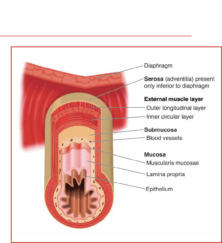
Figure 4.2 The walls of the digestive tube are made up of four
layers: the mucosa, the submucosa, the muscularis (external muscle
layer), and the serosa. The layers are illustrated here.

The lamina propria is a layer of connective tissue beneath
the epithelium that supports the absorptive cells. This layer
contains
loose connective tissue with blood and lymphatic
capillaries to remove dietary material from the columnar
cells and transport the material to the body’s tissues. The
muscularis mucosae has a thin layer of
smooth muscle around
the lamina propria. This layer helps move food through the
digestive tube.
The second major layer of the digestive tube wall is
the submucosa. This layer, similar to the lamina propria
but thicker, has connective tissue and blood vessels. The
submucosa also has some nerves to assist in regulating the
digestive process,
lymph nodules to screen for foreign material
that may cause
antibodies to be made, and sometimes glands,
depending on the part of the tube. These adaptations to the
submucosa will be discussed in the following chapters.
The third layer of the digestive tube wall is the muscu-
laris. This layer is similar to the muscularis mucosa, but is
much thicker and has two layers of smooth muscle. The
inner layer of muscle is arranged in a circular pattern
around the tube. The outer layer of muscle cells runs
parallel to the tube. Both layers of muscle propel the
digestive contents through the tube via a process called
peristalsis (Figure 4.3). The inner layer nudges the material
along with constrictions of the rings of muscle. The outer
layer pushes digestive contents through the tube. The
parallel arrangement causes waves of constriction that
press on the tube, pushing the material. The muscularis
has nerves between the two layers of smooth muscle that
assist in regulating peristalsis.
The last and outermost major layer of the digestive tube
is called the serosa or adventitia. On the esophagus, the outer
covering is called the adventitia. At the end of the digestive
tube, the covering is called the serosa. This covering is also
called the visceral peritoneum, meaning the connective tissue
35
Digestion, Absorption, and Elimination
DIGESTION AND NUTRITION36
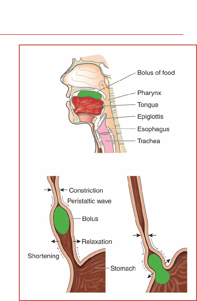
Figure 4.3 After food enters the mouth, it is pushed down the
esophagus through a process called peristalsis. The walls of the
esophagus constrict and relax to move the bolus of food toward the
stomach.

covering of the visceral organs in the peritoneal cavity. This
layer is made of dense, fibrous connective tissue throughout
the tube. The only difference is the name given to the covering,
based on the location of that part of the tube. This serosa/
adventitia of either beef or pigs is used commercially as the
outer covering or casing on sausages, kielbasa, and certain
types of hot dogs.
SURVEY OF THE DIGESTIVE PROCESS
AND COMPONENTS
The process of taking food into the mouth is called ingestion.
The mouth receives the ingested food, breaks it up into smaller
pieces, mixes it with saliva, and sends the food as a bolus to the
pharynx, then into the esophagus. In addition to the physical
digestion of breaking the food into smaller pieces, some
chemical digestion begins in the mouth, especially for
starches. Then the esophagus transports the bolus of food to
the stomach. A detailed description of this part of the process
can be found in Chapter 5. The stomach acts as a blender,
mixing the food with digestive juices secreted by specialized
cells in the stomach lining. One of the digestive chemicals
produced in the stomach is hydrochloric acid at a concentra-
tion strong enough to eat away shoe leather. A large amount
of mucus present in the stomach protects the lining cells from
this acid.
The contents of the stomach are squirted into the small
intestine at regular intervals. Locally produced hormones
control this process. The material at this time is called
chyme
and consists of a combination of ingested food, saliva, and
stomach juices. The material in the small intestine will go
through the rest of the digestive process and be absorbed
into the lining cells of that part of the tube. Additional
digestive juices are brought into the small intestine from
the pancreas and gallbladder. The pancreas contributes
additional enzymes to break down what is left of starch,
37
Digestion, Absorption, and Elimination

DIGESTION AND NUTRITION
protein fragments, and triglycerides. The final breakdown
of the ingested food, including disaccharides, occurs at the
surface of the columnar cells lining the tube, and is then
absorbed into the lining cells. Nearly all the absorption of
nutrients occurs in the small intestine. When nutrients leave
the digestive tract, they go either to the body’s tissues or to
the liver. The liver is an accessory organ to the digestive tract
that regulates much of what goes out to the body through
the bloodstream. A specific description of this part of the
process can be found in Chapter 5.
Most of the water that enters the digestive tract with food
or from the digestive juices of the stomach and
pancreas is
actively removed from the tube by the large intestine. The
removal of most of the water from the digestive tube creates
the material that will be eliminated from the body in the
form of feces. There are a large number of goblet cells in
this portion of the tube to produce the mucus necessary to
move the feces through the rest of the tract. Whatever has
not been broken down or absorbed in the digestive process
will be eliminated through the rectum and the anus. This
is discussed further in Chapter 6.
CONNECTIONS
Nutrients must be broken down to a size capable of being
absorbed into a microscopic cell. Each type of nutrient has
a basic building block that can be absorbed. For sugars, this
basic unit is a monosaccharide. For proteins, this is an
amino acid. Lipids in the form of triglycerides are broken
into glycerol and fatty acid chains, while cholesterol is
absorbed intact.
The wall of the digestive tract is made up of four major
layers: mucosa, submucosa, muscularis, and a connective
tissue covering called a serosa or adventitia. Each section of
the digestive tube has specific functions. The mouth and
esophagus ingest and transport, and the stomach blends the
38

material with digestive juices. The final breakdown of the
food is completed in the small intestine where nutrients are
absorbed. The large intestine salvages most of the water
from the intestinal contents and prepares the solid waste for
elimination.
39
Digestion, Absorption, and Elimination