Sarkar N. (ed.) Human-Robot Interaction
Подождите немного. Документ загружается.


Posture and movement estimation based on reduced information.
Application to the context of FES-based control of lower-limbs
291
2.1. The experimental procedure
2.1.1. Participants
Four spinal cord injured male subjects, with complete spinal lesions between T6 and T12,
participated in the standing study program. The main selection criteria were the following:
(1) participants show high motivation to the study, (2) post-injury standing experience, (3)
appropriate contractions of the leg muscles in response to electrical stimulation, (4)
sufficient upper body arm support strength to lift oneself up and maintain standing, (5) no
cardiac or respiratory illness, (6) no previous stress fractures of upper and lower extremities,
(7) no excessive body weight, (8) acceptable amount of spasticity and contracture in legs, (9)
no psychological pathology.
2.1.2. Materials and Instrumentation:
For leg muscle stimulation during standing, an eight channel stimulator was used (see
Fig.2). The self-adhesive surface electrodes were placed over the motor point areas of the
quadriceps, the gluteus maximus, the tibialis anterior and the biceps femoris muscles of each
leg. The stimulation device was driven directly in real time through a serial link by a PC.
During active standing, patients were stimulated to predetermined FES constant currents,
set up for each channel, in order to ensure safe standing. A video motion analysis system
which included four infrared cameras was used to acquire kinematics data. The reaction
forces measuring system, comprising two six-axis transducers, was attached to handles on
adjustable supporting parallel bars. The six components of the handle reactions were
measured and displayed throughout a real time implemented force sensor interface
software. The handles height and separation were set to comfort for each patient.
2.1.3. Description of the protocol
In a first session, the subjects have been exposed to daily FES exercises, for up to 1 hour per
day during 5 days, in order to strengthen their quadriceps, gluteal maximus/medius, biceps
femoris and tibialis anterior muscles. In a second session, following a thorough explanation
of the study procedure, the patients, under FES, were instructed to stand up from a chair,
assisted by parallel bars, and stay in standing position and sit back down. The standing
phase was as long as one minute. This training phase has been repeated several times in
order for the participants to become familiar with the testing equipment. At session three,
measurements were performed.
2.2. Modelling the human body and arm support
According to observations from human gait, most of joint movements during locomotion
appear to take place in the sagittal plane. In our study, motion in the frontal plane during
standing occurs at very low velocities. Moreover, stimulation on the different muscle groups
of the lower limbs predominantly generates movement in the sagittal plane. For these
reasons, the design of a two-dimensional model of the human body in the sagittal plane is
sufficient for this study. During FES-standing, stimulation of the quadriceps and the
hamstring locks the knee in extension, and therefore prevents knee movement. During
stance, we consider that the distance between the thigh and the handle is constant, which
allows us to assume that the ankle is immobilized. Hence, the lower limbs are here treated

Human-Robot Interaction
292
as a single rigid link. The human body is thus regarded as a four bar linkage with a three
degrees of freedom dynamic structure defined in the sagittal plane, as shown in Fig. 3.
Figure 2. The experimental arrangement and placement of electrodes
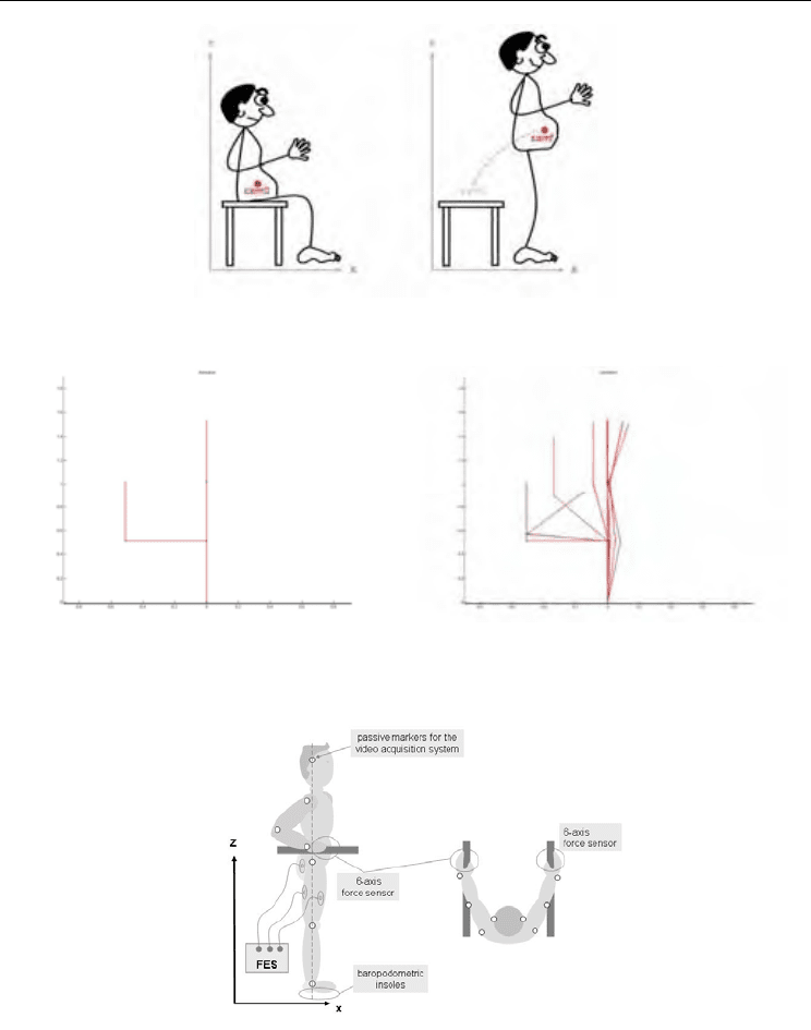
Figure 3. The four bar linkage human model (left). Actual captured image (right)

Posture and movement estimation based on reduced information.
Application to the context of FES-based control of lower-limbs
293
All links are assumed to be rigid bodies. We define q = [q
1
q
2
q
3
]
T
as the joint angle vector,
which is a function of time. It is expressed as a column vector with indices 1, 2 and 3
referring to the hip, the shoulder and the elbow joints respectively. The segments lengths are
denoted by l
j
. In Fig. 3, the variables q
1
and q
2
indicate positive angle directions while q
3
indicates a negative one, with respect to the zero position. Denote P
x
and P
z
the coordinates
of the handle in the sagittal plane. The segmental model is given by (Khalil & Dombre, 2002)
P
x
= l
2
sin(q
1
) + l
3
sin(q
1
+q
2
) + l
4
sin(q
1
+q
2
+q
3
) (1)
and P
z
= l
1
+ l
2
cos(q
1
) + l
3
cos(q
1
+q
2
) + l
4
cos(q
1
+q
2
+q
3
) (2)
During FES-supported movements, paraplegic patients need their arms to maintain balance
and sustain desired movement. Support is taken in charge by two handles, each equipped
with the six axis force/torque sensor, mounted on the supporting frame.
Contact between the human hand and the handle creates a closed chain kinematics linkage.
This interaction is described by the components of the resultant force vector F
c
measured in
the x and z directions. Under the assumption of working in the sagittal plane and
considering that the orientation of the forearm is colinear to the resultant force F
c
, which is
true when the x-axis component of the resultant force satisfies F
x
0 and the z-axis
component satisfies F
z
< 0 (see Fig.3), it is reasonable to write the following hypothesis :
q
1
+ q
2
+ q
3
ï π § arctan(F
x
/F
z
) (3)
2.3. A Set membership identification of posture
Equations (1)-(3) can be re-written as
g(q) = y (4)
where y = [P
x
, P
z
, arctan(F
x
/F
z
)]
T
.
The patient’s posture is given by the q vector, which can be obtained by solving (4). If the
measured quantities y and anthropometric parameters l
j
were known with no uncertainty,
then the problem could be solved analytically through state-of-the-art tools by using inverse
kinematics. Solving (4) when y is subject to uncertainty with classical techniques based on
possibly weighted least squares optimisation for instance, derives reliable results only if the
errors are stochastic and with known probability laws. In fact the measured data are subject
to either stochastic or deterministic uncertainties and it is not easy to derive a reliable
characterization of the probability distribution for these errors. Moreover, the model used
may be based on some simplifying hypotheses for which a full probabilistic description
might not be reliable. Consequently, it is more natural to assume all the uncertain quantities
as unknown but bounded with known bounds and no further hypotheses about probability
distributions. In such a bounded error context, the solution is no longer a point but is the set
of all acceptable values of the q vector, which makes the model output g(q) consistent with
actual data y and prior error bounds.
Denote E a feasible domain for output error and Y = y + E the feasible domain for model
output. The set S to be estimated is the set of all feasible postures:
S = {q ∈ Q | g(q) ∈ Y} (5)
where the set Q is an initial search space for the q vector. Characterizing the set S is a set
inversion problem which can be solved in a guaranteed way using a set inversion algorithm

Human-Robot Interaction
294
based on space partitioning, interval analysis and constraint propagation techniques (see
(Jaulin, et al., 2001) and the references therein). This algorithm explores all the search space
without losing any solution. It makes it possible to derive a guaranteed enclosure of the
solution set S as follows:
S
inner
⊆ S ⊆ S
outer
(6)
The solution set S is enclosed between two approximation sets. The inner enclosure S
inner
consists of the boxes that have been proved feasible. To prove that a box [q] is feasible it is
sufficient to prove that g([q]) ⊆ Y. If, on the other hand, it can be proved that g([q])∩Y=∅,
then the box [q] is unfeasible. Otherwise, no conclusion can be reached and the box [q] is said
undetermined. It is then bisected and tested again until its size reaches a threshold to be
tuned by the user. The outer enclosure S
outer
is defined by S
outer
= S
inner
∪ΔS where ΔS is given
by the union of all the undetermined boxes. The outer enclosure S
outer
contains all the
solutions, if they exist, without losing any of them. It contains also some elements that are
not solution.
2.4. The estimated posture
Posture estimation was done during the standing phase. The subject’s actual posture during
that time interval were measured as :
q
1
§ 0°, q
2
§ 192°, q
3
§ -36° (7)
representing respectively the hip, shoulder and elbow joint angles. The body segment
lengths were directly measured on the patient and are given by :
l
1
§ 0.954 m, l
2
§ 0.518 m, l
3
§ 0.334 m, l
4
§ 0.262 m (8)
The feasible domain for model output are taken as:
P
x
∈ [ï0.02, 0.02] m
P
z
∈ [0.895, 0.995] m (9)
arctan(F
x
/F
z
)] ∈ [ï18.63,ï15.63]°
The prior search space Q, corresponding to the joints articular motion limit, is taken as:
[ï11, 90]° × [90, 210]° × [ï103, 0]°.
The projections of the computed inner and outer solution sets, S
inner
and S
outer
onto the q
i
× q
j
planes are given in Fig.4. Contrary to any optimization based techniques, there are no
optimal solution, therefore any posture taken within the solution set is an acceptable one.
Extreme postures taken from solution set are also plotted in Fig.5. These figures clearly
show that the solution sets contain the actual posture (see also Table 1).
Joints Projection of inner enclosure Projection of outer enclosure
q
1
q
2
q
3
[-1.35 , 25.52]
[192.5 , 213.66]
[-74.10 , -31.05]
[-4.14 , 28.79]
[190.34 , 215.32]
[-77.81 , -28.28]
Table 1. Projection of solution posture

Posture and movement estimation based on reduced information.
Application to the context of FES-based control of lower-limbs
295
Figure 4. Projection of solution set onto q
1
× q
2
(left) and q
1
× q
3
(right)
Figure 5. Postures taken from the solution sets:
(a) Patient leaning back, (b) Actual patient posture, (c) Patient leaning forward
Indeed, the experimental method introduced in this section is capabale of reconstructing the
posture of a patient but with fairly large uncertainties. This reflects the fact that for a fixed
position of the forearm taken within the feasible domain calculated by force measurements
in the sagittal plane only, the hip, the shoulder and the elbow joints still have the possibility
to reach other positions, while being consistent with the defined geometrical constraints. In
order to further reduce the solution set, and hence have a more precise estimation of
patient’s posture, new constraints has to be introduced by using dynamic modelling and
ground reaction forces measurements, for instance.

Human-Robot Interaction
296
3. Observing valid limbs to detect patient intention
In this section, we propose an approach for the recognition of the "signature" of the postural
task the subject intends to realize (sit-to-stand, object grasping, walking, stair climbing, gait
initiation/termination...) through voluntary movement observation. This detection should
occur as soon as possible after the subject has decided to initiate the task. It is particularly
important to detect the transitions between activity modes as soon as possible after the
patient has taken the decision to modify his functioning mode, in order to allow for optimal
posture preparation and execution.
A good illustration for this is the transfer from sit to stand. In our FES context it is essential
to optimize this task, muscle fatigue being a major issue. Minimizing efforts of rising up
could improve the following activities of the patient. For this reason, classical techniques
consisting of maximum stimulation of knee extensors throughout the rising process are not
suitable and involve an over-use of arm support.
Two approaches are considered in the following to estimate patient attitude: the
instrumentation of the walker and body-mounted micro-sensors.
3.1. Sit to stand dynamics analysis
Assuming that the body structure is rigid, continuous dynamics can be expressed under the
Lagrangian form:
M(q)q”+N(q,q’)q’+G(q)=
Γ
+
Γ
ext
(10)
where: q stands for the parametrization vector of the whole configuration space of the biped
considered as free in 3D,
Γ
is the joint actuation torque, M is the inertia matrix, N is the
matrix of centrifugal, gyroscopic and Coriolis effects, G is the generalized gravity force
vector.
Γ
ext
are torques generated by external forces such as ground contacts, interaction
with a chair, a thrust, etc. They can be expressed as:
Γ
ext
=C(q)
T
λ
(q,q’) (11)
C(q) is the Jacobian matrix of the points of the biped to which the external forces are applied
and
λ
corresponds to the amplitudes of these forces. Biped dynamics are characterized by
the existence of variable constraints resulting from interaction with the ground. Ground
efforts correspond to a set of forces applied to the points of the biped in contact with the
ground (Azevedo et al., 2007a).
Using this framework, we propose to express the sit-to-stand transfer as an optimization
problem, where the posture configuration q minimizes a cost function over a time horizon
h:
J=(H
com
-H
comd
)
T
(H
com
-H
comd
) (12)
where H
com
(t)=[X
com
(t); Y
com
(t); X
com
(t+1); Y
com
(t+1);…; X
com
(t+h); Y
com
(t+h)]
T
is the sequence
of centre of mass positions over the time horizon h, H
comd
=[X
comd
; Y
comd
;…; X
comd
; Y
comd
]
T
is a
vector made of the repetition of the desired position of the centre of mass (standing posture)
over the time horizon h. The solution to this problem is illustrated in Figs.6 & Fig.7-a. The
biped goes directly from seated posture to standing.
Posture and movement estimation based on reduced information.
Application to the context of FES-based control of lower-limbs
297
Figure 6. Illustration of the problem of sit to stand consisting in transferring the centre of
mass projection from seat to feet
a- no constraint b- constrains on torques
Figure 7. Simulation of sit to stand transfer by solving an optimization problem minimizing
distance between actual and desired center of mass position over a sliding time horizon
Figure 8. Description of the experimental protocol
If now some constraints are added to the problem in terms of limitation of joint torques, i.e.
Γ
min
Γ
Γ
max
(13)

Human-Robot Interaction
298
the result is that the system has to use its trunk inertia to achieve the movement (fig.7-b),
upper body bends forward before legs initiate movement. This simulation results explain
clearly the important need of coordination between upper and lower limbs to execute a
transfer from seat to stand when available torques are limited, which is obviously the case
for muscles. Without this coordination additional external efforts are needed, such as arm
support.
Based on these preliminary considerations, we propose two approaches for the detection of
sit-to-stand movement.
3.2. Walker instrumentation
We first investigate the possibility of considering body supportive forces as a potential
feedback source for FES-assisted standing-up control (Azevedo et al., 2007b). The six-
degrees of freedom force sensors were mounted onto handles fixed on parallel bars in order
to record upper limbs efforts and insoles were fitted in the patient’s shoes to record plantar
pressure distribution (Fig.8). Eight volunteer complete paraplegic patients (T5-T12) were
verticalized by means of adapted FES. The same training protocol as presented in the
previous section was used. A video motion analysis system recorded the positions of
passive markers placed on the body allowing us to measure kinematics. The results show
that the transfer (phase 1) is mainly ensured by arm support in all our patients (Fig.9). We
gave instruction to the patients to bend their trunk in preparation to the chair rising. An
important observation when looking at trunk, knee and ankle angles is the low intra-
variability between trials of one given patient (Fig.10). A main difference between valid
subjects and patients is the onset of leg movement in regards to trunk bending (Fig.10). To
be efficient, trunk bending forward should start before and last during knee and ankle
movement. This was never the case in our trials on FES-assisted standing.
Minimizing arm support contribution is possible only if trunk inertia is used. This implies a
good triggering of muscle contraction regarding limb movements. Trunk behaviour could
be indirectly observed by analyzing efforts applied by arm support (Fig.11). Indeed, normal
force decreases (pulling) while momentum around transversal axis increases. From these
results we can say that proper threshold detection based on these signals could be used to
initiate the leg stimulation and improve greatly the sit to stand. The same may be used for
stand to sit as shown in Fig.11.
3.3.1. Body-mounted instrumentation
In parallel to the approach presented in the previous section, we have also worked on
demonstrating the pertinence of observing the trunk using a movement sensor placed on the
back of valid subjects (Azevedo & Héliot, 2005). Indeed, as seen before, the trunk normally
initiates the sit-to-stand transfer. We have placed on the back of 10 valid subjects, at
anatomical C7 level, an accelerometer. Trunk acceleration patterns present low intra and
inter-variability as well as a high temporal reproducibility and are therefore a nice
characteristic “signature” of the sit-to-stand transfer (see Fig.12).
It is possible to apply techniques such as abrupt changes theory (Basseville & Nikiforov,
1993) to detect the pattern of sit to stand “intention”. This technique allows detecting
robustly the transfer initiation with a good sensitivity. The algorithm is able to reject a
“false” sit-to-stand movement involving trunk movements such as grasping an object placed
in front of the person. Indeed, the acceleration pattern signs selectively the motion.

Posture and movement estimation based on reduced information.
Application to the context of FES-based control of lower-limbs
299
Figure 9. Patient #3, trial 6. Total feet and arm support. Phase labelled 1 corresponds to sit to
stand phase, 2+3 corresponds to standing, and 4 to knee flexion
Figure 10. Posture coordination during sit to stand. Top: Patient #1, over 4 trials, Bottom:
valid subject. The red dot indicates the maximum trunk bending
It is important to notice here that the detection of transfer has to occur as soon as possible
before the legs should start moving to displace the body centre of mass from the seat to the
feet (Fig.12). Around 600ms separate the instant when the trunk starts bending forward and
the instant when the legs enter in extension movement. It is also necessary to recall here,
that lower limb muscles start to contract before the legs move in order to prepare the

Human-Robot Interaction
300
motion. These so-called anticipative postural adjustments should take place ideally together
with trunk movement.
In order to apply these results on patient FES-assisted sit to stand it is necessary to train
paraplegic patients in executing an optimal trunk movement in order to benefit from its
inertia in the standing transfer. The detection algorithm should then be able to recognize
patient intention to stand and trigger the proper stimulation sequences.
Figure 11. Correspondence between trunk angle and handle information. Patient #1, Trial 1.
Top: angle, Bottom: right side vertical force and momentum around hip axis
Figure 12. Principle of the detection of sit-to-stand based on the observation of trunk
acceleration