Pecharsky V.K., Zavalij P.Y. Fundamentals of Powder Diffraction and Structural Characterization of Materials
Подождите немного. Документ загружается.

Fundamentals of diffraction
131
Hence, the photographic film vaguely resembles a ratemeter because the
intensity is extracted from the degree of darkening of the spots found on the
film
-
the darker the spot, the higher the corresponding intensity because the
larger number of photons have been absorbed by the spot on the film
surface. The three most commonly utilized types of x-ray detectors today are
gas proportional, scintillation, and solid-state detectors, all of which are true
counters.
Yet another classification of detectors is based on whether the detector is
capable of resolving the location where the photon has been absorbed and
thus, whether they can detect the direction of the beam in addition to
counting the number of photons. Conventional gas proportional, scintillation,
and solid-state detectors do not support spatial resolution and therefore, they
are also known as point detectors.
A
point detector registers only the
intensity of the diffracted beam, one point at a time.
In
other words, the
readout of the detector corresponds to a specific value of the Bragg angle as
determined by the position of the detector relatively to both the sample and
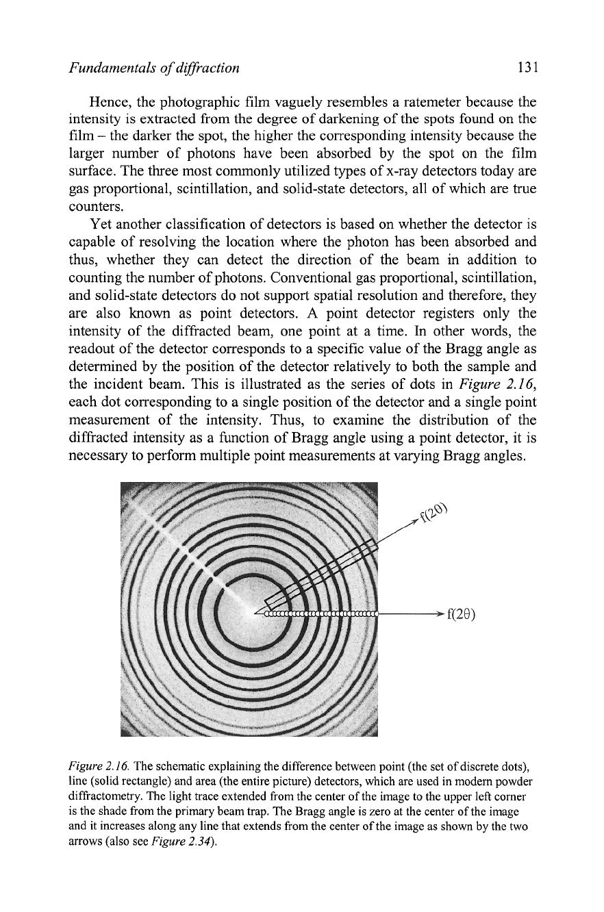
the incident beam. This is illustrated as the series of dots in
Figure
2.16,
each dot corresponding to
a
single position of the detector and a single point
measurement of the intensity. Thus, to examine the distribution of the
diffracted intensity as a function of Bragg angle using a point detector, it is
necessary to perform multiple point measurements at varying Bragg angles.
Figure
2.16.
The schematic explaining the difference between point (the set of discrete dots),
line (solid rectangle) and area (the entire picture) detectors, which are used in modem powder
diffractometry. The light trace extended from the center of the image to the upper left comer
is the shade from the primary beam trap. The Bragg angle is zero at the center of the image
and it increases along any line that extends from the center of the image as shown by the two
arrows (also see
Figure
2.34).
132
Chapter
2
Detectors that support spatial resolution in one direction are usually
termed as line detectors, while those that facilitate resolution in two
dimensions are known as area detectors. Again, photographic film is a
typical example of an area detector because each point on the film can be
characterized by two independent coordinates and the entire film area is
exposed simultaneously. The following three types of line and area detectors
are in common use in powder diffractometry today: position sensitive (PSD),
charge coupled devices (CCD), and image plates
(IPD). The former is a line
detector (its action is represented by the rectangle in
Figure
2.16) and the
latter two are area detectors (the entire area of
Figure
2.16 represents an
image of how the intensity is measured simultaneously). Both line and area
detectors can measure diffracted intensity at multiple points at once and thus,
a single measurement results in the diffraction pattern resolved in one or two
dimensions, respectively.
2.4.3
Point detectors
A
typical
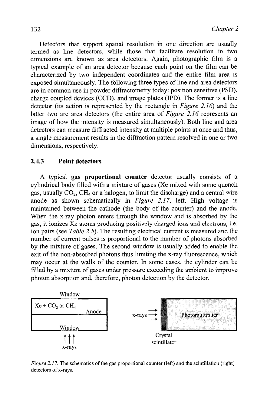
gas proportional counter
detector usually consists of a
cylindrical body filled with a mixture of gases (Xe mixed with some quench
gas, usually C02, CH4 or a halogen, to limit the discharge) and a central wire
anode as shown schematically in
Figure
2.17, left. High voltage is
maintained between the cathode (the body of the counter) and the anode.
When the x-ray photon enters through the window and is absorbed by the
gas, it ionizes Xe atoms producing positively charged ions and electrons,
i.e.
ion pairs (see
Table
2.5).
The resulting electrical current is measured and the
number of current pulses is proportional to the number of photons absorbed
by the mixture of gases. The second window is usually added to enable the
exit of the non-absorbed photons thus limiting the x-ray fluorescence, which
may occur at the walls of the counter.
In
some cases, the cylinder can be
filled by a mixture of gases under pressure exceeding the ambient to improve
photon absorption and, therefore, photon detection by the detector.
Window
-
x-rays
-
+
ttt
Crystal
scintillator
x-rays
Figure
2.17.
The schematics of the gas proportional counter (left) and the scintillation (right)
detectors of x-rays.
Fundamentals of diffraction
133
Gas proportional counters have relatively good resolution, so the heights
of current pulses can be analyzed and discriminated to eliminate pulses that
appear due to
KP
photons and due to low and high energy white radiation
photons. The pulse height discrimination is often used in combination with a
P-filter to improve the elimination of the KP and white background photons.
The lifetime of a proportional detector is limited to about two years
because a fraction of the gas, filling the detector, escapes through the
windows, which are usually made from a thin and low absorbing organic
film to improve quantum efficiency. Another disadvantage of this type of the
detector is its low effectiveness at high photon fluxes and with short wave
(high energy) x-rays, such as Mo (the mass absorption coefficients of Xe are
299 and 38.2
cm2Ig for Cu Ka and Mo Ka radiation, respectively).
A typical scintillation detector employs a different principle for the
detection of x-rays. It is constructed from a crystal scintillator coupled with a
photomultiplier tube as shown in
Figure
2.17,
right. The x-ray photons,
which are absorbed by the crystal, generate photons of blue light. After
exiting the crystal, blue light photons are converted into electrons in a
photomultiplier and amplified, and the resultant voltage pulses are registered
as photon counts.
The crystal scintillator is usually made from cleaved, optically clear
sodium iodide
(NaI) activated with -1% of T1. The crystals are hydroscopic
and thus, they are usually sealed in a vacuum tight enclosure with a thin Be
window in the front (x-rays entry window) and high quality optical glass in
the back (blue light photons exit window). The crystal is usually mounted on
the photomultiplier tube using a viscous fluid that minimizes the refraction
of blue light on the interface between the crystal and the photomultiplier.
Unfortunately, due to the relatively high energy of blue light photons, the
x-ray wavelength resolution of the scintillation detector is quite low.
Although, the extremely short wavelengths
(e.g. cosmic rays) can be
discriminated and eliminated, the KP photons cannot be recognized and
filtered out by the detector. Despite their low resolution, scintillation
detectors are highly stable and effective; especially at high photon fluxes
(see
Table
2.5).
They have very short dead time and therefore, extended
linear range. Because of this, scintillation detectors are by far the most
commonly used detectors in the modem laboratory x-ray powder
diffractometry.
Yet another physical phenomenon is used in solid-state detectors, which
are manufactured from high quality silicon or germanium single crystals
doped with lithium and commonly known as
Si(Li) or Ge(Li) solid-state
detectors. The interaction of the x-ray photon with the crystal (detector)
produces electron-hole pairs in quantities proportional to the energy of the
photon divided by the energy needed to generate a single pair. The latter is
134 Chapter
2
quite low and amounts to approximately 3.7 eV for a Si-based detector. The
electric potential difference applied across the crystal results in the photon-
induced electric current, which is amplified and measured. The current is
indeed proportional to the number of the generated electron-hole pairs.
In
order to minimize noise and Li migration, the solid-state detector
should be cooled, usually to -80 K. This can be done using liquid nitrogen
but it is quite inconvenient to have a cryogenic container mounted on the
detector arm, especially since the container needs to be refilled every few
days. Thus, solid-state detectors coupled with thermoelectric coolers have
been developed and commercialized, and successfully used in powder
diffraction.
The substantial advantage of this type of detector is its high resolution at
low temperature, even when compared with proportional counters (Table
2.5).
Cooled solid-state detectors facilitate excellent filtration of both the
undesirable Kj3 and white radiation, thus resulting in a very low background
without a significant loss of the intensity of the KallKa2 doublets. It is
worth noting that even the highest quality, perfectly aligned monochromator
decreases the characteristic Ka intensity by a factor of two or more. Thus,
with the cooled solid-state detector the monochromator is no longer needed
unless the extreme spectral purity is an issue.
The major drawback of this type of detector, not counting the need for
continuous cooling, is its relatively low linear range
-
only up to about 2x lo4
counts per second. This may result in experimental artifacts when measuring
extremely strong Bragg peaks, similar to those shown in Figure
2.18.
The inset of this figure shows sharp and random spikes peaking at
-1.2~ 10' counts instead of a smooth Bragg peak, due to the highly non-
linear response of the detector when photon flux exceeds 2 to
5x10~ counts
per second. Furthermore, the photon energy resolution-based electronic
monochromatization also fails.
In
addition to the (400) Bragg peak centered
at 20
G
69.2", which is expected for this orientation of the single crystalline
silicon wafer, three additional Bragg peaks are clearly recognizable in the
measured diffraction pattern at 20
z
61.6, 65.9 and 66.3".
A
simple analysis, based on the Braggs' equation, leads to the conclusion
that these three peaks are all due to the reflections from the (400)
crystallographic planes of Si, each representing a different wavelength in a
characteristic spectrum of this particular x-ray tube. The first satellite Bragg
peak (20
z
61.6") corresponds to the incompletely suppressed Kj3
component in the characteristic spectrum of the Cu anode. Obviously this
occurs due to the extremely high intensity of the (400) peak from the nearly
perfect single crystalline specimen. The two remaining Bragg peaks (28
z
65.9 and 66.3") are due to the presence of a
W
impurity deposited on the
surface of the anode (see Figure
2.4)
during the long time operation of the x-
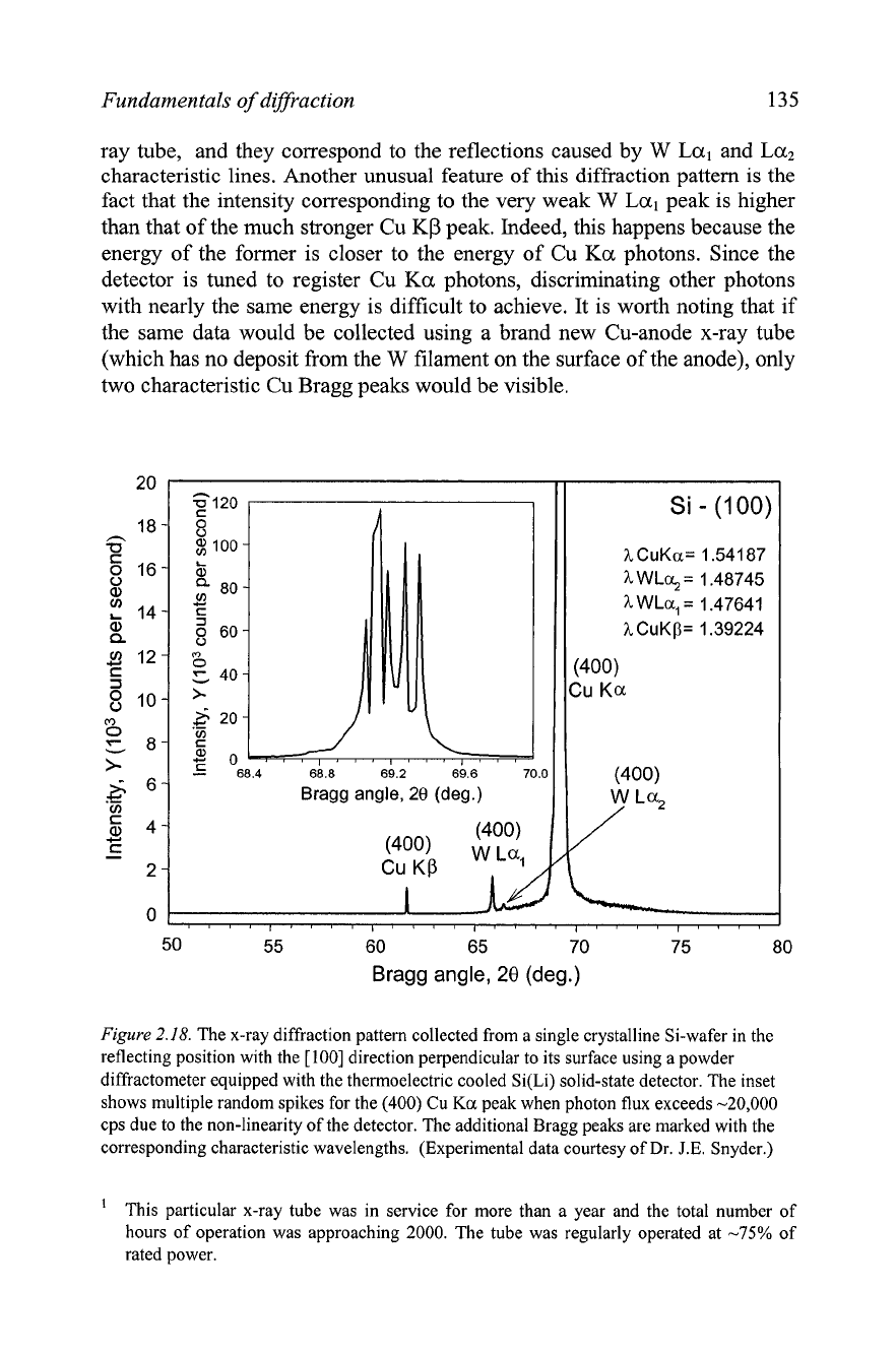
Fundamentals
of
diffraction
135
ray tube, and they correspond to the reflections caused by
W
Lal
and
Laz
characteristic lines. Another unusual feature
of
this diffraction pattern is the
fact that the intensity corresponding to the very weak
W
Lal
peak is higher
than that of the much stronger Cu
KP
peak. Indeed, this happens because the
energy of the former is closer to the energy of Cu
Ka
photons. Since the
detector is tuned to register Cu
Ka
photons, discriminating other photons
with nearly the same energy is difficult to achieve. It is worth noting that if
the same data would be collected using a brand new Cu-anode x-ray tube
(which has no deposit from the
W
filament on the surface of the anode), only
two characteristic Cu Bragg peaks would be visible.
.-
Bragg angle,
28
(deg.)
Bragg angle,
28
(deg.)
Figure
2.18.
The x-ray diffraction pattern collected from a single crystalline Si-wafer in the
reflecting position with the [loo] direction perpendicular to its surface using a powder
diffractometer equipped with the thermoelectric cooled Si(Li) solid-state detector. The inset
shows multiple random spikes for the (400) Cu
Ka
peak when photon flux exceeds -20,000
cps due to the non-linearity of the detector. The additional Bragg peaks are marked with the
corresponding characteristic wavelengths. (Experimental data courtesy of Dr.
J.E.
Snyder.)
'
This particular x-ray tube was in service for more than a year and the total number of
hours of operation was approaching 2000. The tube was regularly operated at
-75%
of
rated power.
136
Chapter
2
2.4.4
Line and area detectors
A
position sensitive detector
(PSD)
employs the principle of a gas
proportional counter, with an added capability to detect the location of a
photon absorption event. Hence, unlike the conventional gas proportional
counter, the PSD is a line detector that can measure the intensity of the
diffi-acted beam in multiple (usually thousands) points simultaneously. As a
result, a powder diffraction experiment becomes much faster, while its
quality generally remains nearly identical to that obtained using a standard
gas proportional counter.'
The basic principle of sensing the position of the photon absorption event
by the PSD is based on the following property of the proportional counter.
The electrons (born by the x-ray photon absorption and creation of Xe ion
-
electron pairs) accelerate along a minimum resistance (i.e. linear) path
towards the wire anode, where they are discharged thus producing the
electrical current pulse in the anode circuit.
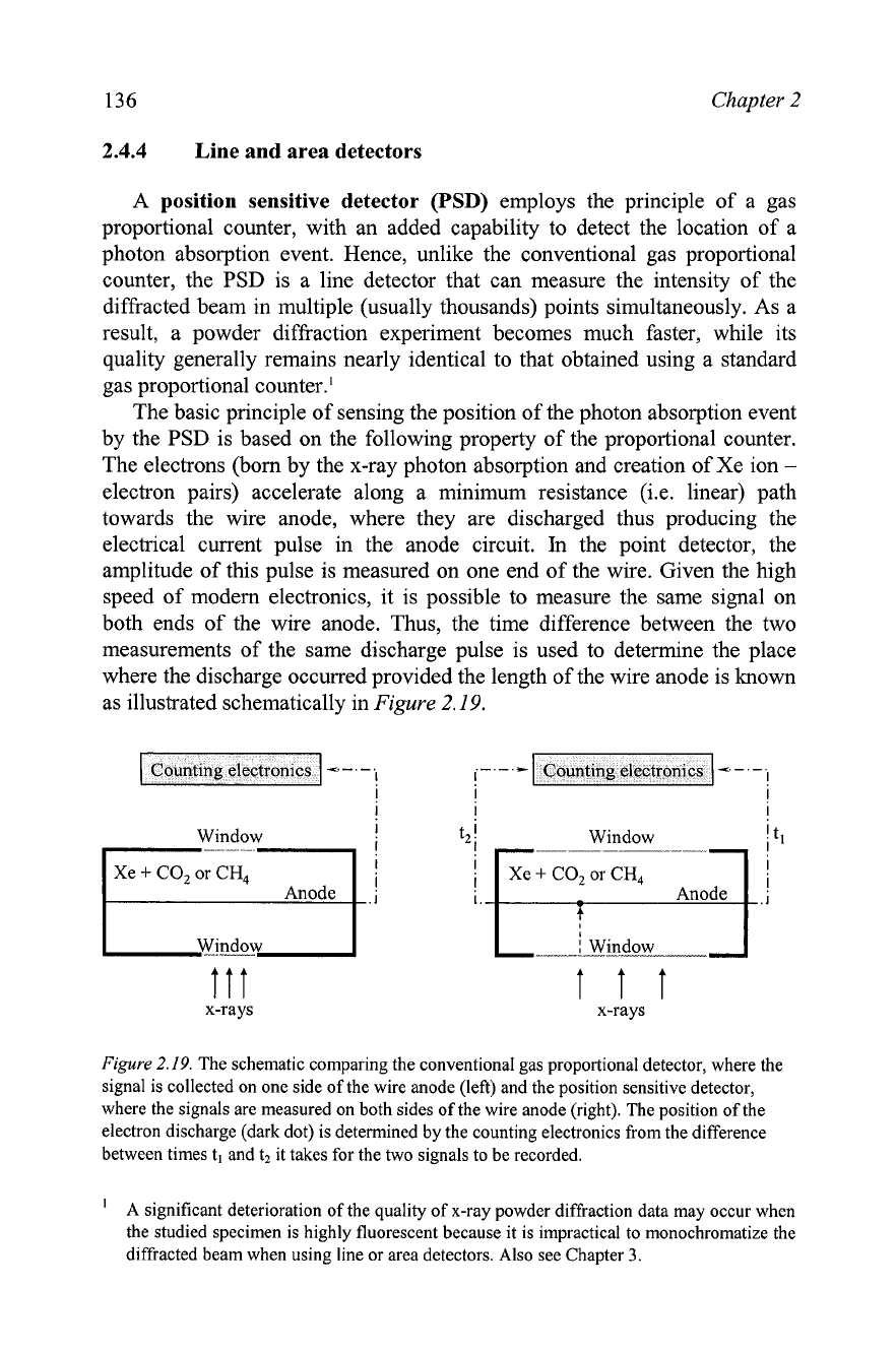
In
the point detector, the
amplitude of this pulse is measured on one end of the wire. Given the high
speed of modern electronics, it is possible to measure the same signal on
both ends of the wire anode. Thus, the time difference between the two
measurements of the same discharge pulse is used to determine the place
where the discharge occurred provided the length of the wire anode is known
as illustrated schematically in
Figure
2.19.
Window
!
Window
-
-
-
1
WiJiw
l
Window
Counting
electronics
Ill
x-rays
I
I
I
x-rays
-
-
-
I
!-
-
*
Figure
2.19.
The schematic comparing the conventional gas proportional detector, where the
signal is collected on one side of the wire anode (left) and the position sensitive detector,
where the signals are measured on both sides of the wire anode (right). The position of the
electron discharge (dark dot) is determined by the counting electronics from the difference
between times
t,
and t2 it takes for the two signals to be recorded.
A significant deterioration of the quality of x-ray powder diffraction data may occur when
the studied specimen is highly fluorescent because it is impractical to monochromatize the
diffracted beam when using line or area detectors. Also see Chapter
3.
Counting electronics
-
-
-
I
Fundamentals of diffraction
The spatial resolution of the PSD's is not as high as that attainable with
the precise positioning of point detectors. Nevertheless, it remains
satisfactory (approaching about 0.01
O)
to conduct good quality experiments.
Yet a minor ,loss of the resolution is a small price to pay for the ability to
collect powder diffraction data in a wide range of Bragg angles, all at once,
which obviously and substantially decreases the duration of the experiment.
A
typical improvement is from many hours using a point detector down to
several minutes or less using a position sensitive detector.
Different models of the PSD's may have different geometry, resolution
and Bragg angle range: short linear PSD's cover a few degrees range
(from
-5
to lo0), while long curved PSD's may cover as much as -120 to 140' 28.
The biggest advantage of the long range PSD's is the considerable
experimental time reduction when compared to short or medium range
position sensitive detectors. Their disadvantage arises from often substantial
differences in the photon counting properties observed at different places
along the detector, for example in the middle vs. the ends of its length. The
large angular spread of long detectors also puts some restrictions on the
quality of focusing of x-rays and usually results in the deterioration of the
shape of Bragg peaks. Relevant discussion about the geometry of powder
diffractometers equipped with PSD's is found in Chapter
3.
Area detectors record diffraction pattern in two dimensions
simultaneously. Not counting the photographic film, two types of electronic
area detectors have been advanced to a commercial status, and are becoming
more frequently used in modem x-ray powder diffraction analysis.
In
a charge coupled device detector, x-ray photons are converted by a
phosphor' into visible light, which is captured using a charge-coupled device
camera.
In
an image plate detector, x-ray photons are also captured by a
pho~phor.~ The excited phosphor pixels, however, are not converted into the
signals immediately. Instead, the information is stored in the phosphor grains
as a latent image, in a way, similar to the activation of silver halide particles
in the photographic film during exposure. When the data collection is
completed, the image is scanned (so to say is "developed") by a laser, which
deactivates pixels that emit the stored energy as a blue light. Visible light
photons are then registered by a photomultiplier in a conventional manner.
'
A
typical
CCD
phosphor is Tb3+ doped
Gd202S,
which converts x-ray photons into visible
light photons.
A
typical image plate phosphor is EU*' doped BaFBr. When exposed to x-rays, Eu2+
oxidizes to Eu3+. Thus produced electrons may either recombine with
EU~+
or they become
trapped by F-vacancies in the crystal lattice of BaFBr. The trapped electrons may exist in
this metastable state for a long time. They are released when exposed to a visible light and
emit blue photons during recombination with
Eu3+ ions, e.g. see
K.
Takahashi, K. Khoda,
J.
Miyahara,
Y.
Kanemitsu,
K.
Amitani, and S. Shionoya, Mechanism of photostimulated
luminescence in B~FX:EU~+ (X
=
C1,
Br) phosphors,
J.
Luminesc.
31-32,266
(1984).
138
Chapter 2
The use of detectors, collimators and monochromators in different
powder diffi-actometers is discussed in Chapter
3,
while the reminder of this
chapter is dedicated to theory of diffraction, understanding the origin of the
powder diffraction pattern, and a brief description of how structural
information can be extracted from powder diffraction data.
2.5
Scattering
by
electrons, atoms and lattices
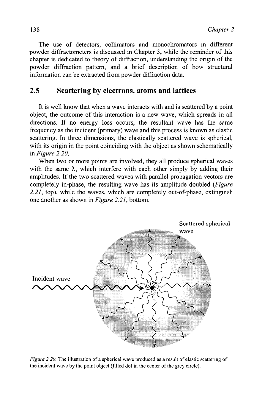
It is well know that when a wave interacts with and is scattered by a point
object, the outcome of this interaction is a new wave, which spreads in all
directions. If no energy loss occurs, the resultant wave has the same
frequency as the incident (primary) wave and this process is known as elastic
scattering.
In
three dimensions, the elastically scattered wave is spherical,
with its origin in the point coinciding with the object as shown schematically
in Figure 2.20.
When two or more points are involved, they all produce spherical waves
with the same
A,
which interfere with each other simply by adding their
amplitudes. If the two scattered waves with parallel propagation vectors are
completely in-phase, the resulting wave has its amplitude doubled (Figure
2.21, top), while the waves, which are completely out-of-phase, extinguish
one another as shown in Figure 2.21, bottom.
Scattered spherical
Figure
2.20.
The illustration of a spherical wave produced as a result of elastic scattering of
the incident wave by the point object (filled dot in the center of the grey circle).
Fundamentals of diffraction
139
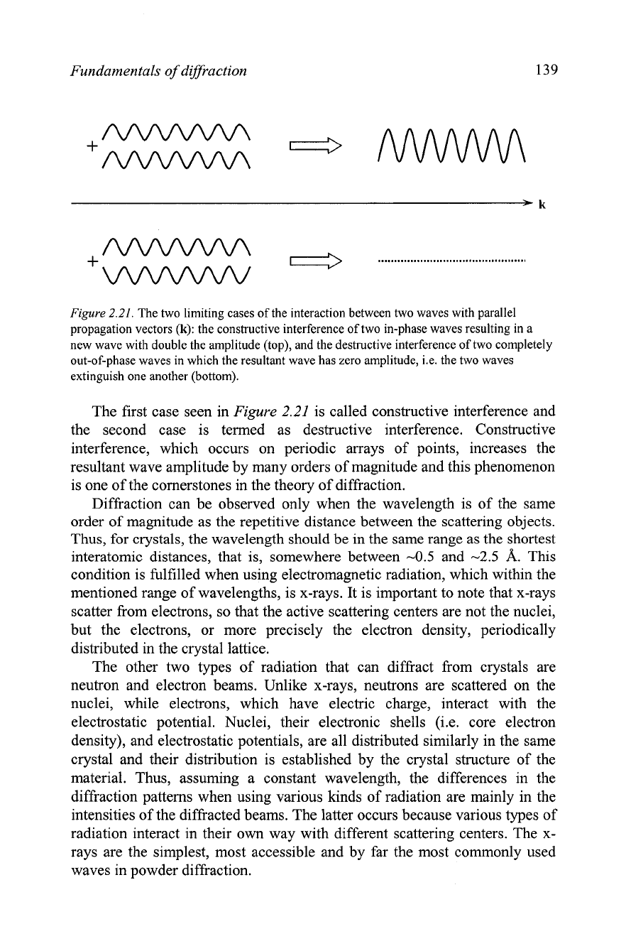
Figure
2.21.
The two limiting cases of the interaction between two waves with parallel
propagation vectors (k): the constructive interference of two in-phase waves resulting in a
new wave with double the amplitude (top), and the destructive interference of two completely
out-of-phase waves in which the resultant wave has zero amplitude,
i.e. the two waves
extinguish one another (bottom).
The first case seen in
Figure
2.21
is called constructive interference and
the second case is termed as destructive interference. Constructive
interference, which occurs on periodic arrays of points, increases the
resultant wave amplitude by many orders of magnitude and this phenomenon
is one of the cornerstones in the theory of diffraction.
Diffraction can be observed only when the wavelength is of the same
order of magnitude as the repetitive distance between the scattering objects.
Thus, for crystals, the wavelength should be in the same range as the shortest
interatomic distances, that is, somewhere between
-0.5
and
-2.5
A.
This
condition is fulfilled when using electromagnetic radiation, which within the
mentioned range of wavelengths, is x-rays. It is important to note that x-rays
scatter from electrons, so that the active scattering centers are not the nuclei,
but the electrons, or more precisely the electron density, periodically
distributed in the crystal lattice.
The other two types of radiation that can diffract from crystals are
neutron and electron beams. Unlike x-rays, neutrons are scattered on the
nuclei, while electrons, which have electric charge, interact with the
electrostatic potential. Nuclei, their electronic shells
(i.e. core electron
density), and electrostatic potentials, are all distributed similarly in the same
crystal and their distribution is established by the crystal structure of the
material. Thus, assuming a constant wavelength, the differences in the
diffraction patterns when using various kinds of radiation are mainly in the
intensities of the diffracted beams. The latter occurs because various types of
radiation interact in their own way with different scattering centers. The
x-
rays are the simplest, most accessible and by far the most commonly used
waves in powder diffraction.
140
Chapter
2
2.5.1
Scattering
by
electrons
The origin of the electromagnetic wave elastically scattered by the
electron can be better understood by recalling the fact that electrons are
charged particles. Thus, an oscillating electric field (see Figure
2.2)
from the
incident wave exerts a force on the electric charge (electron) forcing the
electron to oscillate with the same frequency as the electric field component
of the electromagnetic wave. The oscillating electron accelerates and
decelerates in concert with the varying amplitude of the electric field vector,
and emits electromagnetic radiation, which spreads in all directions.
In
this
respect, the elastically scattered x-ray beam is simply radiated by the
oscillating electron; it has the same frequency and wavelength as the incident
wave, and this type of scattering is also known as coherent scattering.'
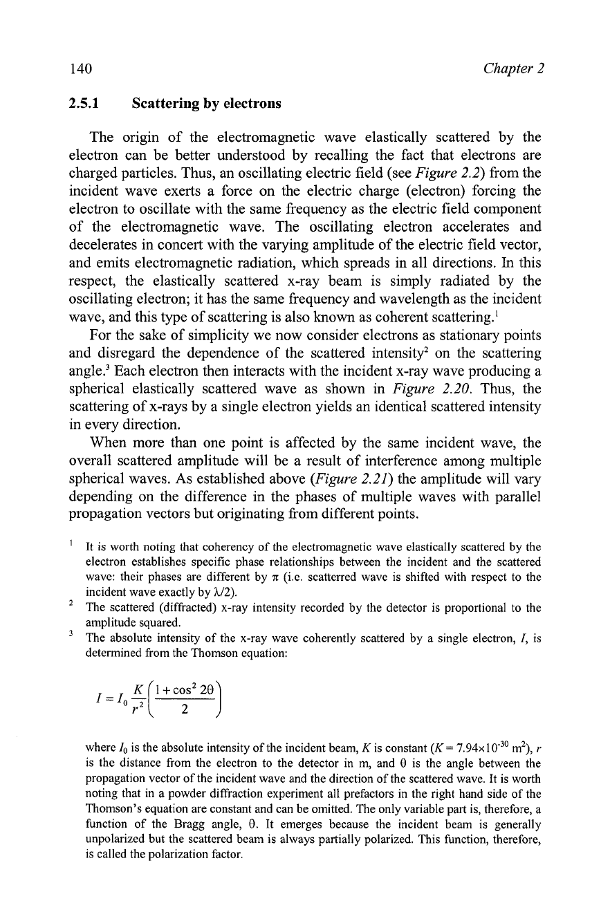
For the sake of simplicity we now consider electrons as stationary points
and disregard the dependence of the scattered intensity2 on the scattering
angle.3 Each electron then interacts with the incident x-ray wave producing a
spherical elastically scattered wave as shown in Figure
2.20.
Thus, the
scattering of x-rays by a single electron yields an identical scattered intensity
in every direction.
When more than one point is affected by the same incident wave, the
overall scattered amplitude will be a result of interference among multiple
spherical waves. As established above (Figure
2.21)
the amplitude will vary
depending on the difference in the phases of multiple waves with parallel
propagation vectors but originating from different points.
'
It is worth noting that coherency of the electromagnetic wave elastically scattered by the
electron establishes specific phase relationships between the incident and the scattered
wave: their phases are different by
n
(i.e. scatterred wave is shifted with respect to the
incident wave exactly by
112).
The scattered (diffracted) x-ray intensity recorded by the detector is proportional to the
amplitude squared.
The absolute intensity of the x-ray wave coherently scattered by a single electron,
I,
is
determined from the Thomson equation:
where
lo
is the absolute intensity of the incident beam,
K
is constant
(K
=
7.94~
m2),
r
is the distance from the electron to the detector in m, and
0
is the angle between the
propagation vector of the incident wave and the direction of the scattered wave. It is worth
noting that in a powder diffraction experiment all prefactors in the right hand side of the
Thomson's equation are constant and can be omitted. The only variable part is, therefore, a
function of the Bragg angle,
0.
It emerges because the incident beam is generally
unpolarized but the scattered beam is always partially polarized. This function, therefore,
is called the polarization factor.
