N?lting B. Methods in Modern Biophysics
Подождите немного. Документ загружается.


10.4 Design details 185
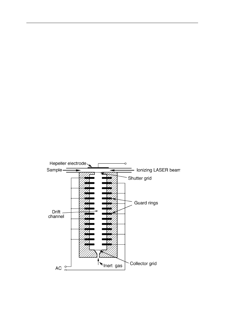
Fig. 10.14
The typical set-up for electrophoresis shows an analogy to ion mobility
spectrometry: in both methods charged molecules are accelerated by an electric field and
slowed down by interaction with molecules of a stationary phase. In electrophoresis, the
stationary phase is generally a gel, and the movement of the sample molecules is often
optically detected. A gas serves as stationary phase in ion mobility spectrometry, and the
movement of the sample ions is electrically detected. A small size and high charge of ions
correlates with a large speed
Ion mobility spectrometry has some similarities to electrophoresis (Fig. 10.14).
Because of the similarities to common chromatography, originally ion mobility
spectrometry was called “plasma chromatography”. However, one has to keep in
mind that in contrast to common chromatography, ion mobility spectrometers have
a narrow linear range due to space charge effects (Bird and Keller, 1976; Blan-
chard and Bacon, 1989; Spangler, 1992b), and show serious matrix interferences
and prolonged memory effects.
Table 10.1
Common methods of sample ionization in ion mobility spectrometry (Lubman
and
Kronick, 1982, 1983; Baim et al., 1983; Leasure et al., 1986; Eiceman et al., 1988; Shu-
mate and Hill, 1989; Begley et al., 1991; Phillips and Gormally, 1992; Davies, 1994; Span-
gler et al., 1994; Carnahan and Tarassov, 1995; Leonhardt, 1996; Lee et al., 1998; Wu et
al., 1998a, 1998b, 2000; Budovich et al., 1999; Döring et al., 1999; Borsdorf et al., 2000;
Megerle and Cohn, 2000; Schnurpfeil and Klepel, 2000; Borsdorf and Rudolph, 2001)
Method of ionization Example
Radioactive isotopes
3
H,
241
Am foil, or
63
Ni foil; Fig. 10.15
Photoionization UV and VUV light from a 30-W krypton or hydrogen
lamp with a MgF
2
-window, or perpendicular to the drift
channel from a frequency-quadrupled Nd:YAG laser at
266 nm (Fig. 10.15). A VUV-absorbing compound may
be added for an increased degree of ionization.
Electrospray Fig. 3.11 in Chap. 3
Laser desorption Fig. 10.15
Electrical (corona) discharge Fig. 10.15
186 10 Ion mobility spectrometry
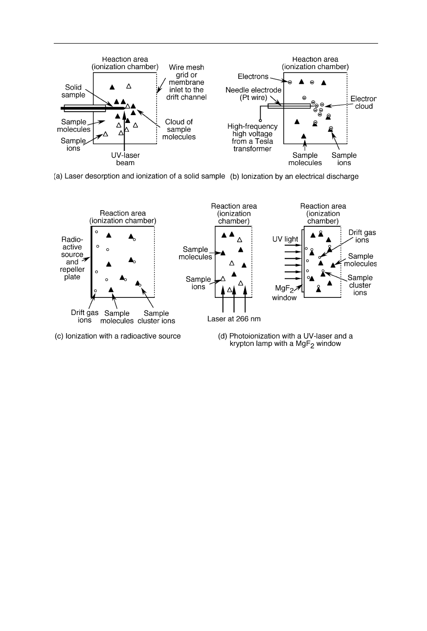
Fig. 10.15
Examples of ionization methods (see also Table 10.1)
Since most of the interesting chemical and biological substances are not
charged, it is necessary to ionize them prior to the drift. Table 10.1 and Figs.
10.15 and 10.16 show methods for ionization in ion mobility spectrometry.
Radioactive isotopes and photoionization are the most common methods. When
using a
63
Ni foil as the source of ionization and air as the drift gas, the primary
ions are mainly short-living N
2
+
, NO
+
, and O
2
–
. These primary ions rapidly react
with traces of water in the drift gas to form clusters of the types N
2
+
(H
2
O)
n
,
NO
+
(H
2
O)
m
, and O
2
–
(H
2
O)
k
. Photoionization with hydrogen plasma discharge
lamps and krypton plasma discharge lamps requires a photon flux of about 10
12
cm
–2
s
–1
. The geometry of the shutter grid is chosen so that most photons cannot
enter the drift channel since this would reduce the resolution. Common radioac-
tive sources in IMS have the advantage of relatively long half-lives of several
years. For immobilization of
3
H, it is gettered in a thin titanium layer.
Ion mobility spectrometry has gained significant importance in the context of
the detection of ultra-trace chemical and biological contaminants (Snyder et al.,
1991a, 1991b, 1996a, 1996b, 1999, 2000; Ogden and Strachan, 1993; Strachan et
10.4 Design details 187
al., 1995; Dworzanski et al., 1997; Smith et al., 1997), explosives (e.g., Fetterolf
and Clark, 1993; Steinfeld and Wormhoudt, 1998; Fig. 10.17), illicit drugs (e.g.,
Miki et al., 1997, 1998; Keller et al., 1998), pesticides, the detection of animals
and animal activity in jungles, and other environmental monitoring.
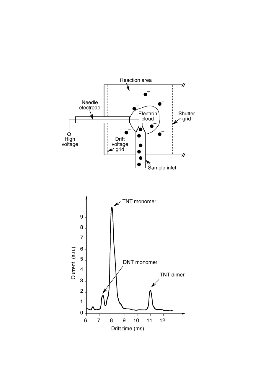
Fig. 10.16
Gas inlet for electrical-discharge (corona discharge) ionization
Fig. 10.17
Ion mobility spectrogram of TNT (trinitrotoluene). From data supplied by the
Institute for Environmental Technologies Ltd., Berlin
188 10 Ion mobility spectrometry
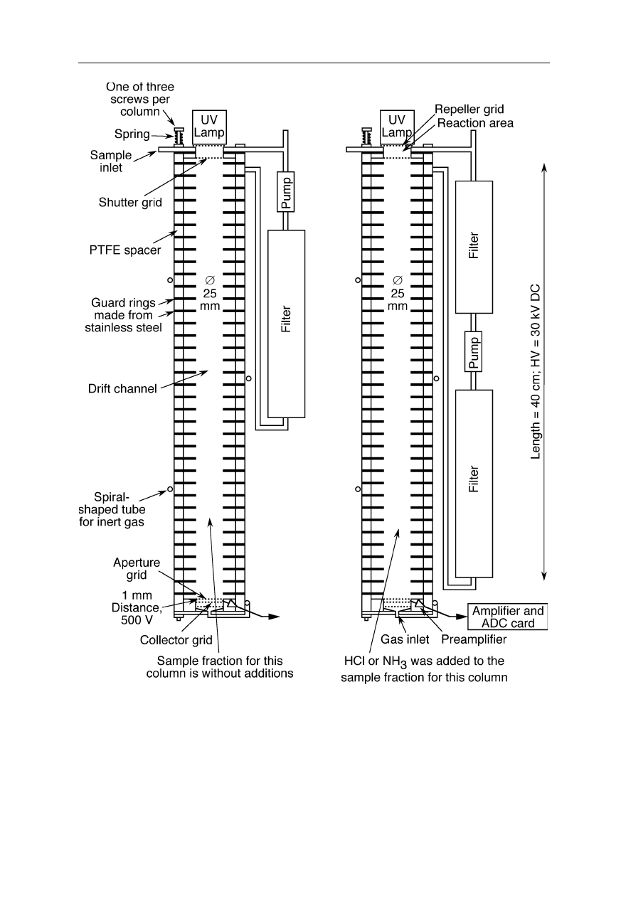
Fig. 10.18
A high-resolution two-channel ion mobility spectrometer. Here the sample is
simultaneously analyzed in two different ways reducing false identifications of agents. In
one of the columns, the sample is chemically and/or physically modified by a chemical
addition
Fig. 10.18 outlines an IMS-based twochannel detector which was designed
within a feasibility study. In this design the differentiation between various
10.4 Design details 189
biological and also some chemical substances is improved by utilizing physical
and chemical modifications of the sample in one of the two columns, e.g., by
adding an acidizing gas: Most proteins display a strong pH-dependency of the
charge (see, e.g., Chap. 2 in Nölting, 2005). Thus, by changing the pH, the charge
state of the protein-containing biological material is altered and consequently its
speed of diffusion in the drift channel is changed. Further, the added gas can
cause chemical changes of some chemical and biological agents. This causes
specific changes of the IMS spectra and thus contributes to a further improvement
of the correct identification of the agents. Multichannel designs are a further
option to reduce false detection rates of IMS. The channels may be operated in
the same way speeding up the measurement of slowly drifting substances.
Alternatively the sample may simultaneously be distributed over different
channels which are operated in various different modes which can improve the
resolution of the method.
IMS with an oscillating electric field allow the application of large field
strengths without need for a very high voltage (Fig. 10.19). Since very large
macromolecules have low mobilities, their fast detection with high resolution
requires a high electric field strength in the drift channel. In the common design
this may cause safety problems and increases the price of the IMS.
Fig. 10.19
IMS with an oscillating electric field of the guard rings instead of a constant
electrostatic field. Only ions of which the movement is in phase with the oscillating
electric field can pass through the drift channel and reach the collector. For further details
on frequency-domain IMS see, e.g., Martin et al., 1998
190 10 Ion mobility spectrometry
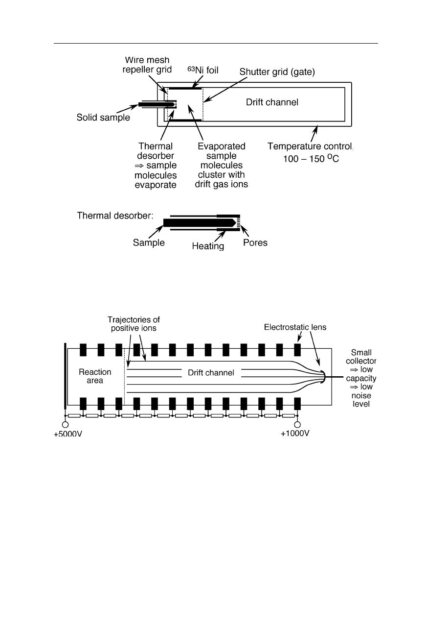
Fig. 10.20
Modified IMS for the detection of chemicals contained in solid samples and for
the identification of different solid materials, e.g., wood (Lawrence et al., 1991; Matz and
Schröder, 1997; Schröder et al., 1998)
Fig. 10.21
Noise reduction with a smaller collector grid: The collector and the guard ring
in front of the collector act together as an electrostatic lens. The smaller collector causes
less noise and, thus, a higher sensitivity
Measurements on solid samples require special sample inlets with a heating and
a temperature-resistant porous membrane (see, e.g., Fig. 10.20) and possibly a
higher temperature of the IMS.
An electrostatic lens focusing the ions towards the collector can allow the
reduction of the collector size and capacity (Fig. 10.21). This decreases noise and
can improve the sensitivity.
10.4 Design details 191
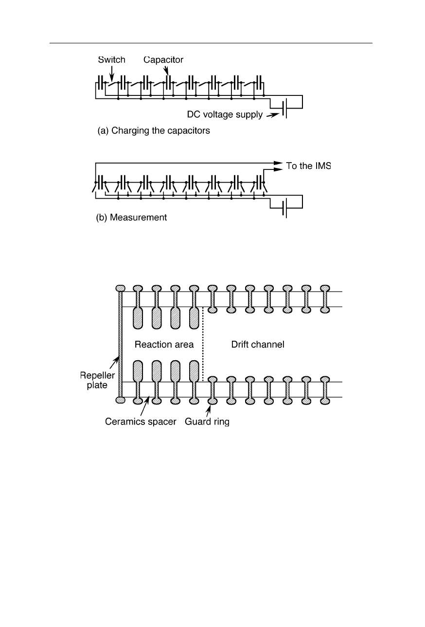
Fig. 10.22
Generating the high voltage for an IMS (Goebel and Breit, 2000). (
a
) A set of
n capacitors is charged with the voltage V. (
b
) The capacitors are connected together in
series for generating the high voltage n
.
V
Fig. 10.23
Improved mechanical stability of the drift channel. For a similar design see
also (Karl, 1994)
A technique of high voltage generation which does not necessarily require
much weight is illustrated in Fig. 10.22: a set of capacitors is charged in parallel
and then connected in series.
Parameters which are important for a high reproducibility of IMS spectra are a
constant degree of humidity, constant electric field strength, constant source of
ionization, efficient removal of previous samples, mechanical stability. The latter
can be improved by a special shape of the guard rings (Fig. 10.23). Figs. 10.24
–
10.26 display some further important innovations in ion mobility spectrometry.
192 10 Ion mobility spectrometry
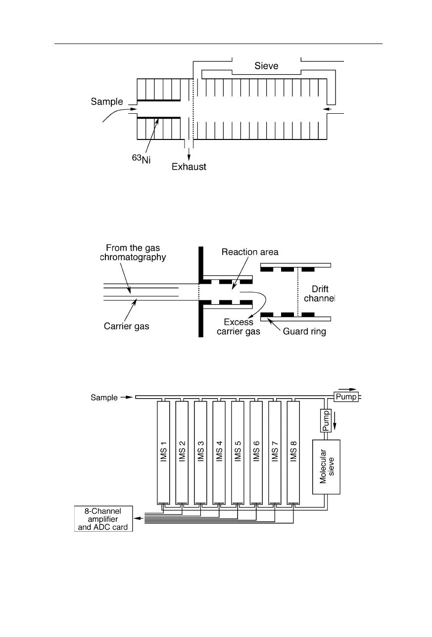
Fig. 10.24
Improved purgeability of the reaction area (see, e.g., Snyder et al., 1993) and
improved homogeneity of the electric field the ions experiencing in the drift channel: the
sample outlet is located close to the shutter grid, and the diameter of the reaction area is
smaller than that of the drift channel
Fig. 10.25
Injection of the output of a gas chromatograph or of the vapor from a solid
sample into the IMS with the help of a gentle stream of carrier gas
Fig. 10.26
A multichannel ion mobility spectrometer. For an 8 times higher sampling rate
than a single-channel spectrometer, all channels are operated in the same way. For
decreased rate of false identifications, one sample may be distributed over different
channels that are operated in different modes (see, e.g., Turner, 1993)
10.5 Detection of biological agents 193
10.5 Detection of biological agents
Unfortunately, many biological agents are too large to be detected directly: The
velocity,
v
, of a large spherical particle depends on its charge,
z
, its radius,
r
, the
Fig. 10.27
Setup for IMS-detection of biological agents (see, e.g., Snyder et al., 2000).
The virtual impactor selects a certain size range of particles, e.g., 1–10
µ
m, and transfers
the selected particles into the pyrolysis tube. Within a few seconds, particles of biological
origin are partially decomposed in the pyrolysis reaction at, e.g., 350
o
C. In the subsequent
analysis of the pyrolysis reaction products, a short gas chromatography and an IMS are
combined for enhanced resolution. The operating temperature of the GC/IMS is typically
80
–150
o
C. For the principle of operation of the virtual impactor see also Fig. 3.20. Due
to the highly dispersed 2D-spectra, Py-GC/IMS can potentially much safer unambiguously
identify traces of biological agents than a measurement of particle size distribution alone
194 10 Ion mobility spectrometry
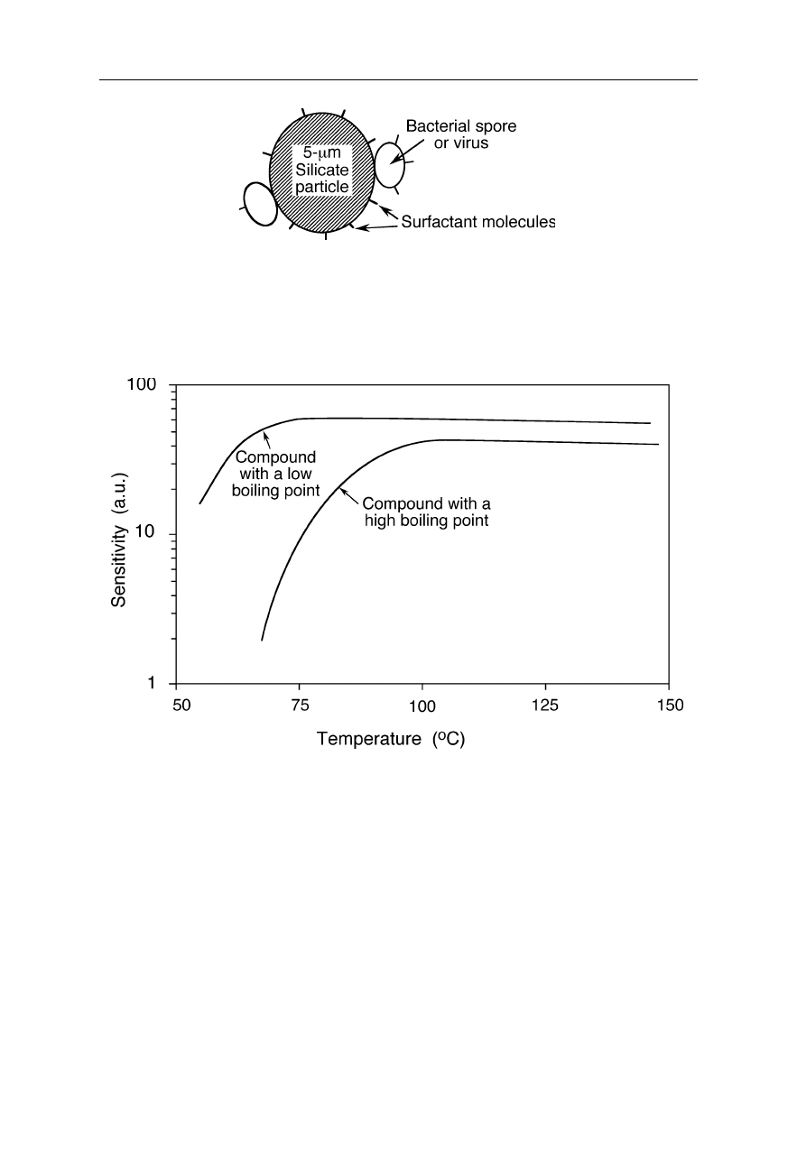
Fig. 10.28
Example of a particle of a dangerous biological contaminant. Light and fluffy
composites of bacterial spores or viruses to dust-forming particles of about 1–5
µ
m
diameter can drift in dry air for 100 miles, and can be sucked into the deepest sacs of the
lung (Preston, 1998)
Fig. 10.29
Typical sensitivity of IMS detection of compounds with different boiling points
and vapor pressures
electric field strength,
E
, and the viscosity of the medium,
η
:
v
=
zE
(6
π
η
r
)
–1
(10.3)
Small chemical compounds typically travel in an IMS with several m s
–1
, but a
single-charged dust particle (
z
= 1.6
×
10
–19
C) with a radius of 1
µ
m travels in air
(
η
air
= 1.8
×
10
–5
N s m
–2
) at a field strength of
E
= 300 V mm
–1
only with about
0.14 mm s
–1
. At higher gating frequencies of the IMS, successive spectra of
slowly moving agents would superimpose. Also, just average size and size distri-