Мир М.Афзал Атлас клинического диагноза
Подождите немного. Документ загружается.

1
ATLAS
OF
CLINICAL
DIAGNOSIS
22
Table
1.4
Causes
of
ptosis
Type
Cause
Congenital Levator muscle/oculomotor nerve
maldevelopment
may be
unilateral
or
bilateral
Local
disease Dehiscence
of
levator aponeurosis,
inflammatory
(e.g.
chalazion, stye),
infiltrative
(e.g.
amyloidosis, lymphoma,
etc.)
Myopathic
Myasthenia, botulism, myotonic dystrophy,
chronic progressive external
ophthalmoplegia
Neuropathic Horner's syndrome, oculomotor paresis,
tabes
dorsalis,
Guillain-Barre
syndrome,
mid-brain
lesion,
facial paresis, frontal
lobe lesions
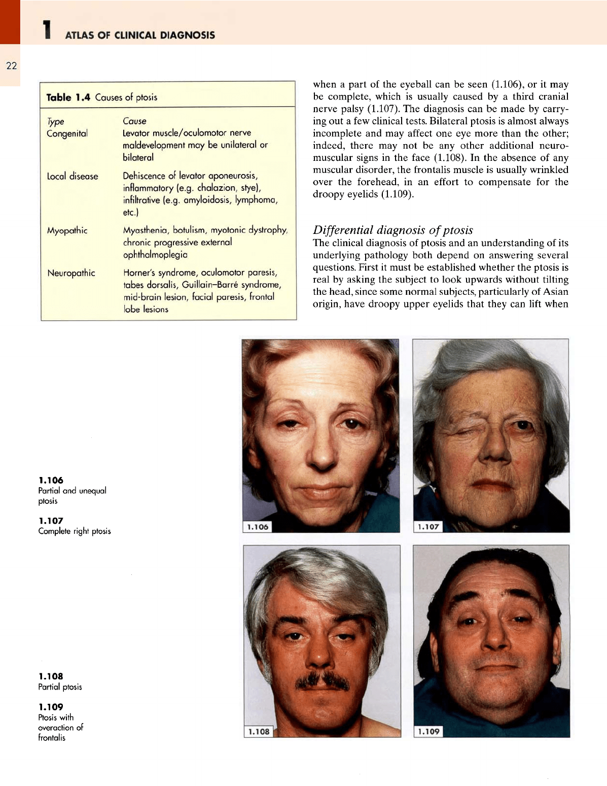
when
a
part
of the
eyeball
can be
seen (1.106),
or it may
be
complete, which
is
usually caused
by a
third cranial
nerve
palsy (1.107).
The
diagnosis
can be
made
by
carry-
ing
out a few
clinical tests. Bilateral ptosis
is
almost always
incomplete
and may
affect
one eye
more than
the
other;
indeed, there
may not be any
other additional neuro-
muscular
signs
in the
face
(1.108).
In the
absence
of any
muscular
disorder,
the
frontalis
muscle
is
usually wrinkled
over
the
forehead,
in an
effort
to
compensate
for the
droopy eyelids (1.109).
Differential
diagnosis
of
ptosis
The
clinical diagnosis
of
ptosis
and an
understanding
of its
underlying
pathology both depend
on
answering several
questions. First
it
must
be
established whether
the
ptosis
is
real
by
asking
the
subject
to
look upwards without tilting
the
head, since some normal subjects, particularly
of
Asian
origin,
have droopy upper eyelids that they
can
lift
when
1.106
Partial
and
unequal
ptosis
1.107
Complete
right
ptosis
1.108
Partial ptosis
1.109
Ptosis
with
overaction
of
frontalis
THE
FACE
1
23
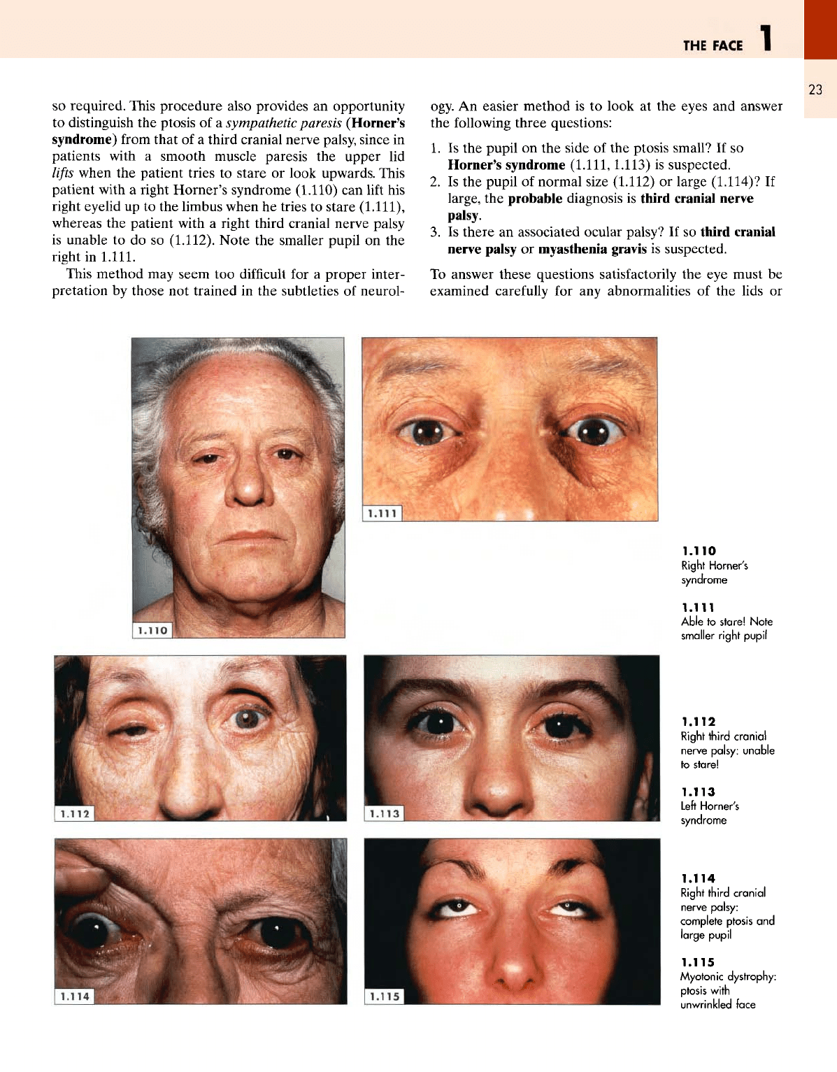
so
required.
This
procedure
also
provides
an
opportunity
to
distinguish
the
ptosis
of a
sympathetic paresis (Horner's
syndrome)
from
that
of a
third cranial nerve palsy, since
in
patients with
a
smooth muscle paresis
the
upper
lid
lifts
when
the
patient tries
to
stare
or
look upwards. This
patient with
a
right
Horner's
syndrome
(1.110)
can
lift
his
right
eyelid
up to the
limbus when
he
tries
to
stare
(1.111),
whereas
the
patient with
a
right third cranial nerve palsy
is
unable
to do so
(1.112).
Note
the
smaller pupil
on the
right
in
1.111.
This method
may
seem
too
difficult
for a
proper
inter-
pretation
by
those
not
trained
in the
subtleties
of
neurol-
ogy.
An
easier
method
is to
look
at the
eyes
and
answer
the
following three questions:
1.
Is the
pupil
on the
side
of the
ptosis small?
If so
Horner's syndrome
(1.111,1.113)
is
suspected.
2.
Is the
pupil
of
normal size
(1.112)
or
large
(1.114)?
If
large,
the
probable diagnosis
is
third
cranial
nerve
palsy.
3. Is
there
an
associated
ocular palsy?
If so
third cranial
nerve
palsy
or
m\asthenia
gravis
is
suspected.
To
answer
these
questions satisfactorily
the eye
must
be
examined
carefully
for any
abnormalities
of the
lids
or
1.110
Right
Horner's
syndrome
1.111
Able
to
stare!
Note
smaller
right
pupil
1.112
Right
third
cranial
nerve
palsy:
unable
to
stare!
1.113
Left
Horner's
syndrome
1.114
Right
third
cranial
nerve
palsy:
complete ptosis
and
large
pupil
1.115
Myotonic
dystrophy:
ptosis
with
unwrinkled
face

1
ATLAS
OF
CLINICAL
DIAGNOSIS
24
globe
and for any
ocular palsies (see Table 1.1). Finally,
one
needs
to
consider whether
the
ptosis
is a
part
of a
neuro-
muscular
disorder such
as
myasthenia
gravis,
myotonia
dystrophica
and
mitochondria!
myopathy.
Patients with
a
myotonic dystrophy tend
to
have
a
long,
lean, expressionless
face
and
they
can
neither
lift
their
upper eyelids
nor
create
any
wrinkles
on
their forehead
(1.115).
Bilateral ptosis with overaction
of
the
frontalis
may
be
congenital
and be
present since birth,
as in
this patient
(1.116),
or
acquired
as in
tabes dorsalis
(1.117).
The
ptosis
of
myasthenia gravis tends
to get
worse
as the day
pro-
gresses
or if the
patient
is
asked
to
open
and
close
the
eyes
repeatedly;
in
this situation
it
improves
after
an
intra-
venous injection
of
edrophonium
(1.118,1.119).
Ocular
palsies
Complete
or
partial
ptosis
associated with
a
dilated pupil
on the
affected
side
is
always
the
result
of a
third cranial
nerve palsy
(1.120).
Deviation
or
strabismus
of the
eyeball
can
also
be
caused
by a
fourth
or
sixth cranial nerve palsy
as
in
this patient
(1.121),
whose right
eye is
turned inwards
because
of the
weakness
of the
right lateral rectus muscle,
thereby permitting unopposed adduction
by the
medial
1.116
Congenital
ptosis
1.117
Acquired
ptosis
with
overaction
of
frontalis
1.118
Before
and 30 s
after
intravenous
edrophonium
1.119
Before
and 1 min
after
intravenous
edrophonium
1.120
Right
third
cranial
nerve palsy
1.121
Right
sixth
cranial
nerve
palsy
THE
FACE
1
25
rectus.
Apart
from
a
strabismus,
one
should
ascertain
whether
the
patient experiences diplopia
and
whether
there
is any
proptosis.
The
former
confirms
that there
is an
ocular palsy
and its
direction
is
suggestive
of the
particu-
lar
cranial nerve involved.
Proptosis
suggests
the
presence
of
a
local pathology.
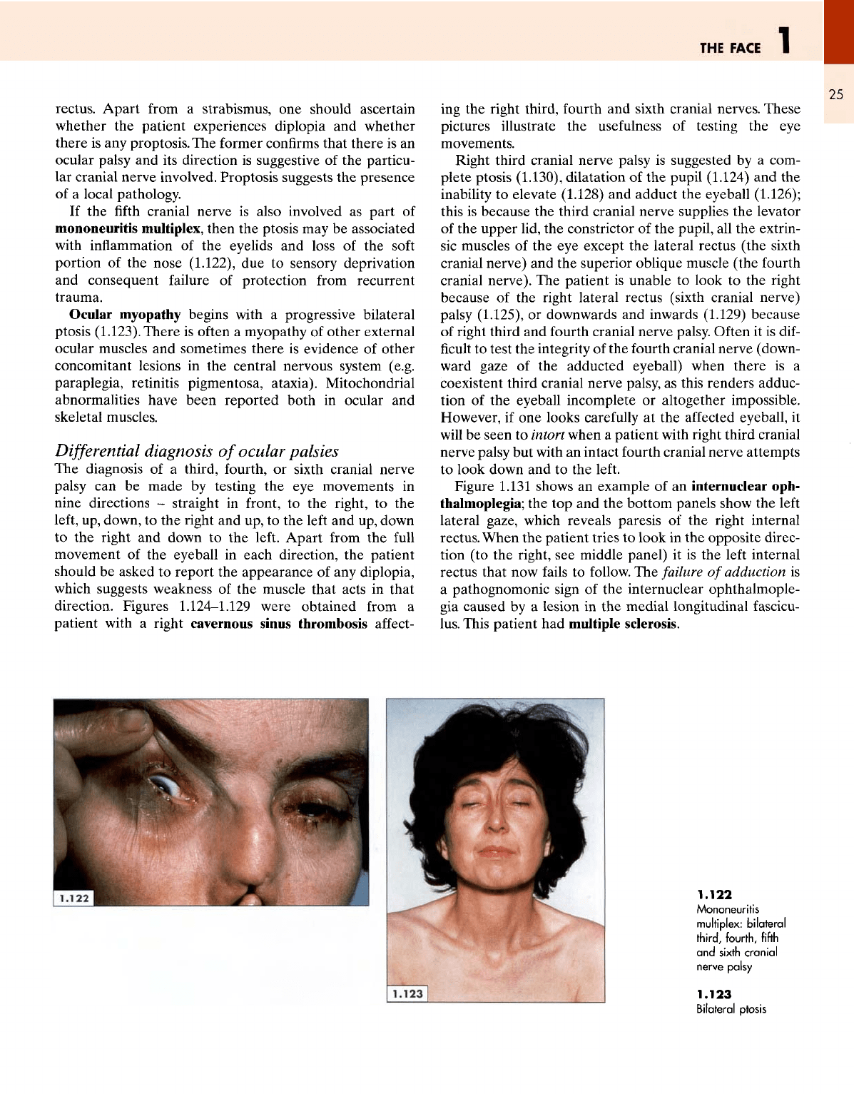
If
the fifth
cranial nerve
is
also involved
as
part
of
mononeuritis
multiplex, then
the
ptosis
may be
associated
with
inflammation
of the
eyelids
and
loss
of the
soft
portion
of the
nose (1.122),
due to
sensory deprivation
and
consequent failure
of
protection
from
recurrent
trauma.
Ocular
myopathy begins with
a
progressive bilateral
ptosis
(1.123).
There
is
often
a
myopathy
of
other external
ocular muscles
and
sometimes there
is
evidence
of
other
concomitant lesions
in the
central nervous system
(e.g.
paraplegia, retinitis pigmentosa, ataxia). Mitochondrial
abnormalities have been reported both
in
ocular
and
skeletal muscles.
Differential
diagnosis
of
ocular palsies
The
diagnosis
of a
third, fourth,
or
sixth cranial nerve
palsy
can be
made
by
testing
the eye
movements
in
nine directions
-
straight
in
front,
to the
right,
to the
left,
up,
down,
to the
right
and up, to the
left
and up,
down
to the
right
and
down
to the
left.
Apart
from
the
full
movement
of the
eyeball
in
each direction,
the
patient
should
be
asked
to
report
the
appearance
of any
diplopia,
which
suggests weakness
of the
muscle that acts
in
that
direction.
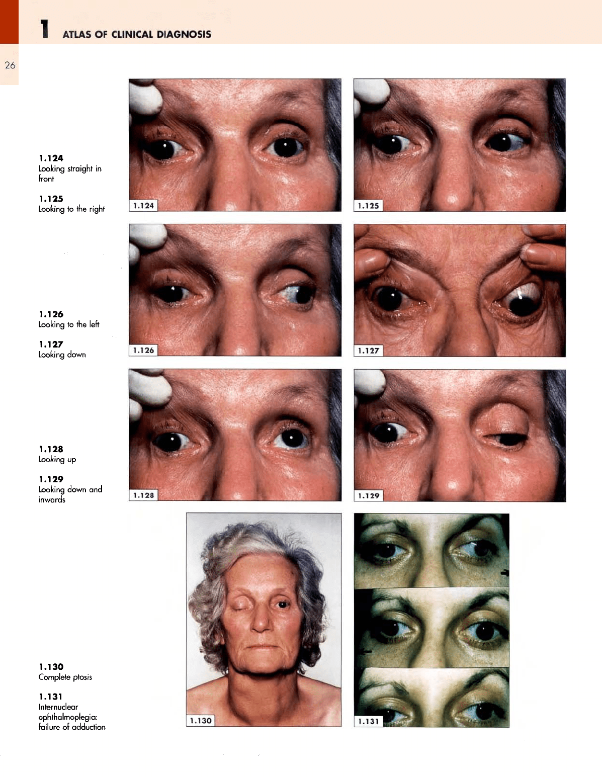
Figures
1.124-1.129
were
obtained
from
a
patient with
a
right cavernous sinus thrombosis
affect-
ing
the
right third, fourth
and
sixth cranial nerves.
These
pictures illustrate
the
usefulness
of
testing
the eye
movements.
Right third cranial nerve palsy
is
suggested
by a
com-
plete
ptosis
(1.130),
dilatation
of the
pupil
(1.124)
and the
inability
to
elevate
(1.128)
and
adduct
the
eyeball (1.126);
this
is
because
the
third cranial nerve supplies
the
levator
of
the
upper lid,
the
constrictor
of the
pupil,
all the
extrin-
sic
muscles
of the eye
except
the
lateral rectus
(the
sixth
cranial nerve)
and the
superior oblique muscle (the
fourth
cranial
nerve).
The
patient
is
unable
to
look
to the
right
because
of the
right lateral rectus (sixth cranial nerve)
palsy
(1.125),
or
downwards
and
inwards (1.129) because
of
right third
and
fourth cranial nerve palsy. Often
it is
dif-
ficult to
test
the
integrity
of the
fourth cranial nerve (down-
ward
gaze
of the
adducted eyeball) when there
is a
coexistent third cranial nerve palsy,
as
this renders adduc-
tion
of the
eyeball incomplete
or
altogether impossible.
However,
if one
looks
carefully
at the
affected
eyeball,
it
will
be
seen
to
intort
when
a
patient with right third cranial
nerve palsy
but
with
an
intact
fourth
cranial nerve attempts
to
look down
and to the
left.
Figure
1.131
shows
an
example
of an
internuclear oph-
thalmoplegia;
the top and the
bottom panels show
the
left
lateral gaze, which reveals paresis
of the
right internal
rectus. When
the
patient
tries
to
look
in the
opposite
direc-
tion
(to the
right,
see
middle panel)
it is the
left
internal
rectus that
now
fails
to
follow.
The
failure
of
adduction
is
a
pathognomonic sign
of the
internuclear ophthalmople-
gia
caused
by a
lesion
in the
medial longitudinal fascicu-
lus.
This patient
had
multiple sclerosis.
1.122
Mononeuritis
multiplex:
bilateral
third,
fourth,
fifth
and
sixth
cranial
nerve
palsy
1.123
Bilateral ptosis
1
ATLAS
OF
CLINICAL
DIAGNOSIS
26
1.124
Looking
straight
in
front
1.125
Looking
to the
right
1.126
Looking
to the
left
1.127
Looking
down
1.128
Looking
up
1.129
Looking
down
and
inwards
1.130
Complete
ptosis
1.131
Internuclear
ophthalmoplegia:
failure
of
adduction
THE
FACE
1
27
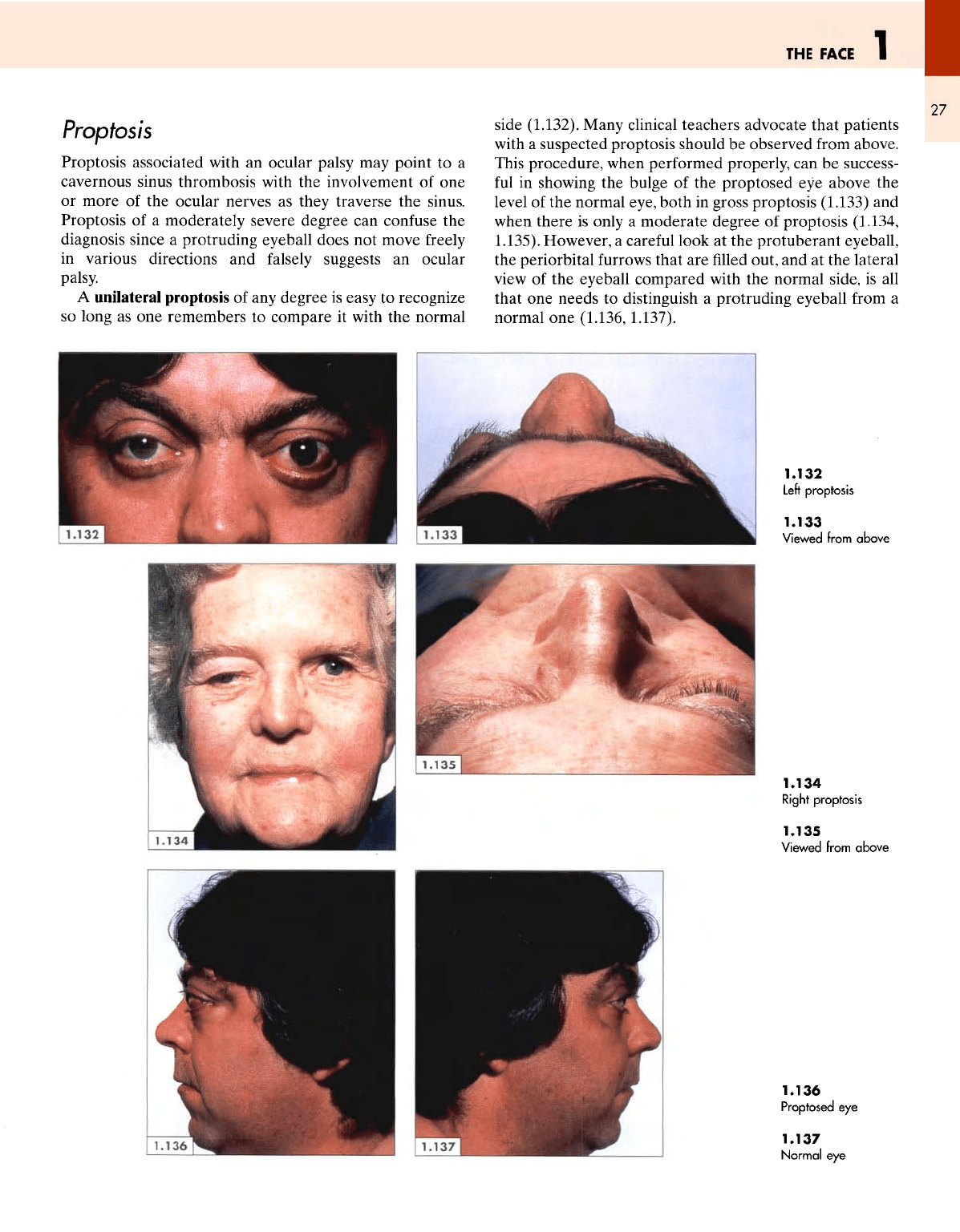
Proptosis
Proptosis associated
with
an
ocular palsy
may
point
to a
cavernous sinus thrombosis with
the
involvement
of one
or
more
of the
ocular nerves
as
they traverse
the
sinus.
Proptosis
of a
moderately severe degree
can
confuse
the
diagnosis since
a
protruding eyeball does
not
move
freely
in
various directions
and
falsely
suggests
an
ocular
palsy.
A
unilateral proptosis
of any
degree
is
easy
to
recognize
so
long
as one
remembers
to
compare
it
with
the
normal
side
(1.132). Many clinical teachers advocate that patients
with
a
suspected proptosis should
be
observed
from
above.
This procedure, when performed properly,
can be
success-
ful
in
showing
the
bulge
of the
proptosed
eye
above
the
level
of the
normal eye, both
in
gross proptosis
(1.133)
and
when
there
is
only
a
moderate degree
of
proptosis (1.134,
1.135). However,
a
careful
look
at the
protuberant eyeball,
the
periorbital
furrows
that
are filled
out,
and at the
lateral
view
of the
eyeball compared with
the
normal side,
is all
that
one
needs
to
distinguish
a
protruding eyeball from
a
normal
one
(1.136,1.137).
1.132
Left
proptosis
1.133
Viewed
from
above
1.134
Right
proptosis
1.135
Viewed
from
above
1.136
Proptosed
eye
1.137
Normal
eye

1
ATLAS
OF
CLINICAL
DIAGNOSIS
28
This
procedure
can be
successful
even
when
there
is
only
a
modest degree
of
proptosis, which
can be
suspected
first
by
looking
at a
patient's
face
(1.138).
Comparing
the
eyes with each other
and by
looking
at
each
eye
carefully,
the
superior
periorbital
sulcus clearly
seen
above
the
right
eye
(1.139)
is
obscured
by the
bulging eyeball
on the
left
side
(1.140).
The
lower eyelid also looks
a
little
fuller
on
the
left
side
and the
left
eye
does
not
look
so
comfortably
housed
in the
orbit, unlike
the
right
eye
with room
to
spare.
The
examination
of
proptosis should
be
completed
by
determining
its
direction, whether
it is
pulsatile
and
reducible
(by
gently pressing
on the
eyeball)
as it was in
this
patient
with
a
carotid-cavernous
fistula
(1.138),
or
whether
it is fixed and
irreducible
as in
Figure
1.132.
Pupillary abnormalities
Pupillary
abnormalities
may be
bilateral,
as in
neu-
rosyphilis,
or
unilateral,
as in the
Holmes-Adie
syndrome
(myotonic pupil).
A
clinician's task
is not
only
to
spot
the
asymmetry
but
also
to
identify
the
abnormal side. Often
there
are
associated abnormalities
in the
eyeball
or
eyelids
such
as a
partial ptosis
in
Horner's
syndrome
(1.141)
or
complete ptosis
in a
third cranial nerve palsy (1.142).
1.138
Left
proptosis
1.139
Normal
orbital
contours
1.140
Obscured
superior
orbital
furrow
1.141
Left
Horner's
syndrome
1.142
Left
third
cranial
nerve palsy

THE
FACE
1
29
The
absence
of any
ocular abnormalities associated with
a
dilated pupil
(1.143)
suggests
a
diagnosis
of the
myotonic
pupil,
a
condition usually seen
in
young women
and
often
associated with absent ankle
and
knee jerks. Such pupils
are
always regular
but
react
poorly
to
light
and on
accom-
modation.
The
Argyll Robertson pupils
are
small (1.144),
often
irregular
and
unequal (although these changes
may
be
very subtle),
and
their characteristic feature
is
that they
are
either non-reactive
to
light
or
react poorly
but
respond
better
on
accommodation.
Their
reaction
to
light
and
accommodation
is
sometimes
difficult
to
determine
be-
cause
of
their small size but,
on
repeated testing, some
reaction
on
accommodation
can be
detected
in the
pupils
that
are
non-reactive even
to
intense light.
Facial
muscles
Facial muscles make
a
major contribution
to the
contour
and
shape
of the
face.
Any
weakness
or
wasting
of one or
more muscles
is
betrayed
by a
resulting asymmetry
and
distortion
of the
facial
features. Atrophy
of one
side
of the
face
-
facial
hemiatrophy
-
may
occur
in
otherwise healthy
subjects
(1.145).
This
is a
rare disorder
and is
often
her-
alded
by the
appearance
of a
dimple, which progressively
enlarges and, within
a few
years, invovles
all the
structures
on one
side
of the
face.
The
abnormal side
(1.146)
with
its
infrazygomatic
pit, thinned nose
and
hollow periorbital
spaces shows
a
striking contrast
to the
normal side
(1.147),
which
looks
full
and fleshy.
1.143
Holmes-Adie
syndrome
1.144
Argyll
Robertson
pupils
1.145
Facial
hemiatrophy
1.146
Hollow
side
1.147
Normal
side

1
ATLAS
OF
CLINICAL
DIAGNOSIS
30
Myopathic facies
are
characteristically unwrinkled,
ex-
pressionless
and
sad, with
lax and
pouting lips,
as
seen
in
this patient with myotonia dystrophica
(1.148).
This long,
lean
and
expressionless
face
with gaping lips
can be
detected even
in
young subjects with
the
condition (1.149).
An
expressionless face
may
also
be
seen
in
association
with
the
facioscapulohumeral muscular dystrophy
(1.150).
There
is
characteristic pouting
of the
lips, which, together
with
poverty
of
expression,
is
seen even
in
those patients
in
whom muscular wasting
of the
face
is not
very marked
and
in
whom there
is no
ptosis (1.151). This form
has an
autosomal dominant inheritance
but
there
is
substantial
variation
in the
severity
in
affected members
of the
same
family.
In
contrast, profound muscular wasting
of the
entire
face,
as in
this patient with bulbospinal muscular atrophy
(1.152),
gives
the
face
a
characteristic appearance with
wide,
noncommunicative eyes,
lax and
open lips,
an
unlined
face
and a
sagging lower jaw. Facial features
are
much less
marked
in the
early stages
of
myasthenia gravis when
ptosis
may be the
only abnormality
(1.153).
1.148
Myopathic
facies:
bilateral
ptosis
with
a
smooth,
unlined
face
1.149
Long,
lean face
with
pouting
lips
1.150
Facial
muscular
atrophy:
unwrinkled,
expressionless
face
1.151
Mild
facial muscular
wasting
1.152
Severe facial
muscular
wasting:
wide
open
eyes
and
a
lax,
partially
open
mouth
1.153
Ptosis
in
myasthenia
gravis
1.154
Right
Bell's
palsy:
wide
open
right
eye,
smooth
right
face

THE
FACE
1
31
Facial nerve palsy
is by far the
commonest abnormal
neuromuscular facies encountered
in
clinical practice.
In
its
complete form,
as in
Bell's palsy
(1.154),
there
is
paraly-
sis
of the
upper
and
lower parts
of the
face,
the
wrinkles
on the
affected
side
of the
forehead
and the
nasolabial
fold
are
smoothed
out,
both
the
eyelids look
lax and
somewhat
retracted
and the
corner
of the
mouth appears
to
droop.
In
the
upper motor neurone muscular weakness (e.g.
lesion
in the
internal capsule),
the
paralysis
is
less well
marked
and
spares
the
upper part
of the
face
if the
lesion
is
supranuclear since there
is
bilateral innervation
of the
forehead
from
the
corticobulbar
fibres.
This
fact
tends
to
be
overemphasized
by
clinical
teachers
since, despite
the
bilateral
innervation, there
is
often
some widening
of the
eyebrows
on the
affected
side. Emotional
and
involuntary
movements such
as
spontaneous
smiling
may be
much less
affected.
Voluntary
effort
to
force
a
smile
and
retract
the
angles
of the
mouth reveals
the
weakness
on the
right side
in
this patient with weakness
of the
facial
muscles
of
central origin
(1.155).
1.155
Right
hemiparesis
1.156
Myotonia
dystrophica
1.157
Facioscapulohumeral
muscular
atrophy
1.158
Attenuated
facial
and
neck
muscles