Lallart M. Ferroelectrics: Characterization and Modeling
Подождите немного. Документ загружается.

Part 1
Characterization: Structural Aspects
1
Structural Studies in
Perovskite Ferroelectric Crystals Based
on Synchrotron Radiation Analysis Techniques
Jingzhong Xiao
1,2
1
CEMDRX, Department of Physics, University of Coimbra, Coimbra,
2
International Centre for Materials Physics,
Chinese Academy of Sciences, Shenyang,
1
Portugal
2
China
1. Introduction
Perovskite oxide materials, having the general formula ABO
3
, form the backbone of the
ferroelectrics industry. These materials have come into widespread use in applications that
range in sophistication from medical ultrasound and underwater sonar systems, relatively
mundane devices to novel applications in active and passive damping systems for sporting
goods and automobiles [1-3]. Recent developments in regard to relaxor-based single crystal
piezoelectrics, such as Pb(Zn
1/3
Nb
2/3
)O
3
–PbTiO
3
(PZNT), Pb(Fe
1/2
Nb
/1/2
)O
3
–PbTiO
3
(PFNT) and Pb(Mg
1/3
Nb
2/3
)O
3
–PbTiO
3
(PMNT) have shown superior performance
compared to the conventional Pb(Zr,Ti)O
3
(PZT) ceramics[4, 5]. Particularly, their ultrahigh
piezoelectric and electromechanical coupling factors in the <001> direction can reach to
d
33
>2000pC/N and k
33
≈94%, which have attracted tremendous interests and still make these
materials very hot.
However, the origin and structural nature of their ultrahigh performances remains one
inquisitive but obscure subject of recent scientific interest.To better understand the
structural nature of the outstanding properties, it is very important for investigating the
ferroelectric domain structure in these materials. In ferroelectrics, according to the electrical
and mechanical compatibility conditions, domain structures of 180
o
and non-180
o
will form
with respect to crystal symmetry. There is a closely relationship between the domain
structure and the crystal symmetry. Through the observation on ferroelectric domain
configurations, the crystal structures can be confirmed. Ferroelectric domains are
homogenous regions within ferroelectric materials in which polarizations lie along one
direction, that influence the piezoelectric and ferroelectric properties of the materials for
utilization in memory devices, micro-electromechanical systems, etc. Understanding the role
of domain structure on properties relies on microscopy methods that can inspect the domain
configuration and reveal the evolution or the dynamic behaviour of domain structure.
It is also well known that the key to solve this issue of exploring the origin of the excellent
properties is to reveal the peculiar complex perovskite crystal structures in these materials.
Through study in structure behavior under high-pressure and local structure at atomic level
will be helpful for better understanding this problem.

Ferroelectrics - Characterization and Modeling
4
2. Synchrotron radiation X-ray structure investigation on ferroelectric
crystals
Pb(Zn
1/3
Nb
2/3
)O
3
-PbTiO
3
(PZNT) and Pb(Fe
1/2
Nb
/1/2
)O
3
–PbTiO
3
(PFNT) crystal are model
ABO
3
-type relaxor ferroelectric materials (or ferroelectrics), which demonstrates excellent
performance in the filed of dielectrics, piezoelectrics, and electrostriction, etc. To explore the
common issues of structural nature and the relationship between structure and performance
of ultrahigh-performance in these materials, in this chapter, the novel X-ray analysis
techniques based on synchrotron radiation light, such as synchrotron radiation X-Ray
topography, high-pressurein situ synchrotron radiation energy dispersive diffraction, and
XAFS method, are utilized to investigate the domain configuration, structure and their
evolution behavior induced by temperature changes and external field.
2.1 Application of white beam synchrotron radiation X-ray topography for in-situ
study of ferroelectric domain structures
Ferroelectric domains can be observed by various imaging techniques such as optical
microscopy, scanning electron microscopy (SEM), transmission electron microscopy (TEM),
X-ray imaging, and etc. Imaging is normally associated with lenses. Unlike visible light or
electrons, however, efficient lenses are not available for hard X-rays, essentially because they
interact weakly with matter. Comparatively as an X-ray imaging method, X-ray topography
plays a vital role in providing a better understanding of ferroelectric domain structure.[6] X-
rays are more penetrating than photons and electrons, and the advent of synchrotron
radiation with good collimation, a continuous spectrum (white beam) and high intensity has
given X-ray topography additional powers. The diffraction image contrast in X-ray
topographs can be accessed from variations in atomic interplanar spacings or interference
effects between X-ray and domain boundaries so that domain structure can be directly
observed (with a micrometer resolution). Especially, via a white beam synchrotron radiation
X-ray diffraction topography technique (WBSRT), one can study the dynamic behaviour of
domain structure and phase evolution in ferroelectric crystals respectively induced by the
changes of sample temperature, applied electric field, and other parameter changes.
In this chapter, a brief introduction to principles for studying ferroelectric domain structure by
X-ray diffraction imaging techniques is provided. The methods and devices for in-situ
studying domain evolution by WBSR are delineated. Several experimental results on dynamic
behavior of domain structure and induced phase transition in ferroelectric crystals accessed at
beam line 4W1A of the Beijing Synchrotron Radiation Laboratory (BSRL) are introduced.
2.1.1 Principle of synchrotron radiation X-ray topography
a. X-ray topography approach
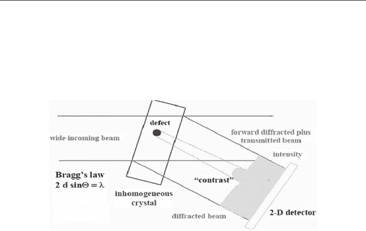
X-ray diffraction topography is an imaging technique based on Bragg diffraction. In the last
decades, X-ray diffraction topography to characterize crystals for the microelectronics
industry were developed and completely renewed by the modern synchrotron radiation
sources. [6]
Its images (topographs) record the intensity profile of a beam of X-rays diffracted by a
crystal. A topograph thus represents a two-dimensional spatial intensity mapping of
reflected X-rays, i.e. the spatial fine structure of a Bragg spot. This intensity mapping reflects
the distribution of scattering power inside the crystal; topographs therefore reveal the
Structural Studies in Perovskite Ferroelectric
Crystals Based on Synchrotron Radiation Analysis Techniques
5
irregularities in a non-ideal crystal lattice. The basic working principle of diffraction
topography is as follows: An incident, spatially extended X-ray beam impinges on a sample,
as shown in Fig.1. The beam may be either monochromatic, or polychromatic (i.e. be
composed of a mixture of wavelengths (white beam topography)). Furthermore, the incident
beam may be either parallel, consisting only of rays propagating all along nearly the same
direction, or divergent/convergent, containing several more strongly different directions of
propagation.
Fig. 1. The scheme of basic principle of diffraction topography.
A homogeneous sample (with a regular crystal lattice) would yield a homogeneous intensity
distribution in the topograph (a "flat" image). Intensity modulations (topographic contrast)
arise from irregularities in the crystal lattice, originating from various kinds of defects such
as cracks, surface scratches, dislocations, grain boundaries, domain walls, etc. Theoretical
descriptions of contrast formation in X-ray topography are largely based on the dynamical
theory of diffraction. This framework is helpful in the description of many aspects of
topographic image formation: entrance of an X-ray wave-field into a crystal, propagation of
the wave-field inside the crystal, interaction of wave-field with crystal defects, altering of
wave-field propagation by local lattice strains, diffraction, multiple scattering, absorption.
Theoretical calculations, and in particular numerical simulations by computer based on this
theory, are thus a valuable tool for the interpretation of topographic images. Contrast
formation in white beam topography is based on the differences in the X-ray beam intensity
diffracted from a distorted region around the defect compared with the intensity diffracted
from the perfect crystal region. The image of this distorted region corresponds to an
increased intensity (direct image) in the low absorption case.
To conduct a topographic experiment, three groups of instruments are required: an x-ray
source, potentially including appropriate x-ray optics; a sample stage with sample
manipulator (diffractometer); and a two-dimensionally resolving detector (most often X-ray
film or camera). The x-ray beam used for topography is generated by an x-ray source,
typically either a laboratory x-ray tube (fixed or rotating) or a synchrotron source. The latter
offers advantages due to its higher beam intensity, lower divergence, and its continuous
wavelength spectrum. The topography technique combinning with a synchrotron source, is
well adapted to in-situ experiments, where the material, in an adequate sample environment
device, is imaged while an external parameter (temperature, electrical field, and etc) is
changed.
Ferroelectrics - Characterization and Modeling
6
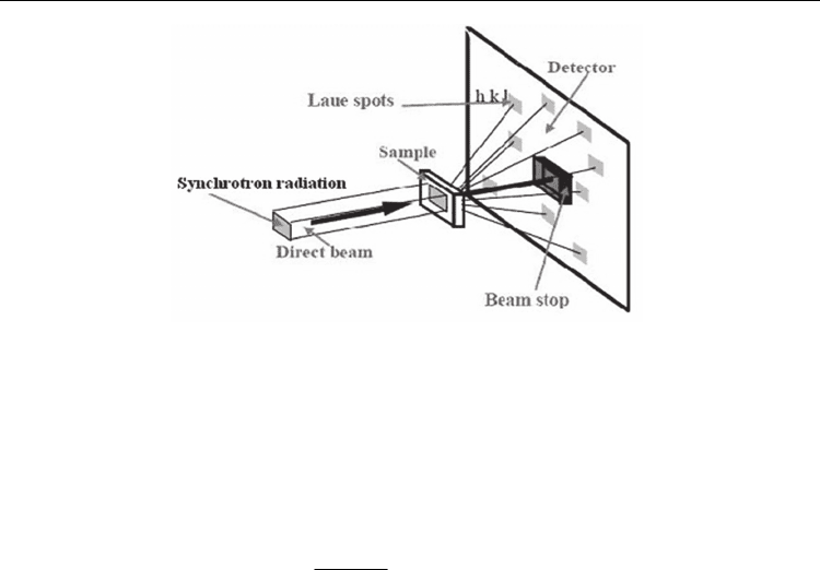
Fig. 2. Experimental arrangement for synchrotron radiation white beam Laue topography.
b. White beam X-ray topography
A simple way to understand the creation of X-ray topographic images is to consider a Laue
photograph (Fig. 2). A polychromatic (white) X-ray beam, containing X-ray energies from
about 6 keV to 50 keV (X-ray wavelengths from approximately 2 Å to 0.25 Å), impinges on a
crystal. [6] The beam is diffracted in many directions, creating Laue spots. The positions of
the diffraction spots appear according to the Bragg equation:
2sin
B
hc
E
d
θ
= , or
2sin
B
d
λθ
=
(1)
where E is the incident X-ray energy (and λ is the incident wavelength) selected by crystal
planes with spacing d, h is Planck’s constant, c is the speed of light, and θ
B
is the Bragg angle.
Each spot contains uniform intensity if the crystal is perfect.
If, however, the crystal is strained, streaks appear instead of spots due to variations in lattice
spacing. In fact, each Laue spot contains a spatial distribution of diffracted intensity
attributable to the presence of defects in the crystal. This distributed intensity is difficult to
see because Laue spots are typically the same size as the X-ray beam pinhole, and the
incident X-ray beam is divergent, but each tiny Laue spot is actually an X-ray topograph. At
synchrotron radiation facilities, a collimated white X-ray beam can be used to illuminate a
sample crystal, and spots with the much larger cross section of the synchrotron X-ray beam
are recorded. The resulting data are an array of Laue spots, as shown in Fig. 2, each of which
is an X-ray topograph arising from a different set of atomic planes.
c. Ferroelectric domain characterizations
The existence of antiparallel 180° domains is one of the fundamental properties of
ferroelectrics and direct observation of these domains is invaluable to the understanding of
the polarization reversal mechanism of ferroelectric structures. Among the techniques of
visualizing antiparallel domains, conventional x-ray topography is an important and
efficient method although its application is limited by the only available characteristic
radiations. However, this limitation is easily overcome by the white-beam synchrotron
radiation topography (WBSRT). A unique aspect of WBSRT is the opportunity it affords to
select out of the continuous spectrum any intended wavelengths to activate strong
anomalous scattering. And, with the WBSRT, it is possible for several diffraction images

Structural Studies in Perovskite Ferroelectric
Crystals Based on Synchrotron Radiation Analysis Techniques
7
with anomalous dispersion effect to be activated simultaneously so that the contrast reversal
of 180° domains can be directly observed.[7-9] The natural collimation and high intensity of
the radiation also make the real-time observation of domain dynamics feasible. As shown in
Fig.3, when working with a coherent x-ray beam, and if the phases of the structure factors
are different, the 180° ferroelectric domains can be revealed.
Considering the mechanical and electrical compatibility conditions, allowed domains in
ferroelectrics are the 180o or non-180o ones with the different planes as the domain walls.[8,
9] The extinction condition for a domain wall is:
0PgΔ• = (2)
where g is the reciprocal vector of the diffracting plane,
12
PP PΔ= − is the difference of the
polarization vectors across the domain wall. Non-180
o
domain structure is illustrated in Fig.4.
Fig. 3. (Colour on the web only) Scheme of 180
o
domain.
Fig. 4. Scheme of non-180
o
domain.
2.1.2 In-situ topography measurements
White beam synchrotron radiation topography not only overcomes the drawback of long
exposure time for the conventional x-ray topography, but also extends the scope of
topography study. The excellent collimation and high intensity of the synchrotron radiation
makes the possibility of enlarging the distance between the light source and sample, to
improve the image resolution and enlarge the distance between the sample and the detector.
These allow ones able to install the samples inside large environmental chambers with
changes of temperature, electric field, or other parameters, to carry out the in-situ
topography studies. [10]
Ferroelectrics - Characterization and Modeling
8
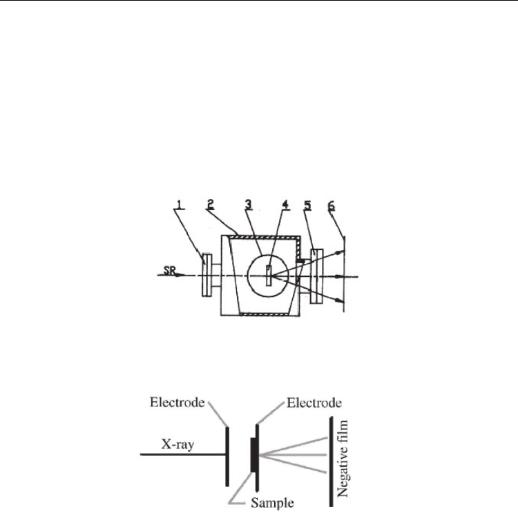
a. High temperature condition
A high-temperature chamber was used for in situ topography study. The cylindrical furnace
in use was made of pure graphite. Two beryllium windows were used for incident and
outgoing x-rays. Two thermocouples attached close to the specimen were used to monitor
and control the temperature. A digital control power supply can ramp the electric current
smoothly when starting to heat. The temperature control system is based on the Eurotherm
controller (model 818) and solid state relay (SSR). By using PID control and time proportion
method, the temperature stability is about 0.05°C when holding and 0.1°C when ramping.
The on-line PC can set and monitor the temperature via RS-232 interface. A sketch of this
chamber is shown in Fig. 5.
Fig. 5. Sketch of the high temperature sample chamber: 1-entrance Be window; 2-water
cooling environment chamber; 3-furnace; 4-specimen; 5-exit Be window; 6-film.
Fig. 6. The experimental arrangement for applying electric field.
b. DC electric field
The change of the ferroelectric domain structure due to the applied DC field can also be
observed by white beam synchrotron radiation topography. Fig. 6 is the experimental
arrangement of applying the DC field. The distance between the two electrodes was 4 mm
and the applied DC voltage ranged from 0 to 4400 V. The samples can be installed on the
surface of plastic plates. The DC electrical field can be applied on two Al electrodes covering
on the surface of two X-ray transparent organic materials.
2.1.3 In-situ topography study in ferroelectric crystals
The in-situ experiments are performed at Beijing Synchrotron Radiation Facility (BSRF). The
x-ray topography station and attached 4W1A beamline are part of the BSRF. The 4W1A is a
45m long white/monochromatic wiggler beamline. When the BEPC is operated at the