Haug H., Koch S. Quantum theory of the optical and electronic properties of semiconductors
Подождите немного. Документ загружается.


January 26, 2004 16:26 WSPC/Book Trim Size for 9in x 6in b ook2
Optical Properties of a Quasi-Equilibrium Electron–Hole Plasma 289
Even though Eq. (15.17) has the same form as the free-carrier suscepti-
bility in Chap. 5, the single-particle energies in Eq. (15.17) are the renor-
malized energies calculated in the quasi-static approximation of Chap. 9.
Hence, Eq. (15.17) includes the band-gap renormalization effects due to
the electron–hole plasma. Next, we introduce a vertex function Γ
k
which
describes the deviations of the full susceptibility χ from χ
0
:
χ
k
=Γ
k
χ
0
k
. (15.19)
The vertex function obeys the equation
Γ
k
=1+
1
d
cv,k
k
χ
0
k
¯
V
s,k,k
Γ
k
. (15.20)
vertex integral equation
The integral equation (15.20) will now be solved by various methods.
The integral over k
is approximated by a discrete sum
k
→
dk
2π
→
i
∆k
i
2π
. (15.21)
In numerical evaluations, one typically obtains converging solutions if
around 100 terms are included in the summation. Here, the k
i
can be taken
equidistantly, however, better accuracy is obtained if the k
i
are taken as
the points of support of a Gaussian quadrature. At low densities the angle-
averaged potential becomes singular for k
= k. This singularity has to be
removed before the numerical matrix inversion is performed. One adds and
subtracts a term
k
F
k,k
¯
V
s,k,k
Γ
k
χ
0
k
+
k
¯
V
s,k,k
&
χ
0
k
Γ
k
− F
k,k
χ
0
k
Γ
k
'
, (15.22)
where F
k,k
is chosen so that F
k,k
=1and that the sum in the first bracket
can be evaluated analytically, e.g., in 3D
F
k,k
=
2k
4
k
2
(k
2
+ k
2
)
. (15.23)
The bracket of the difference term in (15.22) vanishes at k = k
where
¯
V
s,k,k
is singular, so that the total term is a smooth function at k = k
.With

January 26, 2004 16:26 WSPC/Book Trim Size for 9in x 6in b ook2
290 Quantum Theory of the Optical and Electronic Properties of Semiconductors
-2
0
2
4
6
8
10
12
-4
-2 0 2
n
0
n
1
n
2
n
3
Absorption (10 cm )
4-1
3
3.2
3.4
3.6
3.8
4
-4 -2 0 2
n
0
n
1
n
2
n
3
(h - E )/Ew
g0
(h - E )/Ew
g0
Refractive Index
a) b)
Fig. 15.1 Computed absorption (a) and refractive index spectra (b) for bulk GaAs at
T =10K using the matrix-inversion procedure. The used parameters are: m
e
=0.0665,
m
h
=0.457,
o
=13.74,
∞
=10.9, a
0
=125Å, E
0
=4.2 meV, and the densities are 0
(1), 5 · 10
15
cm
−3
(2), 3 · 10
16
cm
−3
(3), and 8 · 10
16
cm
−3
(4), respectively. The damping
is γ =0.05E
0
such a procedure, compensation terms can be found for the plasmon–pole
approximation both in 3D and 2D.
Fig. 15.1 and Fig. 15.2 show the optical spectra which are obtained by
numerical matrix inversion for the examples of bulk GaAs and bulk InSb
for various plasma densities. The resulting complex susceptibility
χ(ω)=
1
L
3
k
d
∗
cv
χ
k
(15.24)
defines the complex optical dielectric function (ω)=1+4πχ(ω) which in
turn determines the spectra of absorption α(ω), Eq. (1.51) and refraction
n(ω), Eq. (1.50).
At low plasma densities the absorption spectra in Fig. 15.1-a shows well-
resolved 1s and 2s exciton resonances, followed by an ionization continuum
enhanced by excitonic effects. A plasma density of 10
16
cm
−3
is close
to the Mott density where the exciton bound states cease to exist. The
excitonic enhancement still causes the appearance of a maximum around
the original exciton ground-state position. At still higher densities a band-
gap reduction far below the position of the original exciton ground state is
seen, and simultaneously a build-up of optical gain occurs. The vanishing of
the exciton resonance in the absorption spectrum also causes considerable
changes in the refractive index, as shown in Fig. 15.1-b. Particularly, if
the laser beam is tuned below the exciton resonance, where the absorption
is relatively weak, the index of refraction decreases with increasing plasma

January 26, 2004 16:26 WSPC/Book Trim Size for 9in x 6in b ook2
Optical Properties of a Quasi-Equilibrium Electron–Hole Plasma 291
0
2
4
-10 0 10 20 30
n
0
n
1
n
2
n
3
Absorption (10 cm )
3-1
(h - E )/Ew
g0
Fig. 15.2 Computed absorption spectra for bulk InSb at T =77Kusingthematrix-
inversion procedure. The used parameters are: m
e
=0.0145, m
h
=0.4,
0
=17.05,
∞
=15.7, a
0
=644.8 Å, E
0
=0.665 meV, and the densities N are 0 (1), 8 · 10
15
cm
−3
(2), 2 · 10
16
cm
−3
(3), and 3 · 10
16
cm
−3
(4), respectively.
density. This effect is often exploited in optical switching devices which are
based on dispersive optical bistability, see Chap. 16.
The bulk InSb spectra of Fig. 15.2 have been calculated with a damping
γ =2E
0
=1meV, which is twice as large as the exciton Rydberg. This
case is typical for narrow band-gap semiconductors. Here, the exciton is not
resolved in the unexcited medium and causes only a step-like absorption
edge.
The corresponding absorption spectra for ideal 2D GaAs are shown in
Fig. 15.3. Only one hole band was taken into account. Experimentally
-2
0
2
4
6
8
10
-4 -2 0 2
n
0
n
1
n
2
n
3
(h - E )/Ew
g0
Absorption (10 cm )
4-1
Fig. 15.3 Computed absorption spectra for 2D GaAs at T =77Kusingthematrix-
inversion procedure. The used parameters are: a
0
=62, 5 Å, and E
0
=16, 8 meV, all the
other parameters are the same as in Fig. 15.1. The densities N are 0 (1), 1 · 10
11
cm
−2
(2), 5 · 10
11
cm
−2
(3), and 1 · 10
12
cm
−2
(4), respectively.

January 26, 2004 16:26 WSPC/Book Trim Size for 9in x 6in b ook2
292 Quantum Theory of the Optical and Electronic Properties of Semiconductors
in quantum-well structures, one often sees superimposed the contributions
of more than one band, e.g. of the heavy- and the light-hole bands in
GaAs-based materials.
Finally, we discuss the plasma density-dependent absorption spectra of
a quantum wire. As already mentioned in Chap. 8, the screening of the
Coulomb potential by the confined plasma is expected to be of little impor-
tance in a quantum wire, because the field lines passing through the barrier
material cannot be screened. Fig. 15.4-a shows the calculated absorption
spectra for a cylindrical GaAs quantum-well wire and Fig. 15.4-b shows the
corresponding refractive index spectra. The q1D Coulomb potential of a
cylindrical quantum wire, Eq. (7.78), is used in the calculations. The spec-
tra have been obtained by matrix inversion and have been calculated with-
out screening. The spectra calculated with screening (Benner and Haug,
1991) do not differ substantially, showing that for q1D systems the effect
of state filling is the most important source of the optical nonlinearities.
Re X( )w
-10
-9
-8
-7
-6
-4
-3
-2
-1
0
1
2
3
-5
0.6
0.7
0.5
0.4
0.3
0.2
0.1
0.0
-0.1
1
2
3
4
0.6
0.7
0.5
0.4
0.3
0.2
0.1
0.0
-0.1
-10
-9
-8
-7
-6
-4
-3
-2
-1
0
1
2
3
-5
1
2
3
4
Im X( )w
a) b)
(h - E )/Ew
g0
(h - E )/Ew
g0
Fig. 15.4 Computed absorption (a) and infractive index spectra (b) for a cylindrical q1D
GaAs quantum wire at T =300K using the matrix-inversion procedure. The parameters
are the same as in Fig. 15.1. The 1D-densities are na
0
=0(1), na
0
=0.5 (2), na
0
=1
(3), and na
0
=2(4), respectively. Ω is the energy spacing between subbands, and E
0
and a
0
are the bulk exciton binding energy and Bohr radius, respectively.
Studying the absorption spectra in Fig. 15.4-a, one might conclude that
the band-gap reduction is not very strong in quantum wires. Actually,
however, the opposite is true. The band-gap shrinkage in q1D is larger
than in higher dimensional materials, as can be seen in Fig. 15.5.
For the density na
o
=2, ∆E
g
reaches already five exciton binding
energies. The fact, that the absorption peak in Fig. 15.4-a shows only a
slight red shift is thus again a result of the compensation between reduction
of the exciton binding energy and gap shift.

January 26, 2004 16:26 WSPC/Book Trim Size for 9in x 6in b ook2
Optical Properties of a Quasi-Equilibrium Electron–Hole Plasma 293
T=300 K
/E =10
cylindrical wire
W
0
20
10
0
-10
-20
-30
12
3
na
0
E/E
0
mm
eh
00
+
mm
eh
+
DE
g
Fig. 15.5 Band-gap reduction, ∆E
g
, and chemical potentials as functions of carrier
density for the quantum wire of Fig. 15.4
We conclude, that with the reduction of the dimension in mesoscopic
semiconductor microstructures the influence of screening on the quasi-
equilibrium nonlinearities decreases, while the influence of state filling in-
creases.
15.2 High-Density Approximations
The integral equation (15.20) can be solved approximately if the attrac-
tive electron–hole potential is not too strong, i.e., in the high-density
limit, where plasma screening and phase-space occupation have reduced
the strength of the Coulomb potential sufficiently. We rewrite Eq. (15.20)
by introducing a formal interaction parameter σ, which will be assumed to
be small
Γ
k
=1+
σ
d
cv
k
¯
V
s,k,k
χ
0
k
Γ
k
. (15.25)
A power expansion of Γ in terms of σ yields
Γ
k
=
n
q
n
σ
n
, (15.26)
where the first coefficient is determined by
q
1
=
1
d
cv
k
¯
V
s,k,k
χ
0
k
. (15.27)

January 26, 2004 16:26 WSPC/Book Trim Size for 9in x 6in b ook2
294 Quantum Theory of the Optical and Electronic Properties of Semiconductors
In general, one can express Eq. (15.26) as the ratio of two polynomials
P
(N,M)
k
=
N
n=0
r
n
σ
n
M
m=0
s
m
σ
m
, (15.28)
which represents the (N, M)-Pad´e approximation. The coefficients r
n
and
s
n
can be obtained by a comparison with the original expansion (15.26).
We use the simplest nontrivial (0,1)-Pad´e approximation
Γ
k
= P
(0,1)
k
=
1
1 − q
1k
, (15.29)
which results in an optical susceptibility of the form
χ(ω)=
1
L
3
k
d
∗
cv
χ
0
k
1 − q
1k
. (15.30)
Pad´e approximation
The denominator in Eq. (15.30) expresses the influence of the multiple
electron–hole scattering due to their attractive interaction potential V
s
,
which causes the excitonic enhancement.
Another approximation for the integral equation (15.20) is obtained by
inserting a dominant momentum, which we take as the Fermi momentum,
into the angle-averaged screened Coulomb potential. The Fermi momentum
is
k
F
=(3π
2
n)
1/3
in 3D (15.31)
k
F
=(2πn)
1/2
in 2D, (15.32)
see Chaps. 6 and 7. Then the susceptibility integral equation (15.16) be-
comes
χ
k
= χ
0
k
1+
1
d
cv
k
χ
k
¯
V
s,k
F
,k
= χ
0
k
[1 + S(ω)] , (15.33)
and for
S(ω)=
1
d
cv
k
χ
k
¯
V
s,k
F
,k
(15.34)

January 26, 2004 16:26 WSPC/Book Trim Size for 9in x 6in b ook2
Optical Properties of a Quasi-Equilibrium Electron–Hole Plasma 295
we get the self-consistency equation
S(ω)=
1
d
cv
k
χ
0
k
¯
V
s,k
F
,k
1+S(ω)
(15.35)
with the solution
1+S(ω)=
1
1 −
1
d
cv
χ
0
k
¯
V
s,k
F
,k
. (15.36)
Eq. (15.36) yields the optical susceptibility
χ(ω)=
d
cv
χ
0
k
L
3
−
1
d
cv
k
χ
0
k
¯
V
s,k
F
,k
(15.37)
expressing again, in a different approximation, the effect of the excitonic en-
hancement, however, with a simple momentum-independent enhancement
factor 1+S(ω).
-0.4
0
0.4
0.8
1.2
1.6
-10 -5 0 5 10 15 20
(h - E )/Ew
g0
Absorption (10 cm )
4-1
1
2
3
4
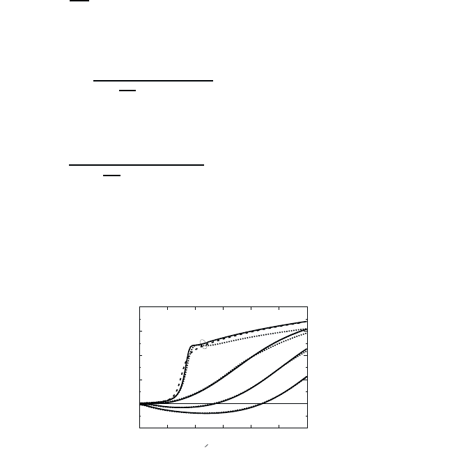
Fig. 15.6 3D absorption spectra for GaAs at 300 K for the densities 1 · 10
16
cm
−3
(1),
1· 10
18
cm
−3
(2), 2·10
18
cm
−3
(3), and 3·10
18
cm
−3
(4), respectively. Matrix inversion (full
lines), Pad´e approximation (dashed lines), high-density approximation (dotted lines).
Taking the spectral representation, Eq. (15.18), only for the imaginary
part, we compare absorption spectra for 3D and 2D semiconductors in
Figs. 15.6 and 15.7 for the different levels of approximations. We show the
results obtained a) by solving Eq. (15.20) by numerical matrix inversion (full
curves), b) by using the (0,1)-Pad´e approximation (15.30) (dashed curves)
and c) using the high-density approximation (15.37) (dotted curves).

January 26, 2004 16:26 WSPC/Book Trim Size for 9in x 6in b ook2
296 Quantum Theory of the Optical and Electronic Properties of Semiconductors
The agreement of both approximations b)andc) with the numerical solu-
tion a) becomes very good, if a pronounced optical gain exists. At lower
plasma-densities, where the gain vanishes, but where the densities are still
above the Mott density, the (0,1)-Pad´e approximation yields a slightly bet-
ter description of the excitonic enhancement than the high-density approx-
imation, particularly in the 2D case. At low densities, where bound states
of the exciton exist, both approximations are unable to give a reliable de-
scription of the bound-state resonances in the absorption spectra.
-0.4
0
0.4
0.8
1.2
1.6
-8 -4 0 4 8
(h - E )/Ew
g0
Absorption (10 cm )
4-1
1
2
3
4
Fig. 15.7 2D absorption and gain spectra for GaAs at 300 K for the densities
1 · 10
10
cm
−2
(1), 1 · 10
11
cm
−2
(2), 2 · 10
12
cm
−2
(3), and 3 · 10
12
cm
−2
(4), respec-
tively. The full lines are the result of the matrix inversion, the dashed line is the Pad´e
approximation and the dotted line represents the high-density approximation.
15.3 Effective Pair-Equation Approximation
For many practical applications, it is very useful to have an approximate
analytical solution for the full density regime, which can then be used in
further studies, e.g., of optical bistability (see Chap. 16) or nonlinear optical
devices. Such an approximation scheme (plasma theory) for 3D systems has
been developed by Banyai and Koch (1986) in terms of an effective electron–
hole–pair equation. The main approximation of this theory is that it ignores
the reduction of the attractive electron–hole Coulomb interaction via occu-
pation of k-states by the plasma, assuming that the dominant weakening
comes through the plasma screening. This assumption is reasonable in 3D
systems at elevated temperatures, but it fails in 2D and q1D where state
filling is more important.

January 26, 2004 16:26 WSPC/Book Trim Size for 9in x 6in b ook2
Optical Properties of a Quasi-Equilibrium Electron–Hole Plasma 297
We start from the Fourier-transformed polarization equation in the form
(ω + iγ) −H
P = −(1 − f
e,k
− f
h.k
)d
cv
E , (15.38)
where the eigenvalue equation of the hermitian Hamilton operator is
Hφ
ν,k
=(
e,k
+
h,k
)φ
ν,k
−
k
V
s,k−k
φ
ν,k
= E
ν
φ
ν,k
. (15.39)
Expanding the polarization components into the eigenfunctions
P
k
=
n
a
ν
φ
ν,k
, (15.40)
the solution of Eq. (15.38) can be written in the form
P
k
= −
ν
φ
ν,k
tanh
β
2
(ω − E
g
− µ)
(ω + iγ) − E
ν
d
cv
E
k
φ
∗
ν,k
, (15.41)
whereweapproximate
1 − f
e,k
− f
h,k
tanh
β
2
(ω − E
g
− µ)
.
The resulting absorption coefficient is then
α(ω)=
4π
2
ω
n
b
c
|d
cv
|
2
tanh
β
2
(ω−E
g
−µ)
ν
|ψ
ν
(r =0)|
2
δ(E
ν
− ω) .
(15.42)
The remaining problem for the analytic evaluation is to obtain the eigen-
functions ψ
ν
and the corresponding eigenvalues for the screened Coulomb
potential. Unfortunately, we do not know an analytic solution for the Wan-
nier equation with the Yukawa potential. However, there is a reasonably
good approximation to the screened Coulomb potential, which is the so-
called Hulth´en potential
V
H
(r)=−
2e
2
κ/
0
e
2κr
− 1
, (15.43)
for which one can solve the Wannier equation (10.35). As in Chap. 10, we
make the ansatz
ψ
ν
(r)=f
nl
(r)Y
lm
(θ, φ) , (15.44)

January 26, 2004 16:26 WSPC/Book Trim Size for 9in x 6in b ook2
298 Quantum Theory of the Optical and Electronic Properties of Semiconductors
where ν = n, l, m represents the relevant set of quantum numbers. We
obtain the radial equation
−
∂
2
f
nl
∂r
2
−
2
r
∂f
nl
∂r
+
l(l +1)
r
2
−
gλ
2
e
λr
− 1
f
nl
=
nl
f
nl
(15.45)
with
λ =2κ, g =
1
a
0
κ
, and
nl
=
E
nl
E
0
a
2
0
.
Since we are only interested in those solutions which do not vanish at the
origin,
f
nl
(r =0)=0 ,
we restrict the analysis of Eq. (15.45) to l =0and drop the index of f and
. For this case, we obtain from Eq. (15.45)
−
∂
2
u
∂r
2
−
gλ
2
u
e
λr
− 1
= u , (15.46)
where we introduced
u(r)=rf(r) . (15.47)
Defining
z =1− e
−λr
,w(r)=u(r)/z(1 − z)
β
(15.48)
with
β =
−/λ
2
(15.49)
and inserting these definitions into Eq. (15.46) yields
z(1 − z)
∂
2
w
∂z
2
+[2− (2β +3)z]
∂w
∂z
− (2β +1− g)w =0 . (15.50)
This is the hypergeometric differential equation, which has the convergent
solution
F (a,b,c; z)=1+
ab
c
z
1!
+
a(a +1)b(b +1)
c(c +1)
z
2
2!
+ ... , (15.51)
where it is required that c =0, −1, −2,... .Inourcase,wehave
