Evans-Martin F.F. The Nervous System
Подождите немного. Документ загружается.


THE NERVOUS SYSTEM
target neuron (usually a dendritic spine or the cell body) to
which the axon sends a nerve signal, there is a tiny gap. It
measures about 10 to 20 nanometers across, and is called the
synaptic cleft. The term
synapse refers to the synaptic cleft
and the areas on the two neurons that are involved in the trans-
mission and reception of a chemical signal. The presynaptic
neuron is the one that sends the message. It releases a neuro-
transmitter into the synaptic cleft. Every neuron produces one
or more kinds of neurotransmitters and stores them inside
spherical-shaped structures in the membrane called synaptic
vesicles until the neuron receives a neural signal. The synaptic
vesicles then move to the presynaptic membrane, bind to
it, and release their contents into the synaptic cleft. Neuro-
transmitters diffuse across the synaptic cleft and bind to a
particular receptor, or membrane protein, found on the surface
of the plasma membrane of the postsynaptic (receiving)
neuron (Figure 1.4). The neurotransmitter fits into the receptor
protein like a key in a lock, and causes an ion channel to open.
As sodium ions enter the postsynaptic neuron through the
activated ion channels, tiny electrical currents are produced.
These currents travel to the place where the cell body meets the
20
THE REFRACTORY PERIOD
An action potential only travels in one direction down the axon.
The reason for this is that there is a
refractory period that
begins immediately after the firing of an action potential. It
lasts for several milliseconds. During the first portion of this
refractory period, called the absolute refractory period, the
neuron cannot fire again, because sodium channels have been
left inactive. As the efflux of potassium ions pushes the voltage
below the threshold potential, a relative refractory period
occurs. During this time, a greater depolarization than usual is
needed to cause an action potential to fire.
CH.YBW.Ner.C01.Final.q 11/30/04 12:04 PM Page 20
axon, a site called the axon hillock.There, the tiny electrical
currents join together. Each neuron receives thousands of
neural signals per second from other neurons. Some of them
are excitatory and open sodium channels. Others are inhibitory
and open chloride or potassium channels. Depending on the
number and type of tiny electrical currents generated as the
neurotransmitter chemicals bind to the receptors of the post-
synaptic membrane, the axon hillock gets a message to fire or
not to fire an action potential. It fires an action potential only
if there are enough currents to open a large enough number of
21
Our Amazing Nervous System
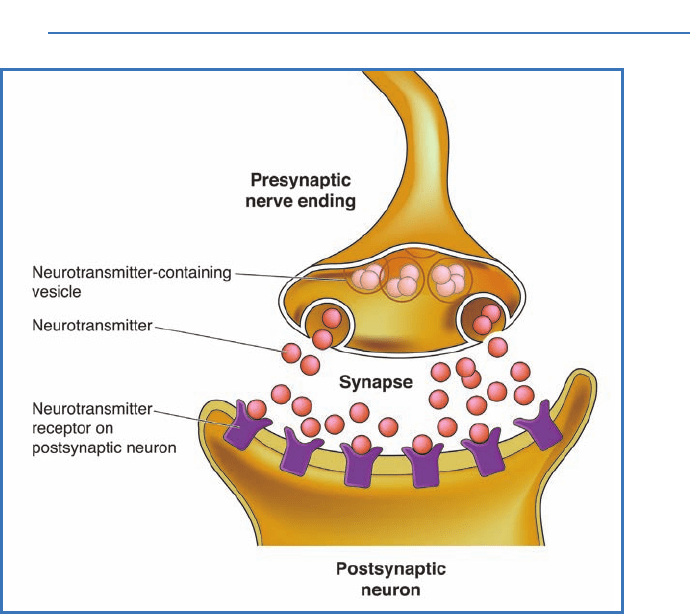
Figure 1.4 The synapse is the tiny space between a nerve ending
and the neuron with which it communicates. Neurotransmitters carry
the nerve signal as a chemical message across the synapse from the
first (presynaptic) neuron to the second (postsynaptic) neuron. They
bind to receptors on the postsynaptic cell membrane.
CH.YBW.Ner.C01.Final.q 11/30/04 12:04 PM Page 21

THE NERVOUS SYSTEM
voltage-gated sodium channels to make the membrane over
the axon hillock reach its threshold potential.
As the action potential travels down the axon, away from
the cell body, it makes the voltage of the area near the axonal
membrane more positive. In turn, this opens voltage-gated
ion channels. As the voltage of the adjoining intracellular
membrane drops to its threshold potential, another action
potential fires. This process continues until a series of action
potentials travels the length of the axon.
Some axons, especially those that have to travel longer
distances, are myelinated.
Myelin is a covering of glial exten-
sions that wrap around and around the axon of a neuron in
layers. This covering forms what is called a myelin sheath.
The layers of myelin provide additional electrical insulation.
This extra insulation lets nerve impulses travel very fast in
a myelinated axon—up to 120 meters (more than the length of
a football field) per second. The extra insulation provided by
the myelin sheath also helps an action potential travel much
farther in a myelinated axon. In the brain and spinal cord, each
oligodendrocyte may wrap its processes around segments of up
to 50 axons. In the nerves outside the brain and spinal cord,
Schwann cell processes wrap around one part of the axon of
just one neuron. An unmyelinated axon has only the lipid
bilayer of its own plasma membrane for electrical insulation.
Each myelinated segment measures about 0.1 to 0.5
micrometers in length. Between these segments are tiny
unmyelinated gaps called the
nodes of Ranvier.At these
nodes, sodium ions enter through voltage-gated ion channels
to propagate, or reproduce, the action potential. As a new
action potential is generated at each node of Ranvier, the
neural signal appears to “jump” from one node to the next.
CONNECTIONS
The nervous system is an intricate network of neurons (nerve
cells) and their connections. Surrounding the neurons are
22
CH.YBW.Ner.C01.Final.q 11/30/04 12:04 PM Page 22

glia, which play many supportive roles in the nervous system.
Neurons receive and process chemical messages from other
neurons and then send electrical signals down their axons to
trigger the release of neurotransmitters, chemical messengers
that go out to other neurons. The electrical current that travels
down the neuronal axon is made up of action potentials, which
are generated by the opening of voltage-gated sodium channels
in the axonal membrane.
23
Our Amazing Nervous System
CH.YBW.Ner.C01.Final.q 11/30/04 12:04 PM Page 23

24
Development of the
Nervous System
2
Considering that our brains help us do just about everything in our
lives, it should come as no surprise that the brain itself grows at an
incredibly fast rate well before we are born. The first visible signs of
the developing nervous system show up during the third week after
conception. At this point, the embryo consists of three layers of cells:
an outer layer called the ectoderm,a middle layer called the mesoderm,
and an inner layer called the endoderm.The ectoderm will develop
into the nervous system as well as the hair, skin, and nails that
cover our bodies. The mesoderm will develop into muscle, bone,
and connective tissue as well as some of the internal organs, includ-
ing the heart and blood vessels. From the endoderm, the digestive
and respiratory tracts and additional internal organs develop.
Around day 16 of development, a thickened layer of cells, called
the neural plate, appears in the midline of the
dorsal surface of the
ectodermal layer. (Since we walk upright, the term dorsal corresponds
to the
posterior,or backside, in human beings. The term
ventral
refers
to the opposite, or
anterior,surface—the front side of a person.)
As the neural plate develops, the cells at its edges multiply faster
than the rest. This makes the plate’s edges curve upward to form a
neural groove in the center. By day 21 of development, the edges of
the two sides of the neural plate meet and join to form the
neural
tube.This fusion begins at the place where the neck region will
eventually be located. It then continues to join
rostrally (toward
CH.YBW.Ner.C02.Final.q 11/30/04 12:07 PM Page 24

the head end) and caudally (toward the tail end) until the
whole dorsal surface of the tube is fused. Finally, the rostral
and caudal ends of the neural tube close on day 24 and day 26,
respectively. This process of forming the neural tube is known
as primary neurulation (Figure 2.1).
The adult spinal cord can be divided into five regions,
from the neck down: cervical, thoracic, lumbar, sacral, and
coccygeal. The cervical, thoracic, and lumbar segments of the
spinal cord develop from the neural tube. The sacral and
coccygeal segments, on the other hand, develop from the
caudal eminence, a cell mass located caudal to the neural
tube. It appears around day 20 and grows larger, then forms
a cavity before it joins the neural tube. This process, called
secondary neurulation, is completed by day 42.
As the neural tube closes, cells separate from the upper
edges, or crests, of the neural folds to form the neural crest.
From the neural crest, parts of the
peripheral nervous system
will develop. (The peripheral nervous system includes all the
nerves and neurons outside the brain and spinal cord.) Cells
from the neural crest move to a position on either side of the
neural tube. Sensory neurons, the
adrenal medulla,peripheral
neurons and glia of the
autonomic nervous system, along
with the two inner layers of the protective lining, or
meninges,
that cover the brain, all develop from neural crest cells.
The outer layer of the meningeal covering of the brain
forms from the mesoderm.
By the sixth week after conception, the nervous system
has already developed to its basic form. The major structures
are all recognizable by the tenth week. All brain structures
are present in an immature form by the end of the first
trimester (first three months). During the first three months
of fetal development, the vertebral column and spinal cord
grow at about the same rate. The nerves from the spinal cord
exit directly through openings in the vertebral column called
intervertebral foramina.After this point, however, the vertebral
column grows faster than the spinal cord. This leaves a space
called the
lumbar cistern in the lower part of the vertebral
25
CH.YBW.Ner.C02.Final.q 11/30/04 12:07 PM Page 25

THE NERVOUS SYSTEM26
Figure 2.1 This diagram shows the neural tube just after neurula-
tion. Notice that the primary germ layers—the ectoderm, endoderm,
and mesoderm—are still present. Each layer gives rise to a specific
set of structures in the developing body.
CH.YBW.Ner.C02.Final.q 11/30/04 12:07 PM Page 26

canal that is not filled by the spinal cord. Spinal nerves associ-
ated with the foramina in the area of the lumbar cistern travel
down from their origin in the spinal cord through the lumbar
cistern before they leave through their associated foramina.
At birth, the brain weighs 400 grams (0.88 lbs) on average.
By age 3, the weight of the brain has tripled, due to myelination
of axons and development of neuronal processes and synaptic
connections. By the time a person is 11 years old, the brain has
reached its maximum weight, which can vary from 1,100 to
1,700 grams (2.4 to 3.7 lbs). The average human brain weighs
about 1,400 grams (3 lbs). After age 50, people experience a
27
Development of the Nervous System
WHAT IS NEUROGENESIS?
Scientists once thought that a human infant was born with all
the neurons it would ever have and that no new neurons were
produced after birth. You can imagine the ripples in the scien-
tific world in 1998 when Peter S. Eriksson, Fred H. Gage, and
their colleagues announced their discovery of
neurogenesis—
the production of new neurons in the adult brain. These
scientists injected bromodeoxyuridine, a thymidine analog
(molecule with a similar structure) that is incorporated into newly
formed DNA, into terminally ill patients and examined their
brains after they died. He and his fellow researchers found
neurons in the hippocampus that were stained by this molecular
marker, which indicated that they had been produced after the
injection. Later research has detected the migration of stem cells
from the subventricular zone (SVZ) to sites in the cerebral cortex.
The SVZ is a layer of cells that lies underneath the ependymal
layer in the walls of the lateral ventricles. Related studies in
rodents have shown that exercise, enriched environments, and
learning enhance neurogenesis and that stress and inflammation
reduce it. Scientists hope that neurogenesis research will even-
tually yield answers that will help restore or regenerate brains
afflicted with neurodegenerative disease.
CH.YBW.Ner.C02.Final.q 11/30/04 12:07 PM Page 27

THE NERVOUS SYSTEM
gradual decrease in brain weight, which may cause a slow
decline in some cognitive, or thinking, functions.
DEVELOPMENTAL NEUROLOGICAL DISORDERS
Approximately 40% of all infant deaths before the first
birthday happen because something goes wrong with the
development of the central nervous system. A leading cause
of death shortly after birth are neural tube defects. In fact,
problems with neural tube development are the leading cause
of infant deaths (second only to heart defects). If the neural
tube does not close properly, the nervous system may not be
correctly formed. This occurs in about 1 out of every 1,000 live
births. Most fetuses with major nervous system malformations
die before or within the first year after birth.
Spina bifida is a birth defect that results when the neural
tube does not close completely at the caudal (tail) end.
Depending on how severe the condition is, the overlying verte-
brae and tissue may not develop, which lets the meninges and
spinal cord protrude to the surface of the back. Spina bifida
may also cause varying degrees of leg paralysis and problems
with bladder control.
Anencephaly is a birth defect that can result when the
rostral (head) end of the neural tube does not close all the way.
When this happens, the cerebral hemispheres will be partially
absent, and some of the overlying bone and tissue may not
form as well. When a baby is born with this condition, it is
usually blind, deaf, and unconscious. It may also have no
ability to feel pain. Infants with anencephaly almost always die
within hours—or, at most, days—after they are born.
Chromosomal abnormalities can cause problems in brain
development. One example is Down’s syndrome, which occurs
in 1 out of 700 infants. The children of mothers who are
over age 45 at the time of birth are more likely to suffer from
Down’s syndrome—the chances are 1 in 25 as compared to
1 in 1,550 for mothers under the age of 20. Babies born with
28
CH.YBW.Ner.C02.Final.q 11/30/04 12:07 PM Page 28

Down’s syndrome have an extra copy of chromosome 21.
Because of this, the disorder is sometimes called trisomy 21.
Symptoms of Down’s syndrome include mental retardation,
flattened facial features, stubby hands, short stature, an open
mouth, and a round head.
Fragile X syndrome is an inherited developmental disor-
der that results from a mutant gene on the X chromosome.
Symptoms include mental retardation, an elongated face with
a large jaw, enlarged testes (in males), and flared ears.
Other developmental abnormalities can result from mal-
nutrition or from exposure to radiation, environmental toxins,
drugs, and some pathogens (organisms that cause infections).
Viruses (such as rubella and cytomegalovirus), bacteria (such
as the spirochete bacterium that causes syphilis), and protozoans
(such as Toxoplasma,which is found in garden dirt and cat
feces) can all lead to nervous system defects. Drugs used to
treat epilepsy can cause defective neural tube development.
Neonatal exposure to lead or mercury can lead to neurological
problems. If the mother smokes, drinks alcohol, or takes cocaine
or other drugs of abuse during her pregnancy, it can also cause
problems in neurological development. There is evidence that
cocaine, for example, interferes with the myelination of axons
in adults.
Because there is an intimate relationship between the
nervous system and the structures of the skin, bone, muscles,
and meninges, when someone has a defect in nervous system
development, he or she usually has problems in other areas as
well. Defects in facial features often accompany problems in
brain development. This is particularly true in cases of fetal
alcohol syndrome, which can occur if the mother drinks alcohol
while she is pregnant. Children with fetal alcohol syndrome
often have slit-like eyes, a thin upper lip, and a small face. They
may also have behavioral and cognitive problems as well as
other birth defects, such as hearing impairments, heart defects,
and speech impediments.
29
Development of the Nervous System
CH.YBW.Ner.C02.Final.q 11/30/04 12:07 PM Page 29