Elsevier Encyclopedia of Geology - vol I A-E
Подождите немного. Документ загружается.

measured by collecting counts sequentially at charac-
teristic X-ray peak positions and at predetermined
background positions, if possible, on either side of
the peak.
The energy-dispersive spectrometer system The de-
tector consisting of lithium-drifted silicon, Si(Li), is a
wafer made from a single crystal of silicon having a
surface area of 10–30 mm
2
. The electrons in the outer
atomic orbitals are shared by several neighbouring
atoms, forming an essentially covalent bond. The
energies of these electrons in the ‘valence band’ orbit-
als are about 1.1 eV lower than the semiconductor’s
‘conduction band levels’. However, it takes about
3.8 eV to promote an electron from the valence to
the conduction band. When an X-ray enters the crys-
tal, it may be absorbed in an interaction with an
electron of one of the silicon atoms, producing a
high-energy photoelectron. This photoelectron dissi-
pates its energy in interactions that promote valence
band electrons to the conduction band, leaving holes
in the once-filled valence band. The number of elec-
tron-hole pairs formed is proportional to the energy of
the X-ray: for example, Ca Ka (3.691 keV) would yield
on average some 970 electron-hole pairs. A bias of
300–1500 V, depending on the thickness of the Si(Li)
chip, is applied and the electrons are swept to the rear,
where they enter the preamplifier as a weak pulse, the
amplitude of which is proportional to the energy of the
X-ray photon. Photo-optic feedback around a field-
effect transistor is used in the preamplifier. Room tem-
perature thermal excitation is sufficient to promote
electrons from the valence to conduction band, so
both the detector and the preamplifier are operated at
liquid nitrogen temperatures. Sophisticated electronics
are necessary to process the weak pulses in order that
they may be stored accurately in a multichannel ana-
lyser. The whole X-ray spectrum is stored simultan-
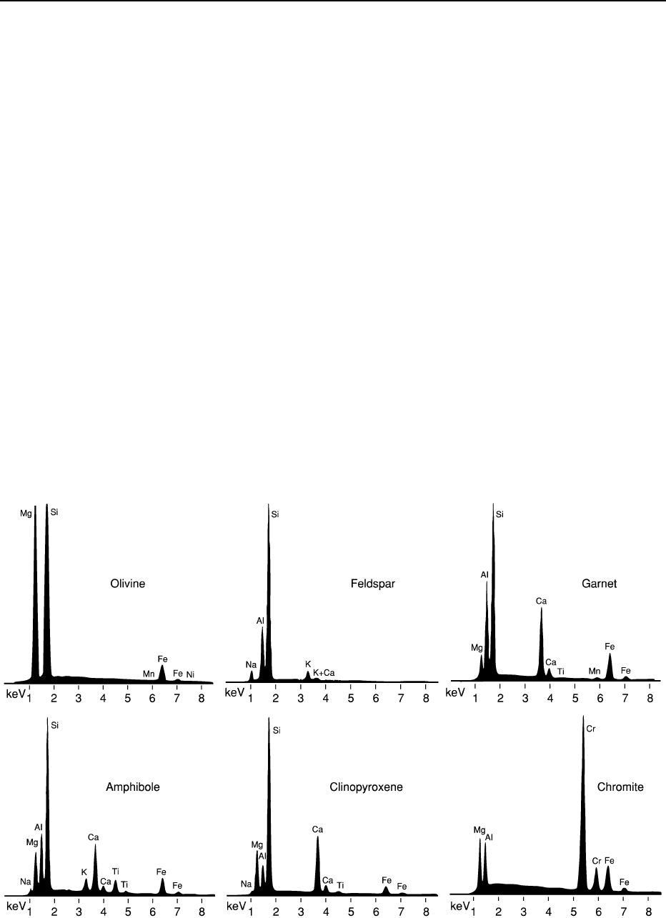
eously, resulting in an X-ray histogram. Examples of
EDS spectra are given in Figure 4; using these spectra as
fingerprints, identification of minerals is possible with
counting times as short as 100 ms. Within the EDS
histogram, it is not always possible to measure back-
ground on either side of the peaks, and for quantitative
analysis, spectrum deconvolution methods are neces-
sary. Commercial software uses either background
modelling or filter fitting.
Energy resolution The resolution of an EDS is
quoted as the full-width at half-maximum (FWHM)
Figure 4 Scanning electron microscope/wavelength-dispersive spectrometer spectra of minerals (15 kV, 1 nA, 20 s, 14 000 counts s
1
).
ANALYTICAL METHODS/Mineral Analysis 111

of the Mn Ka peak. This measure is chosen because
readily available
55
Fe is a source of this X-ray line.
For the Si(Li) detector, the FWHM at energy E is
given by FWHM
E
¼ðFWHM
2
0
þ 21:1FEÞ
0:5
, where
FWHM
0
is the resolution at energy 0 and F is the
Fano factor, which is a measure of the statistical
fluctuations in the ionization and charge collection
processes. Table 3 lists the performance of a range of
X-ray spectrometers.
Other energy-dispersive spectrometer technology Ger-
manium detectors have properties similar to those of
Si(Li) detectors and are preferred for use at higher
energies. They are found on AEM, PIXE, and syn-
chrotron X-ray fluorescence (SXRF) instruments. The
silicon drift detector (SDD) is based on charge-
coupled semiconductor technology and can provide
energy resolution similar to that of the monolithic Si-
crystal EDS, but at a count rate of 500 kHz. Further-
more, an energy resolution of 140 eV can be achieved
at only 13
C. The detector area can be made as
large as 400 mm
2
so that low currents can be used
for high count rates. It is possible to count at more
than 1 MHz, but the resolution degrades as the count
rate increases. This makes the detector unsuitable for
quantitative analysis but ideal for mapping of mineral
grains.
X-Ray bolometry has been developed using thin-
film Ag microcalorimeters, transition edge sensors,
and superconducting quantum interference devices.
Such detectors have energy resolutions down to 2 eV
and count rates of only 1 kHz. In theory, arrays of
these millimetre-sized devices could be constructed
giving a high overall count rate. The operating tem-
perature is 70–100 mK and it is possible to achieve
this using multistage Peltier cooling and an adiabatic
demagnetization refrigerator.
Matrix Corrections
X-Ray intensities are measured in units of counts
per second per nanoampere of beam current. The
weight percent concentration of an element in a
sample, C
samp
, is related to the characteristic X-ray
intensity, I
samp
, by the equation C
samp
¼ C
std
(I
samp
/
I
std
)([MATRIX]
samp
/[MATRIX]
std
), where [MATRIX]
denotes the effect of the chemical composition of the
matrix on the X-ray intensity and ‘std’ refers to a
standard of known composition.
There are four approaches to matrix corrections:
1. Empirical methods assume that each element lin-
early influences the X-ray intensity of each other
element. A table of coefficients, analysed element
against matrix element, is drawn up using extra-
polations from measurements of binary alloys and
solid solution series. These are known as alpha
coefficients.
2. The ZAF corrections separately compute the
effects of atomic number (Z), absorption (A), and
secondary fluorescence (F): ZAF ¼ R/Sf(w)(1 þ g),
where R is the back-scattering fraction and S is
the X-ray generation factor due to stopping power;
both of these are functions of atomic number. The
function of the mass attenuation coefficient, f(w),
corrects for the absorption of the X-rays as they
pass through the sample towards the detector. The
additional contribution when a matrix X-ray
fluoresces an analysed element (E
m
> E
c,a
)is
represented by g.
3. The f(rz) methods: f is defined as the ratio of the
X-ray intensity from a thin layer, dz, of sample at a
mass depth (rz) to the X-ray intensity of a similar
layer isolated in space. The f(rz) procedures inte-
grate this X-ray intensity ratio function, corrected
for multicomponent systems, from the surface to a
Table 3 X-ray spectrometer devices
Crystal/device
a
Dispersion method
b
Resolution FWHM (eV)
c
Max practical c
ount rate (kHZ) Energy range (keV) Collection area (mm
2
)
LiF WDS 5–10 50 4–12 300–1200
PET WDS 4–8 50 1.5–6 300–1200
TAP WDS 3–5 50 0.8–2.2 300–1200
WSi
x
WDS 4–6 40 0.2–0.9 300–1200
Si(Li) EDS 100–200 20 (0.3) 1–20 10–30
Ge EDS 90–190 20 1–40 10–30
SDD EDS 110–250 500 1–20 100–400
Bolometer EDS 2–10 1 0.2–8 0.5–1
a
LiF, Lithium fluoride; PET, pentaerythritol; TAP, thallium acid phthalate; WSi
x
, tungsten silicides; Ge, germanium; SDD, silicon drift
detector.
b
WDS, Wavelength-dispersive spectrometry; EDS, energy-dispersive spectrometry.
c
FWHM, Full-width half-maximum.
112 ANALYTICAL METHODS/Mineral Analysis

depth where f becomes zero. Work by several
groups spanning 30 years has established f(rz)
methods that give reliable matrix corrections for
nearly all mineral analysis.
4. The quantitative microanalysis procedure deve-
loped by J L Pouchou and F Pichoir, and so
named the PAP procedure, has affinities with
both the ZAF and the f(rz) methods. The depth
distribution of X-ray generation is modelled using
parts of two adjoining parabolas, functions that
are easy to integrate. Stopping power and absorp-
tion are carried out together. Fluorescence and
back scattering are corrected separately.
The EMPA instrument is designed specifically for
X-ray analysis and current designs provide up to five
WDSs, one EDS, and an optical microscope built
round the electron optical column. Scanning-beam
imaging by back-scattered electrons, secondary elec-
trons, and cathodoluminescence is also possible. The
specimen stage and spectrometers are automated and
software has been developed that has transformed
the electron microprobe analyser into a turnkey
instrument.
The Scanning Electron Microscope
Good quality mineral analyses may by obtained by
scanning electron microscopy (SEM) and energy-
dispersive spectrometry. SEM does not incorporate
an optical microscope and the minerals must be lo-
cated using back-scattered electron imaging. The
energy-dispersive spectrometer must be robust and
properly calibrated, requires stable electronics, and
an appropriate spectrum deconvolution method
must be employed. Many systems offer ‘standardless
analyses’; these methods work well over a restricted
energy range but should be used with caution in min-
eral analysis, in which the energy range extends from
Na (1.0 keV) to Fe (6.4 keV). Notwithstanding the
high peak-to-background ratio, EDS analysis, prop-
erly executed, should have a better precision than
WDS has for concentrations greater than 5 wt.%.
The Analytical Transmission Electron
Microscope
In analytical electron microscopy, sufficient current
may be focused on a region as small as 5 nm in
diameter, so that an X-ray flux greater than 1000
counts s
1
measured by an EDS system may be gener-
ated in a 150-nm-thick film. Thus, extremely small
domains of mineral grains may be analysed. The high
value of E
0
helps to improve the peak-to-background
ratios and enables the analysis of X-rays at the upper
energy limits of the EDS detector. The intensity of the
X-ray spectrum is a function of the indeterminate
thickness of the sample. Quantification is attained
by using ratios of peak intensities to that of a common
element (Si for silicates, Fe for opaques) and assuming
a value (usually 100%) for the sum of the analy-
sed components: C
x
=C
Si
¼ k
x;Si
ðI
x
=I
Si
Þ,
P
C
x
¼ 1:0,
where C is concentration and I is intensity. The cali-
bration constants, k
x,Si
, are evaluated empirically
and correct for the difference in EDS efficiency and
resolution at different energies. Matrix corrections
are slight and are often ignored but can be applied
using a rough estimation of the film thickness.
Beryllium-window Si or Ge detectors that do not
detect elements lower than sodium are used, but
the technique of electron energy loss spectrometry
(EELS) can be used in the analytical transmission
electron microscope to determine elements down to
beryllium.
Proton Induced X-Ray Emission
In the proton probe, the protons are typically acceler-
ated through 2.5–3.5 MeV. This is below the thresh-
old of the 8 MeV required for atom smashing yet high
enough to generate a reasonable flux of X-rays. The
mass of the proton is 1837 times that of the electron,
and this large mass, combined with the higher energy,
predicates very low scattering, either elastic or inelas-
tic. The X-ray spectrum has a very low background
and X-rays are generated to high E
c
. The limiting
factor for the use of high-energy characteristic X-
rays is the capability of the X-ray detector. The
protons may be focused down to submicrometre
beams using collimation and a series of quadrupole
magnetic lenses. The penetration of the proton beam
is considerably longer than it is in the electron
microprobe analyser: 3.5 MeV protons have a range
of 100 mm in aluminium. Most of the X-rays are
detected from depths of 20–50 mm, where the protons
penetrate, causing very little damage. However, just
before coming to rest, much of the kinetic energy is
absorbed by the sample and considerable damage to
the crystal lattice results (see Figure 2). The protons
end up forming hydroxyl ions, free hydrogen, and
hydrides.
The proton trajectory in the sample is essentially
linear, with a smooth deceleration of about 100 eV
per collision. The mechanisms of energy loss and ion-
ization are well understood, and the algorithm of inte-
gration of PIXE X-ray yields along the path of the beam
provides the foundation for a standardless microanaly-
tical method. However, unavoidable uncertainties in
ANALYTICAL METHODS/Mineral Analysis 113

mass attenuation coefficients, when applied over the
relatively long distances, can give rise to unacceptable
errors in the analysis of high concentrations of elements
such as Na, Mg, Al, and Si.
X-Ray Fluorescence
A beam of X-rays is not much scattered by solid
matter and is absorbed only when ionization occurs.
Ionization is caused when the energy of the X-ray
waveform resonates with the energy of the electron
orbital, increasing the amplitude of vibration so that
the electron leaves the atom. The probability of ion-
ization increases exponentially as the energy of the
photon approaches the critical excitation energy.
Thus, a primary X-ray beam is an efficient producer
of characteristic X-rays in a sample. There is little
background and the peak-to-background ratios are
even better than is obtained in PIXE.
Commercial X-ray microprobes are available. A
low-voltage X-ray tube and a waveguide focuses the
X-ray beam down to a 2 mm spot. In common with
PIXE, the secondary X-ray intensity is too low for
WDS and the spectrometers used are conventional
Be-window Si(Li) detectors.
The intense ‘white’ radiation in a synchrotron
beam line has been used in mineral analysis since the
mid-1980s; X-ray photon fluxes have since increased
by factors of 10
5
. Simple collimation with fine aper-
tures can create a <10 mm beam that may be used
to detect X-rays up to 40 keV in energy. Focusing
mirrors (the Kirkpatrick–Baez method) produce
a <2 mm spot, but the maximum useful energy
is about 10 keV. Phase zone plates can obtain a nano-
metre focus and are used for X-ray mapping and
microtomography.
Laser Ablation
Laser probing started in the 1970s with ultraviolet
(UV; e.g., 266 nm) lasers focused with multiple-lens
UV optics. The ablated material from a single laser
pulse was ionized by a second laser beam that hori-
zontally flooded the space above the sample, and the
ions were then extracted through a tube in the optics
and into a time-of-flight (TOF) mass spectrometer
(MS). This technology has improved so that spatial
resolution is <0.5 m m and mass resolution is >5000
M/DM. Resonant postionization techniques have
made great improvements in sensitivity. These instru-
ments are dedicated mass microprobes and are useful
in sensitive qualitative applications, but there are
problems in attempting quantitative analysis of re-
fractory elements. Unfortunately, so many of the
trace elements of interest to the mineralogist are
refractory.
Since the mid-1980s, lasers have been used in
conjunction with inductively coupled plasma mass
spectrometers to form very successful laser microp-
robes. Today, the technique of laser ablation in con-
junction with ICP-MS is used for the majority of
published trace element mineral analyses. The pol-
ished sample is inserted in a cell and an inert gas is
passed over the surface. The cell in Figure 5 is of a
sophisticated design; most ablation cells use a cylin-
drical box and only one gas, which serves both as an
ablating and a carrier medium. A UV laser, collimated
to 20–200 mm and pulsing typically at 5 Hz, ablates
the sample and the material is carried by the gas,
usually argon, into a plasma torch, where most of
the material is converted into monatomic cations.
These ions are usually analysed by a quadrupole
mass spectrometer.
The laser ablates the sample to a depth approxi-
mating the radius of the beam. Several hundred pulses
are used and a stream of material enters the plasma
torch over a period of up to several minutes. The
ionized product of the ICP enters the high vacuum
of the MS through a series of metal cones having
small apertures in their tips. As little as 0.01% of
the sample may enter the MS. The MS is cycled to
detect the required isotopes in sequence; a cycle takes
of the order of a second, and usually 10–30 isotopes
are counted. Each cycle is deemed to analyse a ‘slice’
of the sample and the counts of each isotope in each
slice are recorded by the on-board computer. Not-
withstanding the inefficiency of transporting material
into the MS, counts of 10
3
to 10
5
ppm
1
are obtained.
As analysis proceeds, material can build up on the
surfaces within the equipment and isotopes may be
detected when the laser is switched off. The MS is
cycled for a period before starting ablation so that the
background levels may be determined. Some oper-
ators are concerned that the act of running the laser
Figure 5 Laser ablation cell (the HelEx system). ICP, Induct-
ively coupled plasma.
114 ANALYTICAL METHODS/Mineral Analysis

may of itself dislodge material in the system, thus
adding to the background. As a precaution, an ultra-
pure silicon standard may be analysed to quantify this
effect.
The National Institute of Standards and Techno-
logy (US Department of Commerce) prepares and
issues a range of glass standards that contain 61
elements at trace concentration, at approximately
50 and 500 ppm. These standards are commonly
used in LA-ICP-MS and are measured repeatedly
throughout an analytical session, perhaps once to
every 10 or 20 sample measurements. Corrections
for drift in the performance of the equipment with
each sample analysis are applied linearly between
standard measurements.
Quantification of the isotope counts requires
knowledge of the concentration of at least one elem-
ent, used as an internal standard; concentrations of
other elements may be determined from the ratios of
isotopes to that of the internal standard. A convenient
element to use is calcium, which is present as a major
element in most silicates and has five stable isotopes
of widely ranging abundance. It is usually possible to
select a calcium isotope giving a signal similar to that
of the trace elements. Other elements can be used for
example, nickel in olivine, vanadium in oxides, and
titanium in micas. With effort, the EMPA laboratory
can provide such internal standard concentrations to
10 ppm at the 1000 ppm level, but isotopes of major
elements such as Si and Mg can give good results. In
sulphides, the sulphur concentration is usually known
and, being an electronegative element, the cation
signal is weak enough to be comparable with those
of the trace elements. During ablation, much material
condenses in and around the ablation pit, and the
more refractory an element, the less likely it will be
carried away by the gas. Thus fractionation processes
occur even when the laser couples well with the min-
eral, and there is always a crater rim to the ablation
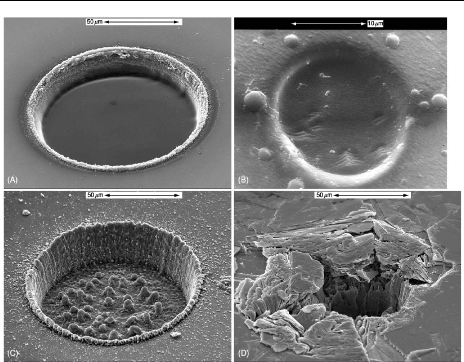
pit (e.g., Figure 6A). In general the worse the coupling
(Figure 6D), the greater the fractionation.
The first stage in quantification is to obtain isotope
ratios corrected for background and fractionation.
The background signal obtained with the laser off is
averaged to give intensity values per slice, and these
are subtracted (together with the values from the
‘null’ pure silicon standard if any) from the isotope
signals measured with the laser on. Then ratios are
calculated for each slice. These ratios, if plotted
against slice number, will have a positive slope if
the unknown undergoes less fractionation than the
internal standard does; if not, then the slope will
be negative. Either way, the plots are regressed to
the point at which the laser is switched on and the
value there is adopted for further calculation. Linear
regression is often adequate; some operators prefer a
polynomial. Anomalous slices, such as those contain-
ing inclusions in the mineral, may be excluded from
the regression. Editing the data is facilitated by a good
graphics computer program, but operations with a
simple spreadsheet are adequate.
Quantification of the isotope ratios is continued by
adjusting them with reference to the changes in ratios
in the glass standard taken before and after the sam-
ple. Finally, the concentration of element x in the
sample, C
x,samp
, is given by the following equation:
C
x;samp
¼ C
int;samp
ðI
x;samp
=I
int;samp
ÞðI
int;std
=I
x;std
Þ
ðC
x;std
=C
int;std
Þ
where ‘std’ denotes the glass standard and ‘int’ is the
internal element.
Differences in the coupling of the laser and hence
the process of ablation between the glass standard
and the sample are responsible for the major source
of error in LA-ICP-MS. Another error is in the failure
to predict overlaps on the analysed isotope. Overlap
of isotopes of different elements and equal mass is
either avoidable or readily quantified, but overlap
from argon-sample dimers and from doubly charged
ions may not be so obvious.
The Ion Microprobe
In secondary ion mass spectrometry (SIMS) and in
sensitive high-resolution ion microprobe (SHRIMP)
analysis (the ‘big brother’ of SIMS), beams of O
,
O
þ
,O
2
,orCs
þ
at 10–20 keV sputter the surface
of the sample, yielding a mixture of ions, molecules,
and plasma. Three types of mass spectrometers are
used: magnetic sector, quadrupole, and time-of-flight.
Although few useful ions are produced, unlike the
LA-ICP-MS, nearly all the ions can be analysed
by the mass spectrometer, and SIMS is a more sen-
sitive technique. Erosion of the sample is usually
1–10 nm s
1
, which is much slower than laser abla-
tion and much less sample is required. In Figure 6, the
volume of material excavated in the SIMS pit is 0.3%
that of the laser pits. SIMS is primarily an isotope
ratio technique, but quantitative elemental analysis at
very low levels is possible.
In contrast to EMPA, a general theory for matrix
corrections in SIMS may never eventuate. Several
specialized methods have been applied; for example,
in the infinite velocity method, emission velocities,
calculated from experimentally measured energy dis-
tributions, are extrapolated to infinite velocity, where
there are no matrix effects. This approach works well
ANALYTICAL METHODS/Mineral Analysis 115
for trace elements implanted in simple matrices such
as high-purity silicon. In mineral analysis, standards
with the same crystal lattice as the unknown are
required. Nevertheless, various laboratories have set
up routine procedures for SIMS analysis in applica-
tions involving rare-earth elements, platinum group
elements, and light elements, including hydrogen.
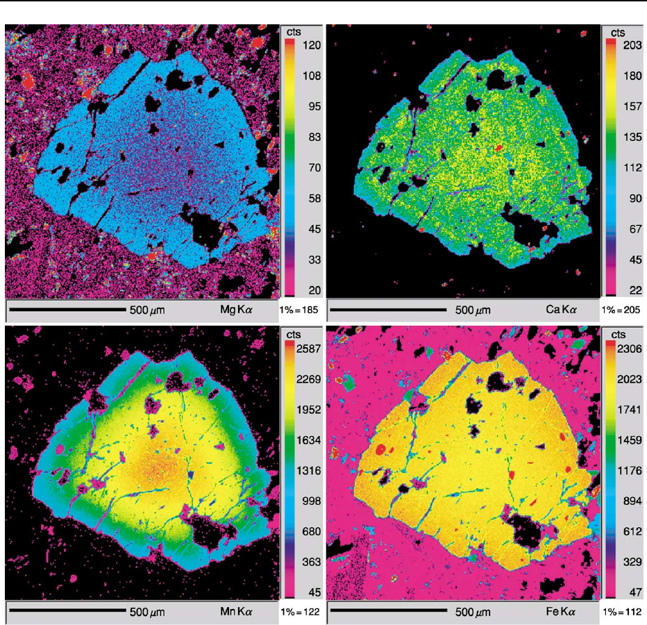
Compositional Mapping
By moving the automated stage under the beam in
any microanalytical instrument, it is possible to build
up an array of data that may be transformed into
false-colour maps of the sort shown in Figure 7. This
has become a routine overnight procedure in many
EMPA laboratories: the X-ray peak intensities may be
recorded for each position together with the back-
scattered electron signal, which furnishes both an
image of the area scanned and a template for the
bremsstrahlung background. For some applications,
it is possible to obtain a full quantitative analysis
at each point (i.e., pixel) with acceptable precision.
Usually the colour scale is calibrated roughly (as in
Figure 7) from the software’s calibration file and
matrix effects are ignored. In addition to EMPA,
compositional maps have been obtained using PIXE,
SXRF, and SIMS. LA-ICP-MS does not have high
spatial resolution but line scans are attempted with
useful results.
Other Mineral Analysis Methods
Analysis of OH
,CO
2
3
, B, Be, and Li and the alloca-
tion of iron between Fe
2þ
and Fe
3þ
pose problems
in mineral analysis by the methods outlined in the
preceding sections. Of the light elements, only F may
be analysed by EMPA with an accuracy comparable
with heavier elements. However, B, Be, and Li may
Figure 6 Scanning electron microscope images of ablation pits, using (A) a 193-nm laser on calcite, (B) secondary ion mass
spectrometry, with O
2
on zircon, and (C) 193-nm and (D) 226-nm lasers on molybdenite.
116 ANALYTICAL METHODS/Mineral Analysis
be determined by LA-ICP-MS, though at a limit of
detection >50 ppm and with indeterminate accuracy
at concentrations corresponding to borates and ber-
yllates. Light elements are readily detected by SIMS
but quantification is beset with uncertainties.
The local atomic environment around the nucleus,
including the electronic, chemical, and magnetic
state, may be studied using Mo
¨
ssbauer spectroscopy.
The Mo
¨
ssbauer effect is the recoilless absorption and
emission of g-rays by specific nuclei in a solid, and the
spectroscopy of
57
Fe has been much studied with
respect to mineral analysis.
Fourier transform infrared spectroscopy using an
optical microscope can give quantitative information
about anions such as OH
and CO
2
3
, but the thick-
ness of the slide must be measured accurately. Mul-
tiple valency may be determined by X-ray absorption
near-edge spectroscopy (XANES), which is per-
formed on the synchrotron, and by X-ray photo-
electric spectroscopy (XPS), which requires an
ultrahigh vacuum and analyses the outer 10 nm of
the sample.
For most silicates, the FeO/Fe
2
O
3
ratio may be
estimated by allocation of Fe to FeO and Fe
2
O
3
so
Figure 7 Electron microprobe analysis/wavelength-dispersive spectrometry maps of garnet (Mg,Ca,Mn,Fe)
3
Al
2
Si
3
O
12
(600 600
pixels, 50 ms pixel
1
, with a 25-kV, 100-nA, 1-mm beam).
ANALYTICAL METHODS/Mineral Analysis 117

that the cation total equals the theoretical amount. A
general equation can be used:
wt:% FeðtrivalentÞ¼ðtotal theoreticalÞ=theoretical
wt:% O 6:98125
In the garnet analysis in Table 1, the cation total is
8.0098 and the theoretical total is 8.0000; the wt.%
oxygen is 39.54%, which is the sum of the oxides,
99.96%, less the sum of the elements. Application of
this formula gives 0.48% Fe
2
O
3
and 33.83% FeO,
and recalculation of the mineral formula gives exactly
8.0000 cations. It is possible to analyse directly for
oxygen with the EMPA, and if this is done with care, a
similar result can be obtained but at the cost of extra
instrument time.
See Also
Analytical Methods: Fission Track Analysis; Geochem-
ical Analysis (Including X-Ray). Minerals: Definition and
Classification; Micas; Olivines; Sulphides.
Further Reading
Cabri LJ and Vaughan DJ (eds.) (1998) Modern Ap-
proaches to Ore and Environmental Mineralogy, Short
Course Series, vol. 27. Ottawa: Mineralogical Associ-
ation of Canada.
Deer WA, Howie RA, and Zussman J (1997) Rock Forming
Minerals (5 vols.). Bath, UK: Geological Society Publ.
House.
Henderson G and Baker D (eds.) (2002) Synchrotron
Radiation: Earth, Environmental and Material Science
Applications, Short Course Series, vol. 30 Ottawa: Min-
eralogical Association of Canada.
Hurlbut CS and Sharp WE (1998) Dana’s Minerals and
How to Study Them, 4th edn. New York: Wiley.
Ireland TR (1995) Ion microprobe mass spectrometry: tech-
niques and applications in cosmochemistry, geochemistry
and goechronology. In: Hyman M and Rowe M (eds.)
Advances in Analytical Geochemistry, vol. 2, pp. 1–118.
Greenwich, CT: JAI Press.
Johansson SAE, Campbell JL, and Malmqvist KG (1995)
Particle Induced X-ray Emission Spectrometry (PIXE).
New York: Wiley.
McCammon C (1995) Mossbauer spectroscopy of min-
erals. In: Ahrens TJ (ed.) A Handbook of Physical
Constants: Mineral Physics and Crystallography, vol. 2,
pp. 332–347. Washington, DC: American Geophysical
Union.
Potts PJ (1992) A Handbook of Silicate Rock Analysis.
London: Blackie Academic & Professional.
Reed SBJ (1993) Electron Microprobe Analysis, 2nd edn.
Cambridge, UK: Cambridge University Press.
Ryan CG (1995) The nuclear microprobe as a probe
of Earth structure and geological processes. Nuclear
Instruments and Methods B104: 377–394.
Schulze D, Bertsch P, and Stucki J (eds.) (1999) Synchrotron
X-ray Methods in Clay Science. Aurora, CO: Clay
Minerals Society of America.
Sylvester P (ed.) (2001) Laser Ablation-ICPMS in the Earth
Sciences. Principles and Applications, Short Course
Series, vol. 29. Ottawa: Mineralogical Association of
Canada.
ANDES
S M Kay, Cornell University, Ithaca, NY, USA
C Mpodozis, SIPETROL SA, Santiago, Chile
V A Ramos, Universidad de Buenos Aires, Buenos
Aires, Argentina
ß 2005, Elsevier Ltd. All Rights Reserved.
Introduction
The Andean mountains are the type example of an
‘Andean’-type subduction zone characterized by sub-
duction of an oceanic plate beneath a continental
margin and uplift of a mountain range without con-
tinental collision. They extend some 8000 km from
Venezuela to Tierra del Fuego and are the major
morphological feature of South America. On the
Earth’s continents, they include the highest active
volcanoes (>6800 m), the highest peaks outside of
the Himalayas (ca. 7000 m), the thickest crust
(>70 km), the second greatest plateau in height and
area (after Tibet), the most important volcanic plat-
eau with the largest Tertiary ignimbrite calderas, and
among the most shortened continental crust, deepest
foreland sedimentary basins, and largest and richest
precious metal (Cu, Au, Ag) and oil deposits. The
central Andes are also the type example of the effects
of shallowly subducting oceanic plates and of non-
accreting margins where continental lithosphere has
been removed by the subduction erosion process. The
evolution of the Andes began in the Jurassic coinci-
dent with the arc system that developed above sub-
ducting oceanic plates along the western margin of
South America during and after the breakup of the
118 ANDES
Mesozoic Pangaean supercontinent. The history of
the Andes is predominantly a story of magmatism,
uplift related to contractional deformation, interven-
ing episodes of oblique extension, collision of oceanic
terranes in the north, formation of sedimentary
basins, mineralization, loss of continental crust by
fore-arc subduction erosion, and removal of the base
of overthickened crust by delamination. The mechan-
isms of uplift and crustal thickening along with the
amount, timing and fate of removed continental
lithosphere are hotly debated topics.
Principal Geological Features of the
Modern Andes
Subducting Oceanic Crust and Distribution and
Character of Andean Magmatism
The morphology and geology of the modern Andes
are strongly influenced by the age, geometry, and
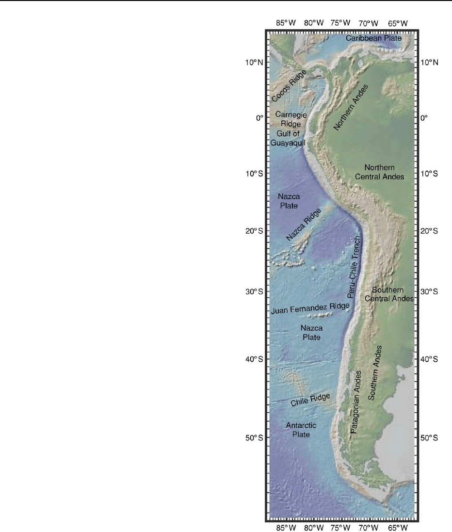
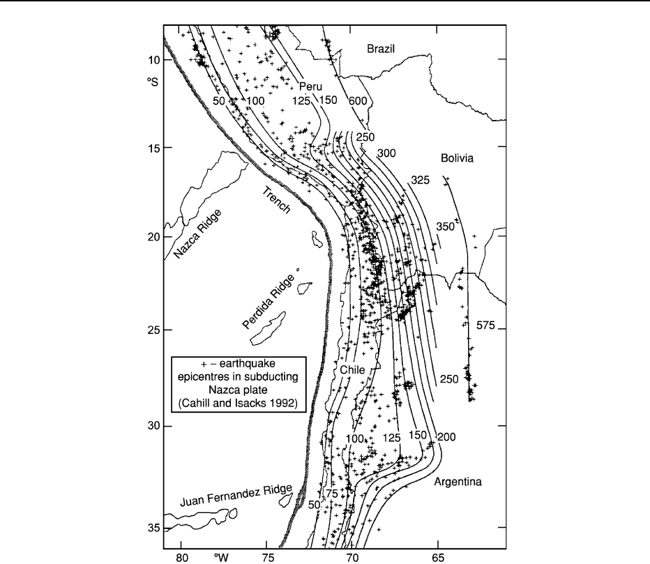
morphology of the subducting oceanic plates (Figures
1 and 2). A first-order feature related to these sub-
ducting plates is the division of the active volcanic
arc into the Northern (NVZ), Central (CVZ), South-
ern (SVZ) and Austral (AVZ) Volcanic zones (Figures
3–6). The NVZ, CVZ and SVZ are underlain by seg-
ments of the Nazca Plate that are subducting at an
angle of 20–30
, and in which the magmas are prin-
cipally generated by hydrous fluxing and melting of
the mantle wedge. CVZ and northern SVZ magmas,
erupted through the thick crust of the Central Andes,
are primarily andesites and dacites, whereas central
and southern SVZ and NVZ magmas, erupted
through thinner crust, are primarily basalts and
mafic andesites. Between these segments are the Peru-
vian and Chilean (or Pampean) amagmatic flat slab
segments under which the subducting Nazca Plate
forms a flat bench at 100 km that can extend up to
300 km east of the high Andes. The near absence of
an asthenospheric wedge accounts for the lack of
volcanism. The origin of the shallowly subducting
segments of the Nazca Plate has been debated and
variously attributed to subduction of the Juan Fernan-
dez and Nazca ridges near their southern margins or
complex interactions between the underriding and
overriding plates. All of the models call for a ‘colli-
sion’ between a shallowly dipping Nazca Plate and the
overriding South American Plate.
Other factors come into play at the northern and
southern ends of the Andes where the geometry of the
subducting plate is less well known. To the north, the
NVZ is flanked by the amagmatic Bucaramanga seg-
ment under which the weakly defined subducting
Caribbean Plate appears to dip at 20
. In the south,
Figure 1 Digital elevation model (DEM) of western South
America and surrounding oceans based on global bathymetry
database at the Lamont Doherty Observatory of Columbia Uni-
versity. The figure shows major features on the subducting
oceanic plate and the correspondence with the division of the
Andes into the Northern Andes bounded to the south by the Gulf
of Guayaquil, the Central Andes bounded to the south by the Juan
Fernandez Ridge, and the Southern Andes.
ANDES 119
the SVZ is separated from the AVZ by a volcanic gap
that coincides with the Chile Triple Junction where the
Chile Rise is colliding with the Chile Trench (Figure 7).
The net convergence rate of the South American Plate
with the Nazca Plate is 9cmyear
1
whereas that with
the Antarctic Plate is 2cmyear
1
.Magmatismis
absent in this region as the subducting slab is too
young and hot to provide the volatiles to flux melting
of the mantle wedge. The andesitic to dacitic ‘adaki-
tic’ magmas of the AVZ are distinctive in that they are
attributed to melting of the young hot subducting
Antarctic Plate. The most convincing slab melt ‘ada-
kites’ erupted anywhere in continental crust are the
14–12 Ma Patagonian adakites (e.g. Cerro Pampa)
that are attributed to melting of the trailing edge of
the subducted Nazca Plate near the time of ridge
collision.
Character of the Ranges, Basins and Faults of the
Northern, Central and Southern Andes
The principal ranges and basins of the Andes reflect
both the geometry of the subducting plate and the
pre-existing continental crust and mantle lithosphere.
The Andes are generally discussed in terms of a north-
ern, a central, and a southern sector. Here the limit
between the Northern and Central Andes is near the
northern boundary of the Peruvian flat slab at 4
S,
and the limit between the Central and Southern
Andes is at the southern margin of the Chilean Flat
Slab near 33
S(Figure 1).
Figure 2 Map showing the Benioff zone geometry of the portion of the Nazca oceanic Plate subducting beneath the Central Andes.
Major north to south changes in distribution and style of volcanism, basin development, and deformational styles can be correlated to
a first order with the shape of the Nazca Plate. (Reproduced with permission from Cahill TA and Isacks BL (1992) Seismicity and shape
of the subducted Nazca Plate.
Journal of Geophysical Research 97: 17 503–17 529.)
120 ANDES
