Chen C.C. (ed.) Selected Topics in DNA Repair
Подождите немного. Документ загружается.

The DNA-Damage Response to Ionizing Radiation in Human Lymphocytes
11
that the level of H2AX phosphorylation was principally correlated to a delayed DSB
resolution rather than apoptosis induction.
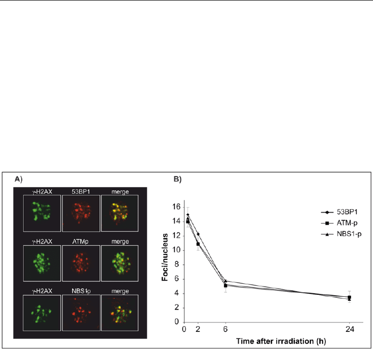
We then tested for the possibility that MMG incubation affects DNA damage response by
altering the recruitment of the signaling proteins, 53BP1, NBS1-p343 and ATM-p1981, which
co-localize with -H2AX foci to DSB sites (Fig. 6A). After irradiation ∼90% of cells became
foci-positive for the three proteins in both gravity conditions (not shown). In contrast to -
H2AX, the fraction of foci-positive cells persisted high up to 24 h after irradiation in 1g and
no differences between the two culture conditions were detected. The number of
foci/nucleus significantly decreased during post-irradiation incubation from 14–16
foci/nucleus at 30 min to 4–5 foci/nucleus at 24 h (Fig. 6B), without differences between
samples in 1g and MMG. The discrepancies with the kinetics of -H2AX foci suggest that
these proteins could represent the remaining scaffold structure used for DSB repair that
persisted after the repair has been completed (Markova et al., 2007, van Veelen et al., 2005).
Fig. 6. Kinetics of 53BP1, ATM-p1981, NBS1-p343 foci in PBL irradiated with -rays and
incubated in 1g. A) Co-localization with -H2AX foci to form the ionizing radiation-induced
foci (IRIF). B) Mean number of foci per nucleus.
3.2 The DNA-damage response of human tumoral lymphocytes cultured in
microgravity after γ-irradiation
We analyzed the DNA damage response to radiation also in human tumoral lymphocytes
(TK6 cells, lymphoblastoid B cells) irradiated with rays (1, 2, 4 Gy) and incubated in 1g or
in MMG during the repair time. In irradiated TK6 cells, we observed a higher survival in
MMG than in 1g, and the difference was significant at 4Gy. In addition, in cells maintained
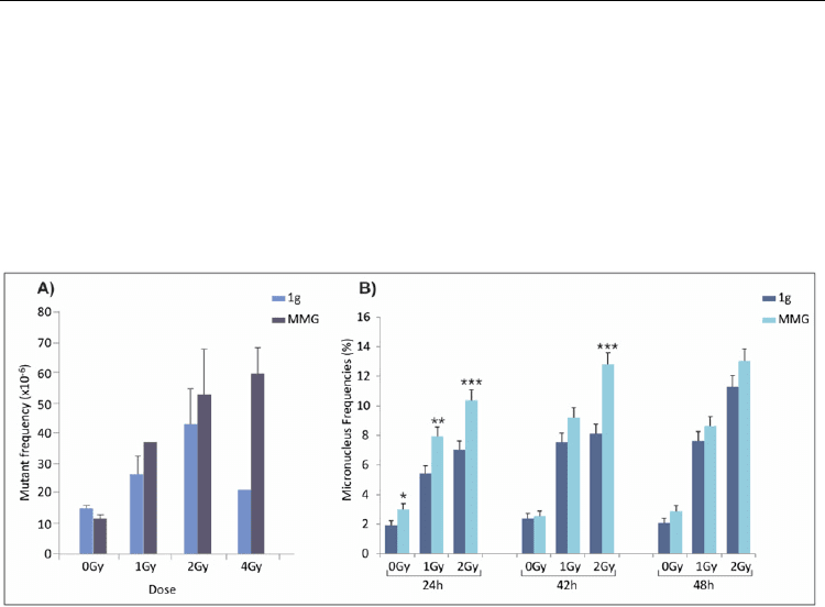
in MMG rather than in 1g after γ-irradiation, higher frequency of HPRT mutants was
observed at all irradiation doses, particularly at 4Gy (Figure 7A). Remarkably, at this dose,
mutant frequency may often be underestimated, since cells with many and severe mutations
are unable to repair DNA damage and die. Instead, in TK6 cells cultured in MMG after
irradiation, mutant frequency increased with doses up to 4Gy (Figure 7A). The frequency of
micronucleated cells was measured in both gravity conditions after irradiation. At the end of
Selected Topics in DNA Repair
12
post-irradiation incubation (24 h time-point), the percentage of micronuclei (MN) was
significantly higher in both non-irradiated and in irradiated cells incubated in MMG
compared with 1g (Fig.7B). Eighteen hours later (42 h from irradiation), the percentage of
MN in cultures incubated in MMG was higher than in 1g only at 2Gy γ-ray dose. At 48 h
time-point, MN frequencies observed in 1g or MMG were comparable. As expected, MN
significantly increased after irradiation in both gravity conditions with respect to non-
irradiated cells; a significant difference was still observed at 48 h after irradiation at both 1
and 2Gy. The significant increase of micronucleated cells in MMG suggested that MMG
itself was able to induce chromosome damage.
Fig. 7. A) Mutant frequency at the HPRT locus of irradiated and non-irradiated TK6 cells
incubated for 24h in 1g or in modeled microgravity. B) Micronucleus frequencies (%) in
irradiated and non-irradiated TK6 cells incubated in 1g of MMG for the first 24h after
irradiation and then cultured in 1g up to 48 h. *P<0.05; **P<0.01; ***P<0.001 (G test).
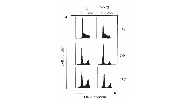
The effect of MMG incubation on cell cycle alteration induced by γ−ray exposure was
assessed by flow cytometry analysis. Figure 8 shows the cell cycle distribution of TK6 cells
at various time-points from irradiation and incubation in MMG or 1g by representative
DNA histograms.-ray irradiation induced an increase in G2/M-phase cells and a reduction
in S-phase cells, both in TK6 maintained in 1g and MMG after irradiation. At the end of
MMG or 1g incubation (24 h time-point), the percentages of cells in G1-phase were higher in
cultures irradiated with 2-4 Gy and incubated in MMG compared with cells maintained in
1g. Moreover, the G2/M block after irradiation was less evident in MMG than in 1g
condition. Also radiation-induced apoptosis was affected in TK6 cells by MMG incubation.
Induction of apoptosis was significantly lower in irradiated TK6 cells incubated in MMG
compared with cells irradiated with the same dose and incubated in 1g. The differences
were more pronounced in cells analyzed at long post-incubation times (72 h time-point).
The observed decrease of apoptotic response in MMG incubated cultures could allow
severely damaged cells, which in 1g condition should be eliminated by selection, to survive,
with negative consequences on genomic integrity. Alterations in cell response to ionizing
radiation due to MMG incubation during the DNA repair period may be caused by the
reduced activity of some proteins, which play a crucial role in damage signaling. Previous
data have shown that absence or reduction of gravity can alter gene expression (Walther et
The DNA-Damage Response to Ionizing Radiation in Human Lymphocytes
13
Fig. 8. Cell cycle distribution of irradiated and non-irradiated TK6 cells at the end of 24h
incubation in 1g or in MMG.
al., 1998; Hughes-Fulford 2001; Kita et al., 2000), which in turn may explain the results
reported here. It remains to be determined if one upstream or several downstream genes
belonging to the pathway of the radiation response are involved in the effects induced by
MMG incubation.
3.3 Gene expression changes in human lymphocytes cultured in microgravity during
the DNA-damage response to radiation
Gene expression changes represent an early bio-indicator of radiation exposure. Given the
increase of HPRT mutants observed in human lymphocytes incubated in modeled
microgravity, we investigated whether this gravity condition can alter the transcription of 14
genes representative of the main DNA repair pathways. The genes analyzed are representative
of the major DNA repair pathways: four genes (Ku70, Ku80, DNA-ligase IV, XRCC4) are
involved in non-homologous end joining processes (NHEJ), three genes (BRCA1, BRCA2,
RAD51) in homologous recombination (HR), four genes (XRCC1, PCNA,GADD45A,
p21Cip1/Waf1) in base excision repair (BER) and two genes (DDB2, XPC) in nucleotide excision
repair (NER). DNA-ligase I, involved in both BER and NER repair pathways, was analyzed too.
Analyses were carried out in three pools of three donor, each by quantitative real time PCR.
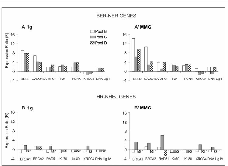
Results show that almost all BER and NER genes were up-regulated in irradiated PBL,
whereas the expression of HR and NHEJ genes was only slightly or not affected by radiation
(Fig.9). Incubation in modeled microgravity after irradiation did not significantly change the
expression of genes involved in DNA repair, suggesting that transcriptional impairment was
not responsible for the increase of mutant frequency observed in irradiated cells incubated in
microgravity in comparison to the static 1 g condition. These findings in agreement with
previous studies on gene expression of non-irradiated space flown and RWV cultured cells,
showing that DNA repair genes were unaffected by low-gravity whereas intracellular
signaling, growth regulatory, cytoskeletal and tumor suppressor genes were altered (Lewis et
al., 2001; Hammond et al., 2000; Pardo et al., 2005).
Selected Topics in DNA Repair
14
Fig. 9. Expression ratios in PBL of pools B–D incubated in 1 g and modeled microgravity
after X-irradiation. (A) R values of BER and NER genes in 1g; (A) R values of BER and NER
in MMG; (B) R values of HR and NHEJ genes in 1g; (B) R values of HR and NHEJ genes in
MMG.
Recently, a new class of important gene modulators has been discovered: microRNAs. They
are a large family of small non-coding RNAs of 18-24 nucleotides that negatively regulate
gene expression levels by binding to microRNA-binding elements in the 3’ untranslated-
region (3’UTR) of target mRNAs thereby triggering decreased protein translation mainly
through mRNA degradation (Guo et al., 2010). A single miRNA may have broad effects on
gene expression networks, such as regulating cell lineage specificity, cellular functions or
stress response. By considering the complexity of the DNA-damage response (DDR),
addressed to maintain genome integrity through cell cycle arrest, DNA repair and/or
apoptosis, it is expected that miRNAs have an important role in this cellular process. Whilst
miRNA-mediated DDR has been studied after UV radiation and hypoxic stress (Pothof et
al., 2009; Crosby et al., 2009) that of radiation combined with microgravity has not been
studied yet and should give important information about risk assessment in space
environment. MicroRNAs profiling were carried out by using the platform “Human miRNA
Microarray kit (V2)” (Agilent), according to the Agilent miRNA protocol. For mRNA
expression profile we used the “Whole Human Genome Oligo Microarray” (Agilent),
consisting of ~41.000 (60-mer) oligonucleotide probes, which span conserved exons across
the transcripts of the targeted full-length genes. Identification of differentially expressed
genes and miRNAs was performed with one and two class Significance Analysis of
Microarray (SAM) program (Tusher et al., 2001) with default settings. Figure 10A shows a
The DNA-Damage Response to Ionizing Radiation in Human Lymphocytes
15
dendrogram relative to some miRNAs differentially expressed following ionizing radiation
in human PBL.
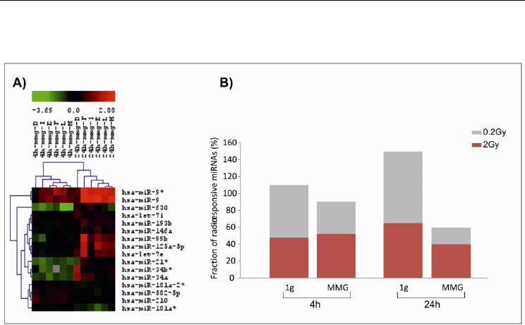
Fig. 10. A) Dendrogram showing several miRNAs differentially expressed in human PBL at
4 and 24h after irradiation with 0.2Gy. Range of expression value is determined as the log2
ratio of irradiated/non-irradiated sample. Down-regulated and up-regulated miRNAs
correspond to green and red boxes, respectively. B) Fraction of radio-responsive miRNAs
(%) in human PBL irradiated with 0.2 and 2Gy and incubated for 4 and 24h- in 1g or in
modeled microgravity (MMG).
MiRNA expression profile was carried out at 4h and 24h after irradiation with 0.2Gy and
2Gy and incubation in 1g and MMG and compared to that of non-irradiated PBL
maintained in parallel conditions. Results showed that in both gravity conditions the
miRNA expression profile was dose-specific, as indicated by the low percentage of common
miRNA responsive to both doses; moreover, the effects of the higher dose predominated at
the late time point. Interestingly, MMG tended to decrease the number of radio-responsive
miRNAs respect to 1g condition, in particular at 24h after irradiation (Figure 10B).
To predict the target genes of differentially expressed miRNAs we first performed a
computational analyses using PITA algorithm available on line (Kertesz et al., 2007).
However, all available software for target prediction are characterized by a large fraction of
false positives, thus to identify the most likely targets, we have integrated mRNA and
miRNA expression data, obtained on the same lymphocyte samples, using MAGIA (MiRNA
And Genes Integrated Analysis) web tool (Sales et al., 2010). We used a non-parametric
index (Spearman correlation coefficient), the most indicated statistical coefficient for a small
number of measures, to estimate the degree of anti-correlation (e.g. up-regulated miRNA
and corresponding down-regulated mRNA target) between any putative pairs of miRNA
and mRNA (Xin et al., 2009; Wang and Li 2009). The anti-correlated transcripts were then
classified according to DAVID (Database for Annotation, Visualization and Integrated
Discovery) web tool (Huang et al. 2009), to determine which Gene Ontology (GO) terms
were significantly enriched in our set of genes. Results of G0 analysis of anti-correlated
Selected Topics in DNA Repair
16
genes showed that in MMG-incubated PBL were not enriched the categories of response to
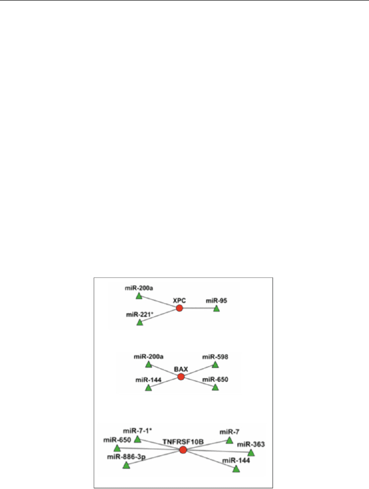
stress, to DNA damage and to apoptosis. miRNA-mRNA anti-correlations of DDR pathway
were visualized by using Cytoscape software package (Shannon et al., 2003; Cline et al.,
2007) (Figure 11). The results showed that, in most cases, the same mRNA was targeted by
different miRNA species according to the different condition of gravity.
Future research is addressed to validate several of the anti-correlations highlighted with our
analyses as important in DDR pathway. In particular, we will perform a functional assay to
demonstrate the regulatory effect of a particular miRNA on its putative target mRNA. The
luciferase assay represents the most efficient approach to evaluate the activity of a miRNA
on its anti-correlated mRNA. This assay allows to demonstrate the activity of a miRNA on
its anti-correlated mRNA by the quantification of the luminescent signal derived from the
luciferase reporter enzyme. Cells are co-transfected with a reporter vector containing the
firefly luciferase gene together with the 3’UTR target sequence predicted for that miRNA
and the miRNA precursor (pre-miRNA) or inhibitor (anti-miRNA), which respectively
mimics and inhibits the endogenous miRNA. The binding of pre-miRNA to the
complementary target sequence will cause the repression of luciferase gene expression,
whereas the binding of anti-miRNA to the endogenous miRNA will induce the expression
of luciferase gene. The quantification of the luminescent signal derived from the luciferase
reporter enzyme thus allows to demonstrate the activity of a miRNA on its putative target
mRNA. In addition to the luciferase assay, it would be interesting to study the role of
selected miRNAs in DDR pathway by a biological approach. Usually, several end points
such as cell survival, DNA repair, cell cycle progression and apoptosis induction are
analyzed in cells over-or under-expressing the miRNA of interest.
Fig. 11. Example of visualization of inversely correlated miRNA-mRNA relationships in
irradiated human PBL. Circles represent transcripts and triangles miRNAs, shown with the
color corresponding to the expression value.

The DNA-Damage Response to Ionizing Radiation in Human Lymphocytes
17
4. The DNA-damage response of human lymphocytes to indirect effect of
ionizing radiation
In addition the cellular effects arising as a direct response to ionizing radiation, in the last
decade it has been suggested that extranuclear or extracellular targets can contribute to the
genetic damage in non-irradiated (bystander) cells. The bystander effect (BE) is the
biological response of non-irradiated cells induced by contact with irradiated cells. The
contact with bystander factors may occur by direct cell–cell interaction or be mediated by
the fluid surrounding the cells. It has been reported that the BE causes cell death, cell cycle
arrest, apoptosis, changes in gene expression, and increases micronucleus induction,
chromosomal aberrations, mutation frequency, and DNA damage in cells neighboring hit
cells. In contrast to DNA damage induced by direct irradiation, bystander cell DNA damage
is still poorly understood. Many data showed that early events of the radiation induced
bystander effect are rapid calcium fluxes and generation of reactive oxygen species in
bystander cells. Mitochondria seem to play a central role in bystander signaling: irradiated
cell conditioned media can cause changes of mitochondrial distribution, loss of
mitochondrial membrane potential, increases in ROS, and increase in apoptosis among the
medium receptor cells, which can be blocked by treatments with antioxidants (Chen et al.,
2008). Experiments carried out in hepatoma cell lines provide evidence that the BE can be
modulated by the p53 status of irradiated cells and that a p53-dependent release of
cytochrome-c from mitochondria may be involved in producing BE (He et al., 2011).
We investigated on the mechanisms of the medium-mediated bystander response induced
by low doses of -rays in human tumoural lymphocytes (TK6 cells), a cell line growing in
suspension, in which gap-junction communications are not involved in transferring
bystander signals and only medium-mediated molecules may be responsible for BE
induction. Cell cultures were irradiated and the culture medium discarded immediately
after irradiation and replaced with a fresh one to eliminate ROS originating during
irradiation. Irradiated cells were incubated for 6h in fresh medium, which, at the end of
incubation time, is referred as conditioned medium (CM) and used to incubate non-
irradiated TK6 cells for different times (2-48 h). In bystander cultures, cell mortality at the
fixed incubation times ranged between 24 and 19%, very similar values to that of directly
irradiated cells (28 and 20%). The mortality percentages for all incubation times were
significantly higher with respect to that of the controls (0Gy and 0Gy CM). The survival
fraction of directly 1Gy irradiated or CM incubated cells was determined by the clonogenic
assay. The data show that both irradiated and bystander TK6 cells had a lower cloning
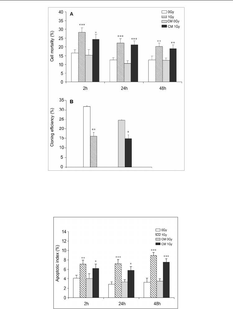
efficiency than their respective controls. Figure 12 reports the results about cell mortality
and survival (given as the ratio of the cloning efficiency of treated vs. untreated control
cells) in TK6 cells exposed directly to IR or to CM. Apoptosis induction was tested by the
presence of fragmented nuclei and apoptotic bodies at 2, 24 and 48h after 1Gy irradiation or
CM incubation. The apoptotic index (A.I.) ranged between 7 and 9 % in irradiated cells and
between 6 and 7.5 % in bystander cells, and was significantly higher than the relative
controls at all times (Figure 13). The induction of apoptosis was also analyzed by the
activation of caspase-3, the principal effector caspase, assayed by the cleavage of the peptide
substrate DEVD-AFC, at 1, 2, 24 and 48h after irradiation or CM incubation. In bystander
cells caspase-3 activation increased from 1.4- to 2.7-fold during the 48h of CM incubation,
suggesting that bystander apoptosis increases after 48h. Bystander apoptosis in TK6 cells
was sensitive to the inhibitor of caspase-8, the Z-IETD-fmk, added during CM treatment or
Selected Topics in DNA Repair
18
Fig. 12. Cell mortality determined by Trypan blue staining (A) and cloning efficiency (B) in
non-irradiated control cells (0Gy), in irradiated cells (1Gy), in cells incubated with
conditioned medium from non-irradiated cells (CM 0Gy) and in cells incubated with
conditioned medium from irradiated cells (CM 1Gy). Results are the means of 3-5
independent experiments ± S.E. (*P< 0.05, **P<0.01, ***P<0.001, t test).
Fig. 13. Apoptotic index (A) in irradiated and bystander cells. Results are the means ± S.E. of
5-8 independent experiments. Significant differences were observed in 1Gy and CM 1Gy vs
0Gy and CM 0Gy, respectively (*P<0.05, **P<0.01, ***P<0.001, t test).

The DNA-Damage Response to Ionizing Radiation in Human Lymphocytes
19
post-irradiation incubation. The presence of the inhibitor significantly decreased the
induction of apoptosis to the control level, but it did not significantly decrease the level of
apoptosis in either irradiated or non-irradiated controls (Figure 14). These results suggest
that caspase-8 activation is triggered by signaling molecules present in the conditioned
medium. The addition of the ROS scavenger Cu-Zn superoxide dismutase and N-
acetylcysteine to the conditioned medium allowed to investigate the involvement of
oxidative stress in inducing bystander apoptosis. ROS scavengers did not significantly
decrease the apoptotic index in CM cultures; by treating non-irradiated TK6 cells with
medium irradiated without cells (IM), we evaluated the contribute of ROS produced by
irradiation in inducing bystander apoptosis.
Fig. 14. Apoptotic index in irradiated cells (1Gy), in irradiated cells incubated with caspase-8
inhibitor (1Gy + Z-IETD-fmk), in bystander cells incubated with caspase-8 inhibitor (CM
1Gy + Z-IETD-fmk), and with ROS scavengers SOD and NAC (CM 1Gy + scav.). Results are
the means of 3-5 independent experiments ± S.E. Significant differences were observed in
CM 1Gy vs CM 1Gy + Z-IETD-fmk at all times. B. Apoptotic index in cells incubated with
irradiated medium (IM 1Gy), with irradiated medium in the presence of caspase-8 inhibitor
(IM 1Gy + Z-IETD-fmk), and ROS scavengers (IM 1Gy + scav.). The values are the means of
3 independent experiments ± S.E. Significant differences were observed at 2 h of incubation
with IM 1 Gy vs IM 1Gy + Z-IETD-fmk (*P<0.05, **P<0.01, ***P<0.001, t-test).
IM incubation for 2h increased the apoptotic index which was totally inhibited by ROS
scavengers and little affected by incubation with the caspase-8 inhibitor, whereas at 24 and
48h no significant differences among samples incubated with IM were observed. DSBs
induced by ionizing radiation can easily be detected by the extensive H2AX
phosphorylation occurring near DNA lesions, forming foci that co-localize with several
repair proteins (Fernandez-Capetillo et al., 2003). 85% of TK6 were -H2AX foci positive at
2h after irradiation with 1Gy, then this percentage decreased to the level of non-irradiated
cells 24h later, fitting DNA repair kinetics. The incubation of cells with CM for 2h
significantly increased the percentage of -H2AX foci positive cells (9-11%) but, when the
CM was kept in contact with bystander cells for 24h the number of positive cells decreased
to control levels, suggesting that DNA lesions induced at the beginning of CM incubation
are repaired and no new damage accumulates later. Data from other human cells show that
-H2AX foci induction in bystander cells persists in time, probably as a consequence of the
Selected Topics in DNA Repair
20
formation of bystander factors that themselves generate ROS, leading to a self-sustaining
system responsible for long-lasting effects (Yang 2005, Sokolov 2005, Kashino 2004, Lyng
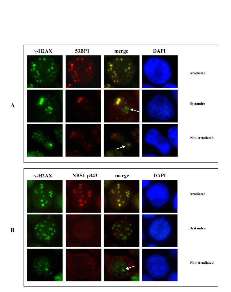
2006). In irradiated TK6 cells both 53BP1 and NBS1p343 proteins co-localized with -H2AX
foci, whereas in bystander cells co-localization was partial or absent (Figure 15).
Fig. 15. Non-irradiated, irradiated and bystander TK6 cells were fixed and co-stained with
anti- H2AX (green), anti-53BP1 and anti-NBS1-p343 (red), at 2 h from irradiation or CM
incubation. The red and green images were merged and subjected to co-localization
analysis. Arrows indicate γH2AX foci without co-localization of 53BP1and NBS1-proteins.
Nuclei were counterstained with DAPI.