Blake A.J.(ed.) Crystal Structure Analysis
Подождите немного. Документ загружается.


52 Space-group determination
Exercises
1. The following unit cell volumes and densities have been
measured for the given compounds. Calculate Z for the
crystal, and comment on how well (or badly) the ‘18 Å
3
rule’ works for each compound:
a) methane (CH
4
) at 70 K: V = 215.8 Å
3
, D = 0.492
gcm
−3
;
b) diamond (C): V = 45.38 Å
3
, D = 3.512 g cm
−3
;
c) glucose (C
6
H
12
O
6
): V = 764.1 Å
3
, D = 1.564 g
cm
−3
;
d) bis(dimethylglyoximato)platinum(II)
(C
8
H
14
N
4
O
4
Pt): V = 1146 Å
3
, D =2.46gcm
−3
.
2. A unit cell has three different axis lengths and three
angles all apparently equal to 90
◦
. What is the met-
ric symmetry? The Laue symmetry, however, does not
agree with this; equivalent intensities are found to be
hkl ≡ hk
l ≡ hkl = hkl
hkl ≡ hkl ≡ hkl = hkl.
What is the true crystal system and its conventional axis
setting?
3. What are the systematic absences for the space groups
I222 and I2
1
2
1
2
1
?
4. Deduce asmuchas you canabout the spacegroupsofthe
compounds for which the following data were obtained.
a) Monoclinic. Conditions for observed reflections:
hkl, none; h0l, h + l even; h00, h even; 0k0, k
even; 00l, l even. Centric distribution for general
reflections.
b) Orthorhombic. Conditions for observed reflec-
tions: hkl, all odd or all even; 0kl, k+l =4n and both
k and l even; h0l, h + l = 4 n and both h and l even;
hk0, h +k = 4 n and both h and k even; h00, h = 4n,
0k0, k = 4n;00l, l = 4n. Centric distribution for
general reflections.
c) Orthorhombic. Conditions for observed reflec-
tions: hkl, none; 0kl, k + l even; h0l, h even; hk0,
none; h00, h even;
0
k0, k even; 00l, l even. Acentric
distribution for general reflections, centric for hk0.
d) Tetragonal. Reflections hkl and khl have the same
intensity. Conditions for observed reflections: hkl,
none; 0kl, none; h0l, none; hk0, none; h00, h even;
0k0, k even; 00l , l = 4n; hh0, none. Acentric distri-
bution for general reflections; centric for 0kl, h0l,
hk0, and hhl subsets of data.

5
Background theory for
data collection
Jacqueline Cole
5.1 Introduction
This chapter takes the reader through the theoretical fundamentals
behind data collection and reduction methods for single-crystal struc-
ture determination. The descriptions are directed towards those using
area-detector diffractometers, since these are now almost the only ones
available commercially and are installed in many laboratories. How-
ever, a large part of the theory may still be relevant to older types of
diffractometers.
A step-wise theoretical journey through an experiment is first pre-
sented, to illustrate the need for alternating real and reciprocal space
thinking at different parts of an experiment. There follows a discussion
of the geometry of X-ray diffraction in both real and reciprocal space, the
sequence of procedures required to determine a unit cell, data-collection
methodologies and strategies, a description of the data-integration pro-
cess for diffractometers housing a standard flat-plate two-dimensional
area detector, and the subsequent data-reduction corrections that are
necessary to afford a list of intensities or structure factors, the starting
point for data solution and refinement.
5.2 A step-wise theoretical journey through
an experiment
The sequence of events that comprise an X-ray diffraction experiment
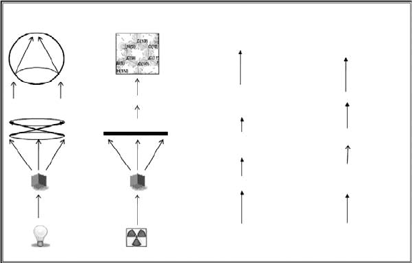
is in many ways analogous to that of a microscopic investigation, as
Fig. 5.1 illustrates.
When a sufficiently intense and collimated source of photons is
directed at a small crystal, an inverted image of the form of that crys-
tal can be collected on a lens or detector placed on the opposite side
of the crystal. The wavelength of the photon source will dictate the
scale of the contents of this image: an optical source (λ ∼ 1 μm) will
53
54 Background theory for data collection
Real space
Optical image
of crystal
Microscope
lenses
Calculations
Image of electron
density in crystal
Detector
Small crystal
Retina
Light X-rays
Eye
Crystal
Structure
Data reduction
Experiment
Sample
preparation
Reciprocal
space
Reciprocal
space
Real space
Real space X-ray Machine
F.T
Fig. 5.1 The components of an optical and ‘X-ray’ microscope, revealing the real and
reciprocal parts of an experiment.
present a microscopic inverted view of the exposed part of the crystal,
whilst an X-ray beam (λ ∼ 10
−10
m = 1 Å) will reveal an inverted image
of the exposed part of the crystal on the Ångstrom scale, i.e. the scale
corresponding to the size of atoms. Where X-ray diffraction (rather than
absorption) occurs, the diffracted image will represent the average view
of the contents of many unit cells in a reciprocal form, given the periodic
nature of single crystals. Such an image would normally comprise many
spots, their relative location on the image providing information about
the unit cell, whilst their intensities contain all of the information about
the relative positions of all atoms within the unit cell.
In the case of an optical microscope, the retina in the human eye pro-
vides a natural image-inversion process so that one can readily obtain a
real-spaceinterpretation of an optically inverted image, simply by view-
ing the optical lens that captured the image via objective lens with an
appropriate magnification.
Realizing a real-space view of the diffracted part of an X-ray image is
an entirely different matter, however; only fictional characters have ‘X-
ray eyes’ and even they could not oblige due to the irradiation damage
that they would sustain from putting their head in the X-ray beam!
An X-ray-sensitive detector, whether this is photographic film or an
electronic device, can be used to obtain the reciprocal diffraction image.
The positions and intensities of diffraction spots can therefore be ascer-
tained. However, with no ‘X-ray eye retina’, this information still needs
to be converted fromreciprocal to realspace.Acalculation,in the form of
a Fourier transformation, is needed to make this conversion. It requires
the ‘structure factor’, F, of each spot on the image. The square of the
structure factor, F
2
, is proportional to the intensity of a spot, which is
measurable, and so the amplitude |F| can be evaluated. The phase of
5.3 The geometry of X-ray diffraction 55
each F (the sign for centrosymmetric structures) cannot be found by a
normal experiment, however, since spot intensities do not directly carry
phase information: there is no way of knowing the phase of an X-ray
beam as it meets the detector. All intensities are positive. The phase of F
is crucial, however, since the Fourier transform is based implicitly on a
summation involving F. Later chapters in this bookconcerning structure
solution explain how one can overcome this so-called ‘phase problem’.
Once solved, the required conversion from reciprocal to real space can
be undertaken to reveal the real-space representation of a unit cell, in
the form of an electron-density map.
This virtual journey through an experiment not only illustrates the
analogy of an ‘X-ray microscope’ but hopefully also shows that one
needs to look at the different stages of an experiment with different
space concepts. The physical experiment itself, comprising the sample
and the diffractometer, obviously exists in real space. The X-rays are
fired at the crystal in real space. The diffraction patterns observed by
the detector represent reciprocal space images of the crystal structure
of the sample. Each spot, or ‘reflection’, in a diffraction pattern can
be identified uniquely by Miller indices, or hkl values. Such indices,
described in detail later, are therefore reciprocal space quantities, as
are their associated structure factors. The goal of all subsequent data
integration and reduction stages is to obtain the magnitudes (ampli-
tudes) of the structure factors of each diffraction spot, via the square
root of their measured intensities, corrected for various defined geo-
metrical and physical effects. With these in hand, one can then consider
the Fourier-transformation calculation to obtain the desired real-space
view of a molecule via an electron-density map.
5.3 The geometry of X-ray diffraction
X-ray waves interact with electrons in a material. X-ray diffraction
occurs when a crystal is oriented towards incoming X-ray waves, of a
suitable wavelength (λ), such that the waves interfere non-destructively
between ordered rows of electronic concentration (layers of atoms) in
the crystal that are of a suitable separation (d ).
The condition for diffraction can be defined mathematically by
Bragg’s law or geometrically by the Ewald sphere construction, in real
or reciprocal space, respectively.
Position of atom in upper plane
relative to lower plane (Å)
Point of
interaction
d = 4 Å
d = 3 Å
d = 2 Å
0
Fig. 5.2 The level of diffraction generated
by interference of two X-ray waves with
two planes of atoms positioned directly
above each other, as a function of distance
between these planes, d [d = 4 Å (top),
3 Å (middle), 2 Å (bottom); θ = 5
◦
and
λ = 0.7 Å throughout]. A fully interactive
and extended version of these snapshots is
given by Proffen and Neder (2008a).
5.3.1 Real-space considerations: Bragg’s law
Bragg’s law states that nλ = 2d sin θ. λ and d have been defined above
and θ is the angle between a plane of atoms and the line of the diffracted
(or incident) beam; n is an integer. Therefore, diffraction occurs only for
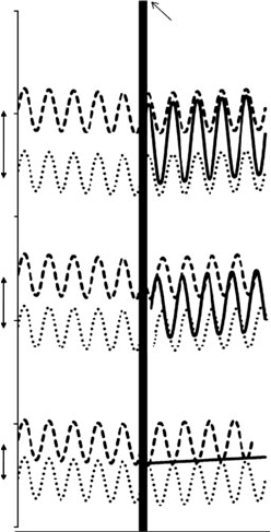
certain combinations of d, λ and θ. The variation of d, whilst keeping λ
and θ approximately constant, results in differingmagnitudes of diffrac-
tion by two lattice planesin one dimension, asillustrated in Fig. 5.2; here,
56 Background theory for data collection
where incident X-ray waves are 100% in-phase (d = 4) the maximum
amount of diffraction occurs, whilst 100% out-of-phase (d = 2) will pre-
clude diffraction; 75% in-phase waves result from d = 3 and so affords a
moderate amount of diffraction.
Two X-ray waves diffracted from a plane will have a difference in
path length, necessarily depending on d. One may notice in Fig. 5.2, for
example, that the 100% in-phase interference of two waves has a path
difference that corresponds to exactly one wavelength. Indeed, there
exists a general relationship that the maximum amount of diffraction
between two points occurs at a value of d where the path difference
between two waves is an integral multiple of the X-ray wavelength, nλ.
This multiplier, n, is the same integer used in Bragg’s law.
Given that there are many parallel planes in a crystal, and that the
diffraction condition needs to be satisfied in three dimensions simul-
taneously rather than in one as presented here, in practice diffraction
is observed only where n is an integer. In nearly all cases of conven-
tional X-ray diffraction, one can consider that n = 1, since one can
always divide d by n if it is greater than 1, in order to obtain a value
of d corresponding to n =1.
The value of d that corresponds to 100% in-phase diffraction, in each
of the three-dimensional basis-set vectors being used, is represented by
vectors that relate to a, b and c and integers h, k and l, respectively,
where a, b and c represent the unit cell parameters and the integers are
the reciprocal-space (hkl ) indices. Therefore, in practice, one can say that
λ = 2d
hkl
sin θ.
In conventional crystal-structure determination, our aim is to collect
as many unique diffraction spots, hkl, as possible within the practical
bounds of time available. The variation of d represents the orientation of
a crystal with respect to the incident X-ray beam, and ideally we would
want to collect diffraction data for all possible orientationsof a crystal for
a three-dimensional crystal-structure determination. In an experiment,
d is therefore usually varied, whilst keeping λ constant (X-ray sources
in a laboratory are monochromatic and λ is selected by the type of X-ray
tube installed on an instrument) and stepping through θ incrementally
to collect the data since 2θ , the net scattering angle, represents the angle
of the detector centre relative to the incident X-ray beam, 2θ = 0
◦
being
the straight-through beam.
5.3.2 Reciprocal-space considerations: the
Ewald sphere
Since the diffraction patterns that we observe in an experiment relate to
reciprocal space, a geometrical construct of a reciprocal space version
of Bragg’s law is often useful in interpreting such patterns. The Ewald
sphere, illustrated in Fig. 5.3(a), provides this construct: the sample lies
at the centre of a sphere whose radius is the reciprocal of the wavelength
(1/λ); the diffraction condition is met when a spot touches the surface of
this sphere. With an incident X-ray beam traversing the horizontal from

5.3 The geometry of X-ray diffraction 57
Diffracted
X-ray beam
Transmitted Incoming
101
X-ray beam X-ray beam
Crystal
(a) (b)
Crystal
2u
1/d
Ewald Sphere.
Radius, r = 1/l
001 101
100
000
101
201001
100
201
Fig. 5.3 The Ewald sphere: (a) definition of parameters; (b) the intersection of the (101) reflection with the surface of the Ewald sphere,
thereby meeting the Bragg condition of diffraction.
one side of the sphere, transmitted X-rays simply continue to the other
side of the sphere, whereupon the beam meets the surface to satisfy the
diffraction condition for the (000) reflection that corresponds to F(000).
This reflection is always calculated rather than measured because the
beamstop necessarily obscures the transmitted X-ray beam from the
detector. Indeed, even if it could be measured, the (000) reflection would
be contaminated with undiffracted direct beam and so such an intensity
measurement would be inaccurate. The (000) reflection is referenced as
the origin of reciprocal space.
Wherediffractionoccurs,one can draw a vector fromthe crystal centre
to the surface of the Ewald sphere that subtends an angle, 2θ, (the net
scattering angle) to the vector representing the transmitted X-ray beam.
The point thus generated on the surface of the sphere corresponds to
a reflection, hkl, and the chord that links this point with the reciprocal
lattice origin has a length 1/d, where d is the lattice spacing for that
reflection.
As the crystal is rotated during a diffraction experiment, different
lattice planes are exposed to the incident X-ray beam, and so different
reflections intersect the surface of the sphere, i.e. diffract. Figure 5.3(b)
shows an illustrative example of a rotating lattice plane of reflections,
where diffraction at that time-captured moment derives from the (101)
reflection.
A very useful demonstration of an Ewald sphere simulation is given
by Proffen and Neder (2008b).
One can use the Ewald sphere construct to interpret geometrically the
patterns of spots appearing on successive frames of diffraction during
data collection.
If one has determined the unit cell parameters of a crystal within the
diffractometer frame of reference (see Section 5.5), one can also calculate
the hkl indices of each spot in a diffraction pattern using the appropriate

58 Background theory for data collection
Table 5.1. Values of 1/d
2
hkl
for each crystal system (Giacovazzo, 1992).
Crystal System 1/d
2
hkl
Triclinic (1 − cos
2
α − cos
2
β − cos
2
γ + 2 cos α cos β cos γ)
−1
h
2
a
2
sin
2
α +
k
2
b
2
sin
2
β +
l
2
c
2
sin
2
γ
+
2kl
bc
(cos β cos γ − cos α) +
2lh
ca
(cos γ cos α − cos β) +
2hk
ab
(cos α cos β −cos γ)
Monoclinic
h
2
a
2
sin
2
β
+
k
2
b
2
+
l
2
c
2
sin
2
β
−
2hl cos β
ac sin
2
β
Trigonal (R)
1
a
2
(h
2
+ k
2
+ l
2
) sin
2
α + 2(hk + hl + kl)(cos
2
α − cos α)
1 + 2 cos
3
α − 3 cos
2
α
Hexagonal/Trigonal (P)
4
3a
2
(h
2
+ k
2
+ hk) +
l
2
c
2
Orthorhombic
h
2
a
2
+
k
2
b
2
+
l
2
c
2
Tetragonal
h
2
+ k
2
a
2
+
l
2
c
2
Cubic (h
2
+ k
2
+ l
2
)/a
2
equation in Table 5.1, thus uniquely identifying each reflection as one
collects data.
The ability to identify each reflection means that one can also predict
where all diffractionspots will appear in frames of data. This enables the
experimenter to manipulate the diffraction geometry, via diffractometer
control programs, to perform various functions: ‘dial-up’ certain reflec-
tions, lattice planes, time-optimize data collection strategies, and so on.
5.4 Determining the unit cell: the
indexing process
There are two principal methods that are used to determine a unit
cell: a real-space method, called ‘autoindexing’; and a reciprocal-space
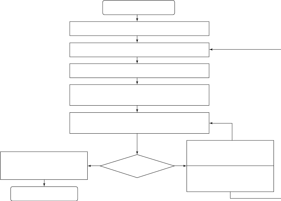
method. Both methods follow a similar common procedure in that they
determine the unit cell using a small portion of diffraction data taken
from different parts of reciprocal space, and their methodology follows
the generic flow-diagram below (Fig. 5.4).
5.4.1 Indexing: a conceptual view
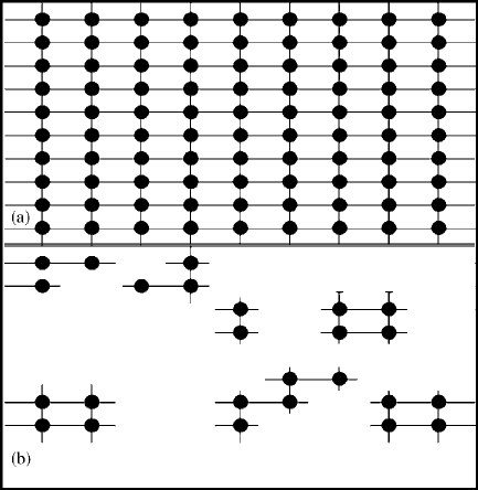
If one possessed data that corresponded to every possible diffraction
spot in a sufficiently large volume of reciprocal space, one could simply
calculate the lattice spacings for each reflection in real space and thence
build up a three-dimensional grid of periodic lattice points. This grid
would contain a repeat motif, which one could identify by observation,
5.4 Determining the unit cell: the indexing process 59
Indexing process: START
Collect data from several small parts of reciprocal space
Generate list of vectors between the reflections collected
Find the three shortest non-coplanar vectors in this list
Identify these three vectors as a trial unit cell: they must be
a (primitive) subset of the cell, if not the correct unit cell
Using this trial unit cell, assign hkl indices to all vectors,
thus generating an initial reflection list
Determine the metric symmetry via
analyzing (hkl) symmetric absences
of least-squares refined reflections.
Unit cell obtained: STOP
YES NO
Are
all hkl indices
integral?
If many indices are simple fractions,
divide appropriate a⬘, b⬘, or c⬘ by
this fraction: generate new trial cell.
Otherwise, collect more data, use
subset of present data, or try other
indexing method: then repeat cycle
Fig. 5.4 Indexing flowchart.
and that would represent the unit cell of the sample in hand (e.g. see
Fig. 5.5a).
Realizing such a situation is not practical when initially indexing a
crystal and so one relies, instead, on utilizing a small portion of data
collected selectively from several small volumes of reciprocal space that
together will afford a representative derivation of a partial lattice in
three dimensions. Whilst such a representation is rather ‘patchy’ (e.g.
see Fig. 5.5b), as long as there are enough parts of the lattice to indicate
all three unit cell parameters and corroborate them, one should be able
to determine the correct unit cell of any subject material, assuming that
the sample is a good-quality single crystal.
This is a rather conceptual view of indexing, but hopefully is a help-
ful one. In practice, one works largely with a comparison of vectors as
described in the next section. That said, one can often gain an insight
into the likely unit cell parameters by simply observing the frames of
data collected for indexing purposes: if many diffraction spots are close
60 Background theory for data collection
Fig. 5.5 Conceptual unit cells.
together along one direction, one would expect a long unit cell parame-
ter in the corresponding real-space dimension. This may indicate a unit
cell of a certain crystal system and thus put constraints upon some of
their parameters.
5.4.2 Indexing procedure
Detailed accounts of each of the two principal indexing methods can be
found in many articles (Hornstra and Vossers, 1974; Clegg, 1984; Sparks,
1976, 1982; Kabsch, 1993). However, it is instructive to consider here
the basic generic procedure behind each method, via a flow diagram
(Fig. 5.4) with an associated description. The first step has already been
discussed in the previous section.
Each of the two indexing methodologies uses a core set of vectors, x,
thatcorrespondto the reciprocal lattice points observed in the diffraction
patterns selectively collected for indexing purposes. In the reciprocal-
space method, this core set of vectors is augmented by summative or
difference vectors generated from combinations of these initial vectors,
x
i
±x
j
; the same difference vectors are often added to the vector set in the
real-spacemethod as well,particularlywhereone anticipates a largeunit
cell. In each case, the three shortest non-coplanar vectors for this list are
then identified and used to define an initial cell, in the form of reciprocal
cell axes or a real-space trial unit cell. Whilst such a trial solution does
not usually correspond to the true unit cell, it will represent a subcell of
the true unit cell.
5.4 Determining the unit cell: the indexing process 61
By assimilating this trial cell to the true unit cell, one can calculate a
trial (hkl) index for each of the core set of vectors, via the usual mathe-
matical equations for a given predicted crystal system, as described in
Table 5.1, Section 5.3.2, or via analogous reciprocal space axis relations.
This affords a trial list of uniquely identified reflections that one can use
to test the validity of the trial unit cell: the true unit cell must gener-
ate only integral values of h, k and l, for all reflections in this list, by
the fundamental nature of Miller indices. One can readily tell if all (hkl)
assignments approximate to integral quantities by simply observing the
list of (hkl) values. In practice, one rarely finds all (hkl) values perfectly
integral due to experimental error, spurious reflections, i.e. reflections
that derive from alien reciprocal lattices, or instrumental artefacts, but
one should expect most h, k and l values to be near-integer quantities if
the trial cell corresponds to the primitive form of the true unit cell.
If the bulk of h, k or l values approximate to simple fractions, this
may indicate that one cell parameter corresponding to this index is a
corresponding multiple of the true cell parameter. In this case, there will
exist a common multiplier (2, 3, 4 ...) that will afford a list of integral
hkl reflections when one multiplies the appropriate one of the trial unit
cell parameters, a, b or c, by this common multiplier. Simple fractions
are often observed when the three unit cell lengths are highly disparate.
If most reflections have non-integral hkl values, however, and there
is no obvious numerical relationship between them, indexing fails with
this core set of vectors. There are various options to consider in this
scenario. Four of the most common approaches are: (i) create a new
core set of vectors by collecting a new set of reflections from the same
crystal and repeat the indexing process; (ii) re-index using a subset of the
existing data set, typically only strong reflections defined by an intensity
threshold; (iii) try another indexing method; (iv) select a new crystal for
the experiment.
Where one is able to generate a list of integral hkl values, the trial
cell can be considered to be the primitive form of the true unit cell,
which will equate to the true unit cell if that is primitive. Alternatively,
the true cell may possess higher metric symmetry. In order to ascertain
the full metric symmetry of the sample, one first performs a full-matrix
least-squares refinement on the reflection list, to remove any spurious
or ill-defined reflections. Then one can determine if systematic absences
are revealed that indicate a centred unit cell. If no centring is found by an
automated procedure, it can be judicious to double-check for centring
by considering the Laue symmetry. The metric symmetry can never be
lower than the Laue symmetry but it can be higher. By checking the Laue
symmetry one can, for example, identify ostensibly centred monoclinic
cells that are really triclinic, or rhombohedral cells masquerading as
monoclinic ones. In cases where one can not be certain if the unit cell is
centred or not, at the indexing stage of data collection, one must always
undertake a data-collection strategy that assumes the lower-symmetry
crystal system. This is because, if higher symmetry is confirmed post-
data collection (Laue symmetry is more easily distinguishable with a
