BD Diagnostic Systems (publ.). Difco Manual (Manual of Microbiological Culture)
Подождите немного. Документ загружается.


546 The Difco Manual
Urea Agar Base, Urea Agar Base Concentrate, Urea Broth & Urea Broth Concentrate Section II
Materials Required but not Provided
Glassware
Distilled or deionized water
Autoclave
Incubator (35°C)
Sterile tubes with closures
Method of Preparation
1. Suspend 38 grams in 1 liter distilled or deionized water.
2. Heat gently to dissolve completely.
3. Autoclave at 121°C for 15 minutes. Cool to room temperature.
4. Store prepared medium at 2-8°C.
Specimen Collection and Preparation
Refer to appropriate references for specimen collection and preparation.
Test Procedure
Substitute Universal Preenrichment Broth for preenrichment
media as specified for Salmonella and Listeria
1,2
and follow
recommended procedures.
Results
Salmonella and Listeria demonstrate good growth and recovery
following preenrichment in this broth.
References
1. Vanderzant, C., and D.F. Splittstoesser (ed.). 1992. Compendium
of methods for the microbiological examination of foods, 3rd ed.
American Public Health Association, Washington, D.C.
2. Association of Official Analytical Chemists. 1995. Bacterio-
logical analytical manual, 8th ed. AOAC International,
Gaithersburg, MD.
3. Bailey, J. S., and N. A. Cox. 1992. Universal preenrichment broth
for the simultaneous detection of Salmonella and Listeria in foods.
J. Food Protect. 55:256-259.
4. Bailey, J. S., D. L. Fletcher, and N. A. Cox. 1990. Efficacy of
enrichment media for recovery of heat-injured Listeria
monocytogenes. J. Food Prot. 53:473-477.
5. Juven, B. J., N. A. Cox, J. S. Bailey, J. E. Thomson, O. W. Charles,
and J. V. Shutze. 1984. Recovery of Salmonella from artificially
contaminated poultry feed in non-selective and selective broth
media. J. Food Prot. 47:299-302.
Packaging
Universal Preenrichment Broth 500 g 0235-17
Bacto
®
Urea Agar Base
.
Bacto Urea Agar Base Concentrate
Bacto Urea Broth
.
Bacto Urea Broth Concentrate
User Quality Control
Identity Specifications
Urea Agar Base
Dehydrated Appearance: Light orange-red to orange-red, homogeneous,
inherently lumpy.
Solution: 2.9% solution, soluble in distilled or
deionized water. Solution is orange, clear.
Prepared Agar Medium: Reddish orange, very slightly opalescent.
Reaction of 2.9%
Solution at 25°C: pH 6.8 ± 0.1
Urea Agar Base Concentrate
Solution: 29% solution is reddish orange, clear liquid.
Reaction of 29%
Solution at 25°C: pH 6.75 ± 0.15
Urea Broth
Dehydrated Appearance: Light orange to light pink, homogeneous,
inherently lumpy.
Solution: 3.87% solution, soluble in distilled or
deionized water. Solution is
orange-yellow, clear.
Reaction of 3.87%
Solution at 25°C: pH 6.8 ± 0.1
continued on following page
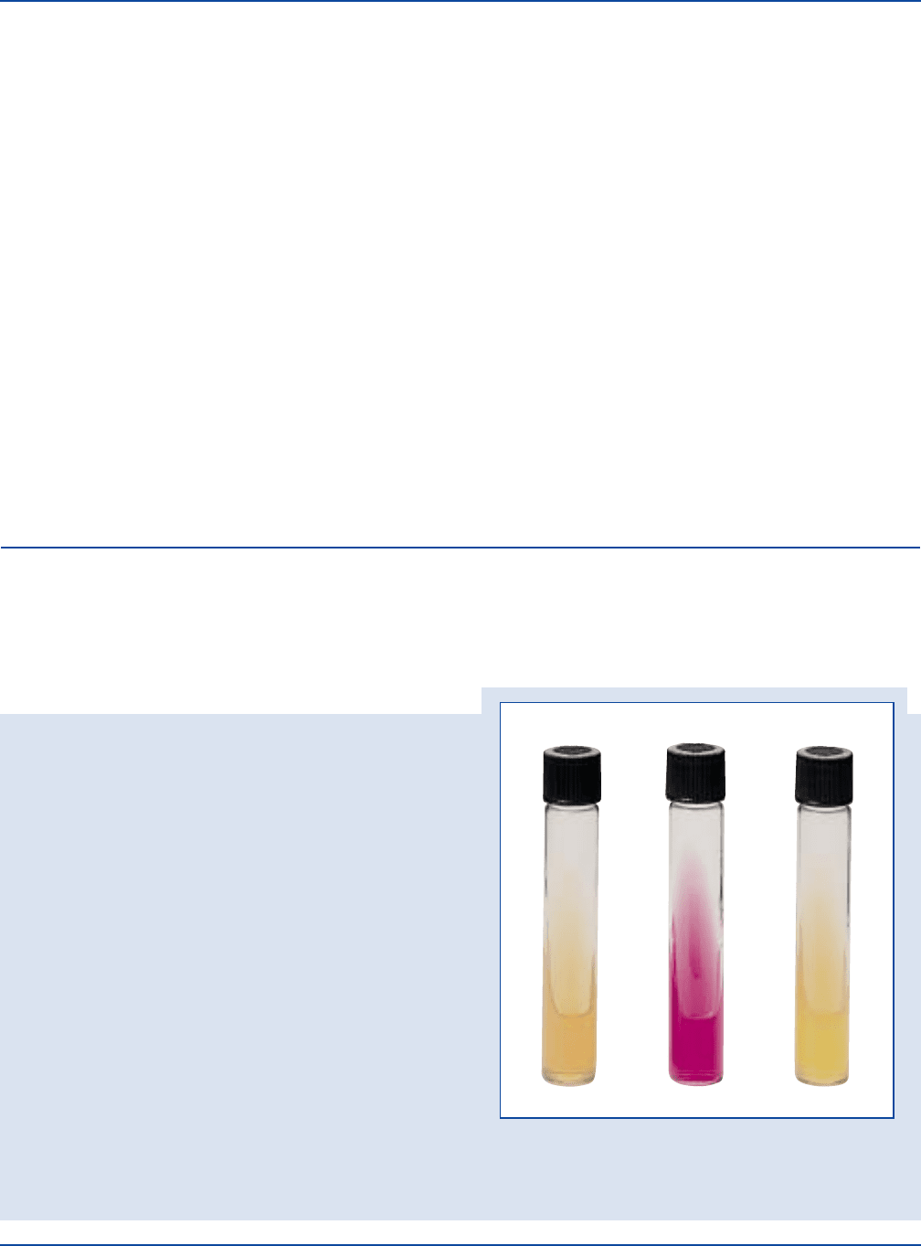
Escherichia coli
ATCC
®
25922
Uninoculated
tube
Proteus vulgaris
ATCC
®
13315

The Difco Manual 547
Section II Urea Agar Base, Urea Agar Base Concentrate, Urea Broth & Urea Broth Concentrate
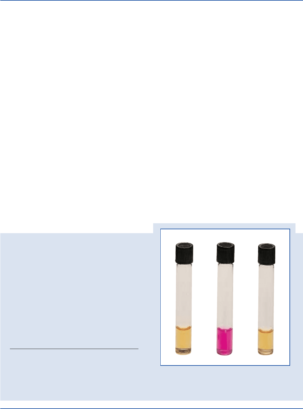
Escherichia coli
ATCC
®
25922
Uninoculated
tube
Proteus vulgaris
ATCC
®
13315
User Quality Control cont.
Urea Broth Concentrate
Appearance: 38.7% solution is reddish-orange,
clear liquid.
Reaction of 38.7%
Solution at 25°C: pH 6.8 ± 0.2
Cultural Response
Urea Agar Base and Urea Agar Base Concentrate
Prepare Urea Agar per label directions. Inoculate and incubate at
35 ± 2°C for 6-48 hours.
Urea Broth and Urea Broth Concentrate
Prepare Urea Broth per label directions. Inoculate and incubate
at 35 ± 2°C for 8-48 hours.
ORGANISM ATCC
®
UREASE PRODUCTION
Escherichia coli 25922* negative, no color change
in the medium
Proteus vulgaris 13315* positive, red or cerise medium
The cultures listed are the minimum that should be used for
performance testing.
*These cultures are available as Bactrol
™
Disks and should be used as directed in Bactrol Disks Technical Information.
Urea Broth
Intended Use
Bacto Urea Agar Base, when combined with Bacto Agar, is used for
differentiating microorganisms based on urease activity.
Bacto Urea Agar Base Concentrate is a sterile 10X solution of Urea
Agar Base which, when combined with Bacto Agar, is used for preparing
Urea Agar.
Bacto Urea Broth is used for differentiating microorganisms, particularly
Proteus species, based on urease production.
Bacto Urea Broth Concentrate is a sterile 10X solution of Urea Broth
ready to use as recommended. It is suggested for laboratories that
require only small amounts of medium.
Also Known As
Urea Agar Base is also known as Urea Agar Base, Christensen or
Christensen’s Urea Agar.
Urea Broth is also referred to as Stuart’s Urea Broth.
Summary and Explanation
Christensen
1
devised a urea agar medium containing peptone and
dextrose that had a reduced buffer content. The medium supported a
more vigorous growth of many of the gram-negative enteric bacilli and
readily permitted observation of urease production.
Ewing
2
used Urea Agar as a differential medium in the examination of
many cultures from stool specimens. Urea Agar may be used as a
screening medium (along with Triple Sugar Iron Agar) for the selection
of Salmonella and Shigella cultures for serologic classification.
3
Qadri
et al.
4
developed a spot test for the rapid detection of urease activity
by applying diluted Urea Agar Base Concentrate to filter paper and
inoculating the paper with a loopful of 24-48 hour culture. Urease-
positive results were obtained within 2 minutes. When combined with
results of other rapid screening methods, Urea Agar is the most
common way to detect the production of urease by yeasts.
5
Urea Agar
Base Concentrate has also been used in differentiating mycobacteria
species.
6
Urea Broth, prepared according to the formula of Stuart, Van Stratum
and Rustigian
7
is a highly buffered urea medium that provides all the
essential growth requirements for Proteus. Stuart et al.
7
noted that by
decreasing the amount of buffer in their standard medium to one-tenth
or one-hundredth of the original concentration, the incubation time for
Proteus could be decreased from 12-48 hours to 2-4 hours. When the
amount of buffer is decreased, however, other organisms capable of
urease production give a positive test. Rustigian and Stuart
8
used urea
decomposition as a limiting characteristic for the identification of
Proteus strains from other members of the family Enterobacteriaceae.
Ferguson and Hook
9
reported that urease production could be used to
differentiate between members of the Proteus and Salmonella groups.
The medium is positive for Proteus, Morganella morganii, Providencia
rettgeri and a few Providencia stuartii strains.
The detection of urease production is an important differential test in
microbiology and is outlined in standard references.
10-16
Principles of the Procedure
Bacto Peptone provides carbon and nitrogen required for good growth
of a wide variety of organisms. Yeast Extract provides vitamins and
cofactors required for growth and as an additional source of nitrogen
and carbon. Dextrose is included as an energy source. Sodium Chloride
maintains the osmotic balance of the medium. Potassium Phosphate,
Monobasic and Potassium Phosphate, Dibasic provide buffering
capability. Urea provides a source of nitrogen for those organisms
producing urease. This is indicated by a color change of the pH indicator,
Phenol Red, from yellow (pH 6.8) to red to pink-red (pH 8.1).

548 The Difco Manual
Formula
Urea Agar Base
Formula Per Liter
Bacto Peptone . . . . . . . . . . . . . . . . . . . . . . . . . . . . . . . . . . . . 1 g
Bacto Dextrose . . . . . . . . . . . . . . . . . . . . . . . . . . . . . . . . . . . 1 g
Sodium Chloride . . . . . . . . . . . . . . . . . . . . . . . . . . . . . . . . . . 5 g
Potassium Phosphate, Monobasic . . . . . . . . . . . . . . . . . . . . 2 g
Urea . . . . . . . . . . . . . . . . . . . . . . . . . . . . . . . . . . . . . . . . . . . 20 g
Bacto Phenol Red . . . . . . . . . . . . . . . . . . . . . . . . . . . . . . 0.012 g
Final pH 6.8 ± 0.1 at 25°C
Urea Agar Base Concentrate
A liquid, 10X concentrate of Urea Agar Base.
Urea Broth
Formula Per Liter
Bacto Yeast Extract . . . . . . . . . . . . . . . . . . . . . . . . . . . . . . . 0.1 g
Potassium Phosphate, Monobasic . . . . . . . . . . . . . . . . . . . 9.1 g
Potassium Phosphate, Dibasic . . . . . . . . . . . . . . . . . . . . . . 9.5 g
Bacto Urea . . . . . . . . . . . . . . . . . . . . . . . . . . . . . . . . . . . . . 20 g
Bacto Phenol Red . . . . . . . . . . . . . . . . . . . . . . . . . . . . . . . 0.01 g
Final pH 6.8 ± 0.1 at 25°C
Urea Broth Concentrate
A liquid, 10X concentrate of Urea Broth.
Precautions
1. For Laboratory Use.
2. Urea Broth: IRRITANT. IRRITATING TO EYES, RESPIRATORY
SYSTEM AND SKIN. Avoid contact with skin and eyes. Do not
breathe dust. Wear suitable protective clothing. Keep container
tightly closed.
FIRST AID: In case of contact with eyes, rinse immediately with
plenty of water and seek medical advice. After contact with skin,
wash immediately with plenty of water. If inhaled, remove to fresh
air. If not breathing, give artificial respiration. If breathing is diffi-
cult, give oxygen. Seek medical advice. If swallowed seek medical
advice immediately and show this container or label.
3. Follow proper established laboratory procedure in handling and
disposing of infectious materials.
Storage
Store the dehydrated media at 2-8°C. The dehydrated media are very
hygroscopic. Keep containers tightly closed. Store the prepared media
also at 2-8°C.
Store Urea Agar Base Concentrate and Urea Broth Concentrate at 2-8°C.
Expiration Date
The expiration date applies to the products in their intact container
when stored as directed. Do not use a product if it fails to meet specifi-
cations for identity and performance.
Procedure
Materials Provided
Urea Agar Base
Urea Agar Base Concentrate
Urea Broth
Urea Broth Concentrate
Materials Required But Not Provided
Glassware
Autoclave
Refrigerator (2-8°C)
Waterbath (50-55°C) (optional)
Incubator (35°C)
Bacto Agar
Filter sterilization apparatus
Method of Preparation
Urea Agar Base
Equilibrate this medium to room temperature before opening.
The presence of urea in this medium renders it inherently lumpy. This
condition will not adversely affect a properly stored medium.
1. Dissolve 29 grams in 100 ml distilled or deionized water.
2. Filter sterilize. DO NOT BOIL OR AUTOCLAVE.
3. Suspend 15 grams of Bacto Agar in 900 ml distilled or deionized
water.
4. Boil to dissolve completely.
5. Autoclave at 121°C for 15 minutes.
6. Cool to 50-55°C.
7. Aseptically add 100 ml of the filter sterilized Urea Agar Base to
the cooled Bacto Agar. Mix thoroughly. DO NOT HEAT THE
COMPLETE MEDIUM.
8. Distribute in sterile test tubes. Slant the tubes to have a butt about
2 cm in depth and a slant about 3 cm in length.
Urea Agar Base Concentrate
If crystals have formed in the concentrate prior to preparing the final
medium, place the tube(s) in a water bath at 40-50°C for a few
moments. Agitate to dissolve the crystals.
1. Suspend 1.5 grams of Bacto Agar in 90 ml distilled or deionized
water.
2. Boil to dissolve completely.
3. Autoclave at 121°C for 15 minutes.
4. Cool to 50-55°C. Aseptically add 10 ml of Urea Agar Base
Concentrate.
5. Mix thoroughly; dispense into tubes and slant.
Urea Broth
Equilibrate this medium to room temperature before opening.
The presence of urea in this medium renders it inherently lumpy. This
condition will not adversely affect a properly stored medium.
1. Dissolve 38.7 grams in 1 liter distilled or deionized water. Mix
thoroughly to dissolve completely.
2. Filter sterilize. DO NOT BOIL OR AUTOCLAVE THE MEDIUM.
3. Aseptically distribute 3 ml amounts into sterile test tubes (14 x
125 mm or equivalent).
Urea Broth Concentrate
If crystals have formed in the concentrate prior to preparing the final
medium, place the tube(s) in a water bath at 40-50°C for a few
moments. Agitate to dissolve the crystals.
Do no heat Urea Broth above 50°C during preparation or sterilization.
Urea Agar Base, Urea Agar Base Concentrate, Urea Broth & Urea Broth Concentrate Section II

The Difco Manual 549
1. To prepare 100 ml of final medium, sterilize 90 ml of distilled or
deionized water at 121-124°C for 15 minutes.
2. Cool to 50-55°C. Aseptically add 10 ml of Urea Broth Concentrate.
Mix thoroughly.
3. Distribute 3 ml amounts into sterile test tubes (14 x 125 mm).
Specimen Collection and Preparation
Refer to appropriate references for specimen collection and preparation.
Test Procedure
Urea Agar
1. Use a heavy inoculum of growth from a pure 18-24 hour culture.
Inoculate by streaking back and forth over the entire slant surface.
Do not stab the butt because it serves as a color control.
2. Incubate tubes with loosened caps at 35 ± 2°C.
3. Observe reactions after 6 and 24 hours and every day thereafter for
a total of 6 days.
1
Longer periods of incubation may be necessary.
Urea Broth
1. Inoculate with a heavy inoculum, using a straight needle or a drop
from an 18-24 hour culture. Shake tube gently to resuspend the
bacteria.
2. Incubate aerobically at 35 ± 2°C.
3. Record reactions after 8, 12, 24 and 48 hours of incubation.
Results
Urea Agar
Positive: The production of urease is indicated by an intense red or
cerise color on the slant which may penetrate into the butt.
Negative: No color change of the medium.
Urea Broth
Positive: The production of urease is indicated by an intense red or
cerise color throughout the broth.
Negative: No color change of the broth.
Limitations of the Procedure
Urea Agar Base
1. The alkaline reaction produced in this medium after prolonged
incubation may not be caused by urease activity. False positive
reactions may occur due to the utilization of peptones (especially
in slant agar by Pseudomonas aeruginosa, for example) or other
proteins which raise the pH due to protein hydrolysis and the
release of excessive amino acid residues. To eliminate possible
protein hydrolysis, perform a control test with the same test
medium without urea.
17
2. Do not heat or reheat the medium because urea decomposes
very easily.
3. Urea Agar detects rapid urease activity of only the urease-positive
Proteus species. For results to be valid for the detection of Proteus,
the results must be read within the first 2 to 6 hours after incubation.
Urease-positive Enterobacter, Citrobacter or Klebsiella, in contrast,
hydrolyze urea much more slowly, showing only slight penetration
of the alkaline reaction into the butt of the medium in 6 hours and
requiring 3 to 5 days to change the reaction of the entire butt.
Urea Broth
1. To rule out false positives due to protein hydrolysis (as opposed to
urea hydrolysis) that may occur in the medium after prolonged
incubation, perform a control test with the same test medium
without urea.
17
2. Do not heat or reheat the medium because urea decomposes
very easily.
3. The high buffering system in this medium masks urease activity in
organisms that are delayed positive. This medium is therefore
recommended for the detection of urease activity in all Proteus
spp., Providencia rettgeri and urease- positive Providencia
stuartii.
1
M. morganii slowly hydrolyzes urea and may require
approximately a 36 hour incubation for a strong urease-positive
reaction to occur.
1
If in doubt as to a result, compare with an
uninoculated tube or incubate for an additional 24 hours.
4. Variations in the size of the inoculum can affect the time required
to reach positive (alkaline, pH 8.1) results. The accepted standard
inoculum is 0.1 ml.
1
References
1. Christensen, W. B. 1946. Urea decomposition as a means of
differentiating Proteus and paracolon cultures from each other and
from Salmonella and Shigella types. J. Bacteriol. 52:461.
2. Ewing, W. H. 1946. An additional Shigella paradysenteriae
serotype. J. Bacteriol. 51:433-445.
3. Ewing, W. H., and D. W. Bruner. 1947. Selection of Salmonella
and Shigella cultures for serologic classification. Am. J. Clin.
Path. 17:1-12.
4. Qadri, S. M. Hussain, S. Zubairi, H. P. Hawley, and E. G.
Ramirez. 1984. Simple spot test for rapid detection of urease
activity. J. Clin. Microbiol. 20(6):1198-1199.
5. Baron, E. J., L. R. Peterson, and S. M. Finegold. 1994. Bailey &
Scott’s Diagnostic Microbiology, 9th edition. Mosby-Year Book,
Inc., St. Louis, MO.
6. Kent, P. T., and G. P. Kubica. 1985. Public health
mycobacteriology - A guide for the level III laboratory. U.S.
Public Health Service, Atlanta, GA.
7. Stuart, C. A., E. Van Stratum, and R. Rustigian. 1945. Further
studies on urease production by Proteus and related organisms.
J. Bacteriol. 49:437.
8. Rustigian, R., and C. A. Stuart. 1941. Decomposition of urea by
Proteus. Proc. Soc. Exptl. Biol. Med. 47:108-112.
9. Ferguson, W. W., and A. E. Hook. 1943. Urease activity
of Proteus and Salmonella organisms. J. Lab. Clin. Med.
28:1715-1719.
10. Vanderzant, C., and D. F. Splittstoesser. 1992. Compendium of
methods for the microbiological examination of foods, 3rd ed.
American Public Health Assoc., Washington, D.C.
11. Marshall, R. T. (ed.) 1993. Standard methods for the examination
of dairy products, 16th ed. American Public Health Assoc.,
Washington, D.C.
12. Holt, J. G., N. R. Krieg, P. H. A. Sneath, J. T. Staley, and S. T.
Williams. 1994. Bergey’s manual of determinative bacteriology,
9th edition. Williams & Wilkins, Baltimore, MD.
Section II Urea Agar Base, Urea Agar Base Concentrate, Urea Broth & Urea Broth Concentrate

550 The Difco Manual
13. Murray, P. R., E. J. Baron, M. A. Pfaller, F. C. Tenover, and
R. H. Yolken. 1995. Manual of clinical microbiology, 6th ed. ASM
Press, Washington, D.C.
14. Bacteriological Analytical Manual, 8th ed. 1995. AOAC
International, Gaithersburg, MD.
15. Oberhofer, T. R. 1985. Manual of nonfermenting gram-negative
bacteria. Churchill Livingstone, New York, NY.
16. Ewing, W. H. 1986. Edwards and Ewing’s Identification of
Enterobacteriaceae, 4th ed. Elsevier Science Publishing Co., Inc.,
New York, NY.
17. MacFaddin, J. F. 1985. Media for isolation-cultivation-
identification-maintenance of medical bacteria. Williams &
Wilkins, Baltimore, MD.
Packaging
Urea Agar Base 100 g 0283-15
500 g 0283-17
Urea Agar Base Concentrate 10X 12 x 10 ml 0284-61
Urea Broth 500 g 0272-17
Urea Broth Concentrate 12 x 10 ml 0280-61
VJ Agar Section II
Bacto
®
VJ Agar
User Quality Control
Identity Specifications
Dehydrated Appearance: Pink, homogenous, free-flowing.
Solution: 6.0% solution, soluble in distilled or
deionized water on boiling. Solution
is red, slightly opalescent with a
white precipitate.
Prepared Medium: Red, slightly opalescent, may have
slight white precipitate.
Reaction of 6.0%
Solution at 25°C: pH 7.2 ± 0.1
Cultural Response
Prepare VJ Agar per label directions with the addition of
Chapman Tellurite Solution, 1%. Inoculate and incubate the plates
at 35 ± 2°C for 18-48 hours.
Staphylococcus aureus
ATCC
®
25923
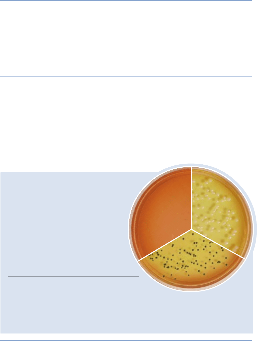
Uninoculated
palate
Staphylococcus aureus
ATCC
®
25923
with Potassium Tellurite
The cultures listed are the minimum that should be used for performance testing.
*These cultures are available as Bactrol
™
Disks and should be used as directed in Bactrol Disks Technical Information.
TELLURITE MANNITOL
ORGANISM ATCC
®
GROWTH REDUCTION FERMENTATION
Escherichia coli 25922* marked to – (no change) – (no change)
complete inhibition
Proteus mirabilis 25933 partial to + (black) – (red)
complete inhibition
Staphylococcus aureus 25923* good + (black) + (yellow)
Staphylococcus 12228* none to fair ± (translucent – (red)
epidermidis to black)
Intended Use
Bacto VJ Agar is used with Bacto Chapman Tellurite Solution 1% for
isolating coagulase-positive, mannitol-fermenting staphylococci.
Also Known As
VJ Agar is also known as Vogel and Johnson Agar, Modification of
Tellurite-Glycine Agar
1
, and Tellurite-Glycine-Phenol Red Agar Base
2
Summary and Explanation
Coagulase-positive staphylococci, primarily Staphylococcus aureus,
are among the microorganisms that can cause spoilage or chemical
changes in cosmetic products.
4
To isolate coagulase-positive, mannitol fermenting staphylococci,
Vogel and Johnson
3
modified Tellurite-Glycine Agar by Zebovitz
et al.
1
by increasing the mannitol content and adding a pH indicator.
Vogel-Johnson (VJ) Agar selects and differentiates the coagulase-
positive staphylococci which ferment mannitol and reduce tellurite.
2
VJ Agar is specified as a standard methods medium for cosmetics,
4,5
pharmaceutical articles
6
and nutritional supplements.
6
Principles of the Procedure
VJ Agar contains Tryptone as a source of carbon, nitrogen, vitamins
and minerals. Yeast Extract supplies B-complex vitamins which
stimulate bacterial growth. Mannitol is the carbohydrate. Chapman

The Difco Manual 551
Section II Veal Infusion Agar & Veal Infusion Broth
Tellurite Solution 1% contains Potassium Tellurite which, along with
Lithium Chloride and Glycine, inhibits most microorganisms except
the staphylococci. Phenol Red is the pH indicator. Bacto Agar is the
solidifying agent.
Formula
Bacto VJ Agar
Formula Per Liter
Bacto Tryptone . . . . . . . . . . . . . . . . . . . . . . . . . . . . . . . . . . 10 g
Bacto Yeast Extract . . . . . . . . . . . . . . . . . . . . . . . . . . . . . . . . 5 g
Bacto Mannitol . . . . . . . . . . . . . . . . . . . . . . . . . . . . . . . . . . 10 g
Dipotassium Phosphate . . . . . . . . . . . . . . . . . . . . . . . . . . . . 5 g
Lithium Chloride . . . . . . . . . . . . . . . . . . . . . . . . . . . . . . . . . 5 g
Glycine . . . . . . . . . . . . . . . . . . . . . . . . . . . . . . . . . . . . . . . . 10 g
Bacto Agar . . . . . . . . . . . . . . . . . . . . . . . . . . . . . . . . . . . . . 15 g
Bacto Phenol Red . . . . . . . . . . . . . . . . . . . . . . . . . . . . . . 0.025 g
Final pH 7.2 ± 0.1 at 25°C
Precautions
1. For Laboratory Use.
2. HARMFUL. IRRITATING TO EYES, RESPIRATORY SYSTEM
AND SKIN. MAY CAUSE HARM TO THE UNBORN CHILD.
Avoid contact with skin and eyes. Do not breathe dust. Wear
suitable protective clothing. Keep container tightly closed.
TARGET ORGAN(S): Blood, Kidneys, Nerves.
FIRST AID: In case of contact with eyes, rinse immediately with
plenty of water and seek medical advice. After contact with skin,
wash immediately with plenty of water. If inhaled, remove to fresh
air. If not breathing, give artificial respiration. If breathing is
difficult, give oxygen. Seek medical advice. If swallowed seek
medical advice immediately and show this container or label.
3. Follow proper established laboratory procedures in handling and
disposing of infectious materials.
Storage
Store the dehydrated VJ Agar below 30°C. The dehydrated medium is
very hygroscopic. Keep container tightly closed.
Expiration Date
The expiration date applies to the product in its intact container when
stored as directed. Do not use a product if it fails to meet specifications
for identity and performance.
Procedure
Materials Provided
VJ Agar
Materials Required but not Provided
Chapman Tellurite Solution 1%
Glassware
Petri dishes
Distilled or deionized water
Autoclave
Incubator (35°C)
Method of Preparation
VJ Agar
1. Suspend 60 grams in 1 liter distilled or deionized water.
2. Heat to boiling to dissolve completely.
3. Autoclave at 121°C for 15 minutes. Cool to 45-50°C.
4. Add 20 ml Chapman Tellurite Solution 1%. Mix well.
Specimen Collection and Preparation
Refer to appropriate references for specimen collection and preparation.
Test Procedure
See appropriate references for specific procedures.
Results
Coagulase-positive strains of S. aureus reduce tellurite and form black
colonies on the medium. These strains typically ferment mannitol and
exhibit yellow halos around the black colonies.
References
1. Zebovitz, E., J. B. Evans, and C. F. Niven, Jr. 1955. Tellurite-
Glycine Agar: a selective plating medium for the quantitative
detection of coagulase-positive staphylococci. J. Bacteriol. 70:686.
2. MacFaddin, J. F. 1985. Media for isolation-cultivation-
identification-maintenance of medical bacteria, vol. 1, p. 846-849.
Williams & Wilkins, Baltimore, MD.
3. Vogel, R. A., and M. Johnson. 1960. A modification of the
Tellurite-Glycine medium for use in the identification of
Staphylococcus aureus. Public Health Lab. 18:131.
4. Hitchins, A. D., T. T. Tran, and J. E. McCarron. 1995.
Microbiological methods for cosmetics, p. 23.01-23.11. In
Bacteriological analytical manual, 8th ed. AOAC International,
Gaithersburg, MD.
5. Curry, A. S., J. G. Graf, and G. N. McEwen, Jr. (ed.). 1993.
CTFA microbiology guidelines. The Cosmetic, Toiletry, and
Fragrance Association, Washington, D.C.
6. United States Pharmacopeial Convention. 1995. The United
States pharmacopeia, 23rd ed. The United States Pharmacopeial
Convention. Rockville, MD.
Packaging
VJ Agar 100 g 0562-15
500 g 0562-17
Bacto
®
Veal Infusion Agar
Bacto Veal Infusion Broth
Intended Use
Bacto Veal Infusion Agar is used for cultivating fastidious microorganisms
with or without added enrichment.
Bacto Veal Infusion Broth is used for cultivating fastidious microorganisms.

552 The Difco Manual
User Quality Control
Identity Specifications
Veal Infusion Agar
Dehydrated Appearance: Very light beige, free-flowing,
homogeneous.
Solution: 4.0% solution, soluble in distilled or
deionized water on boiling. Light to
medium amber, very slightly to
slightly opalescent without
significant precipitate.
Prepared Medium: Light to medium amber, slightly
opalescent without precipitate.
Reaction of 4.0%
Solution at 25°C: pH 7.4 ± 0.2
Veal Infusion Broth
Dehydrated Appearance: Very light beige, free-flowing,
homogeneous.
Solution: 2.5% solution, soluble in distilled or
deionized water, very light amber,
clear to very slightly opalescent.
Prepared Medium: Very light amber, clear to very
slightly opalescent with no more
than very slight precipitation.
Reaction of 2.5%
Solution at 25°C: pH 7.4 ± 0.2
Cultural Response
Prepare Veal Infusion Agar per label directions with and
without 5% sterile defibrinated sheep blood. Inoculate medium
with the test organisms. Incubate inoculated plates at 35 ± 2°C
for 18-48 hours under approximately 10% CO
2
.
Prepare Veal Infusion Broth per label directions. Inoculate
tubes with the test organisms. Incubate inoculated tubes at
35 ± 2°C for 18-48 hours.
INOCULUM
ORGANISM ATCC
®
CFU GROWTH
Neisseria meningitidis 13090* 100-1,000 good
Staphylococcus epidermidis 12228* 100-1,000 good
Streptococcus mitis 9895 100-1,000 good
Streptococcus pneumoniae 6305 100-1,000 good
The cultures listed are the minimum that should be used for
performance testing.
*These cultures are available as Bactrol
™
Disks and should be
used as directed in Bactrol Disks Technical Information.
Summary and Explanation
The nutritive factors of Veal Infusion media permit luxuriant growth of
fastidious microorganisms. Veal Infusion Agar may be used as a base
with blood, ascitic fluid, serum or other enrichments. Veal Infusion
media are specified for use in the examination of food.
1,2
Veal Infusion
Agar is specified in AOAC Official Methods of Analysis for culturing
eggs and egg products, and as a maintenance medium for E. coli.
3
Veal Infusion Broth is recommended for culturing E. coli in the AOAC
procedure for invasiveness of mammalian cells.
3
Principles of the Procedure
Infusion from Lean Veal and Proteose Peptone No. 3 provides the
nitrogen, vitamins, carbon and amino acids in Veal Infusion media.
Sodium Chloride maintains the osmotic balance of the formulations.
Bacto Agar is the solidifying agent in Veal Infusion Agar.
Formula
Veal Infusion Agar
Formula Per Liter
Lean Veal, Infusion from . . . . . . . . . . . . . . . . . . . . . . . . . 500 g
Bacto Proteose Peptone No. 3 . . . . . . . . . . . . . . . . . . . . . . 10 g
Sodium Chloride . . . . . . . . . . . . . . . . . . . . . . . . . . . . . . . . . . 5 g
Bacto Agar . . . . . . . . . . . . . . . . . . . . . . . . . . . . . . . . . . . . . 15 g
Final pH 7.4 ± 0.2 at 25°C
Veal Infusion Broth
Formula Per Liter
Lean Veal, Infusion from . . . . . . . . . . . . . . . . . . . . . . . . . 500 g
Bacto Proteose Peptone No. 3 . . . . . . . . . . . . . . . . . . . . . . 10 g
Sodium Chloride . . . . . . . . . . . . . . . . . . . . . . . . . . . . . . . . . . 5 g
Final pH 7.4 ± 0.2 at 25°C
Precautions
1. For Laboratory Use.
2. Follow proper established laboratory procedures in handling and
disposing of infectious materials.
Storage
Store the dehydrated medium below 30°C. The dehydrated medium
is very hygroscopic. Keep container tightly closed.
Expiration Date
The expiration date applies to the product in its intact container
when stored as directed. Do not use a product if it fails to meet
specifications for identity and performance.
Procedure
Materials Provided
Veal Infusion Agar
Veal Infusion Broth
Materials Required But Not Provided
Glassware
Autoclave
Incubator (35°C)
Waterbath (45-50°C) (optional)
Sterile defibrinated blood (optional)
Sterile Petri dishes
Sterile tubes with closures
Method of Preparation
1. Suspend the appropriate amount of medium in 1 liter distilled or
deionized water:
Veal Infusion Agar 40 g/l
Veal Infusion Broth 25 g/l
2. Heat to boiling to dissolve completely (Veal Infusion Agar).
3. Autoclave at 121°C for 15 minutes.
Veal Infusion Agar & Veal Infusion Broth Section II

The Difco Manual 553
4. OPTIONAL: To prepare blood agar, aseptically add 5% sterile
defibrinated blood to the medium at 45-50°C. Mix well.
5. Dispense as desired.
Specimen Collection and Preparation
Obtain and process specimens according to the procedures established
by laboratory policy.
Test Procedure
For a complete discussion on the examination of fastidious microor-
ganisms in food refer to the procedures outlined in the references.
1,2,3
Limitations of the Procedure
1. Since the nutritional requirements of organisms vary, some strains
may be encountered that fail to grow or grow poorly on this medium.
References
1. Association of Official Analytical Chemists. 1995. Bacteriological
analytical manual, 8th ed. AOAC International, Gaithersburg, MD.
2. Vanderzant, C., and D. F Splittstoesser. (ed.). 1992. Compendium
of methods for the microbiological examination of food, 3rd ed.
American Public Health Association, Washington, D.C.
3. Association of Official Analytical Chemists. 1995. Official
methods of analysis of AOAC International, 16th ed. AOAC
International, Arlington, VA.
Packaging
Veal Infusion Agar 500 g 0343-17
Veal Infusion Broth 500 g 0344-17
10 kg 0344-08
Section II Veillonella Agar
Bacto
®
Veillonella Agar
User Quality Control
Identity Specifications
Dehydrated Appearance: Beige, free-flowing, homogeneous
with small dark particles.
Solution: 3.6% solution, soluble in distilled
or deionized water on boiling.
Prepared Medium: Pink, slightly opalescent without
precipitate.
Reaction of 3.6%
Solution at 25°C pH 7.5 ± 0.2
Cultural Response
Prepare Veillonella Agar per label directions. Using the pour
plate technique, inoculate plates with 1 ml of the diluted test
organisms and 1 ml of the specimen. Pour 20 ml medium per
plate, mix well. Incubate plates at 35 ± 2°C anaerobically for
18-48 hours.
INOCULUM
ORGANISM ATCC
®
CFU GROWTH
Veillonella criceti 17747 100-1,000 good
Veillonella dispar 17748 100-1,000 good
Veillonella ratti 17746 100-1,000 good
Streptococcus pyogenes 19615* 100-1,000 inhibited
The cultures listed are the minimum that should be used for
performance testing.
*This culture is available as Bactrol
™
Disks and should be used
as directed in Bactrol Disks Technical Information.
Intended Use
Bacto Veillonella Agar is used with added vancomycin in isolating
Veillonella.
Summary and Explanation
Veillonella Agar is prepared according to the formula described by
Rogosa
1,2
as modified by Rogosa, Fitzgerald, MacKintosh and
Beaman.
3
Rogosa’s
2
experiments with oral specimens from humans
and rats demonstrated the medium to be highly selective for Veillonella
species. Streptomycin was originally employed as the selective agent.
Later, Rogosa et al.
3
demonstrated vancomycin to be superior to
streptomycin in reducing growth of extraneous organisms without
restricting growth of Veillonella.
Veillonella parvula is part of the normal human fecal flora.
4
V. parvula,
V. atypica and V. dispar are flora colonizing the oral cavity.
4
Veillonella
species have been encountered in patients with bite wound, head,
neck, oral and miscellaneous soft tissue infections.
5
Veillonella species
are anaerobic gram negative diplococci and appear as clumps of
diplococci when stained.
Principles of the Procedure
Tryptone and Yeast Extract provide the nitrogen, vitamins, amino
acids and carbon in Veillonella Agar. Sodium Thioglycollate is
a reducing agent, and lowers the oxidation-reduction potential of the
medium by removing oxygen to maintain a low pH. Basic Fuchsin
and Vancomycin are the selective agents. Sodium Lactate, 60%
provides nutrients and selective properties. Bacto Agar is the
solidifying agent.
Formula
Veillonella Agar
Formula Per Liter
Bacto Tryptone . . . . . . . . . . . . . . . . . . . . . . . . . . . . . . . . . . . 5 g
Bacto Yeast Extract . . . . . . . . . . . . . . . . . . . . . . . . . . . . . . . . 3 g
Sodium Thioglycollate . . . . . . . . . . . . . . . . . . . . . . . . . . . 0.75 g
Bacto Basic Fuchsin . . . . . . . . . . . . . . . . . . . . . . . . . . . . 0.002 g
Sodium Lactate, 60% . . . . . . . . . . . . . . . . . . . . . . . . . . . . . 21 ml
Bacto Agar . . . . . . . . . . . . . . . . . . . . . . . . . . . . . . . . . . . . . 15 g
Final pH 7.5 ± 0.2 at 25°C
Precautions
1. For Laboratory Use.
2. Follow proper established laboratory procedures in handling and
disposing of infectious materials.
Storage
Store the dehydrated medium at 2-8°C. The dehydrated medium is very
hygroscopic. Keep container tightly closed.

554 The Difco Manual
Expiration Date
The expiration date applies to the product in its intact container when
stored as directed. Do not use a product if it fails to meet specifications
for identity and performance.
Procedure
Materials Provided
Veillonella Agar
Materials Required But Not Provided
Glassware
Autoclave
Incubator (35°C)
Waterbath (45-50°C) (optional)
Sterile Petri dishes
Tween
®
80 (optional)
Vancomycin
Method of Preparation
1. Suspend 36 grams in 1 liter distilled or deionized water.
2. Heat to boiling to dissolve completely.
3. OPTIONAL: Add 1 gram Tween
®
80, if desired.
4. Autoclave at 121°C for 15 minutes.
5. Add 7.5 mcg vancomycin per ml of sterile medium at 50-55°C.
Mix thoroughly.
Specimen Collection and Preparation
Anaerobic bacteria are overlooked or missed unless the specimen is
properly collected and transported to the laboratory.
6
Obtain and process
specimens according to the techniques and procedures established by
institutional policy.
Test Procedure
1. Rogosa
1,2,3
recommends that one ml of the diluted specimen be
added to a sterile Petri dish.
2. Pour approximately 20 ml of medium to the Petri dish, and rotate
to mix well with the inoculum.
3. Incubate plates anaerobically at 35 ± 2°C for 40-48 hours; 72 hours
if necessary.
For a complete discussion on Veillonella species from clinical specimens,
refer to the appropriate procedures outlined in the references.
4,6,7
For
the examination of anaerobic bacteria in food refer to standard methods.
8, 9, 10
Limitations of the Procedure
1. Since the nutritional requirements of organisms vary, some strains may
be encountered that fail to grow or grow poorly on this medium.
2. Clinical specimens must be obtained properly and transported to
the laboratory in a suitable anaerobic transport container.
6
3. The microbiologist must be able to verify quality control of the
medium and determine whether the environment is anaerobic.
6
4. The microbiologist must perform aerotolerance testing on each
isolate recovered to ensure the organism is an anaerobe.
6
References
1. Rogosa, M. 1955. Nutrition of the Vellonella. J. Dent. Res.
34:721-722.
2. Rogosa, M. 1956. A selective medium for the isolation and
enumeration of the Veillonella from the oral cavity. J. Bacteriol.
72:533-536.
3. Rogosa, M., R. J. Fitzgerald, M. E. MacKintosh, and
A. J. Beaman. 1958. Improved medium for selective isolation of
Veillonella. J. Bacteriol. 76:455-456.
4. Murray, P. R., E. J. Baron, M. A. Pfaller, F. C. Tenover, and
R. H. Yolken (ed.). 1995. Manual of clinical microbiology, 6th ed.
American Society for Microbiology, Washington, D.C.
5. Summanen, P., E. J. Baron, D. M. Citron, C. Strong,
H. M. Wexler, and S. M. Finegold. 1993. Wadsworth anaerobic
bacteriology manual, 5th ed. Star Publishing Co., Belmont, CA.
6. Isenberg, H. D. (ed.). 1992. Clinical microbiology procedures
handbook. American Society for Microbiology, Washington, D.C.
7. Baron, E. J., L. R. Peterson, and S. M. Finegold. 1994.
Etiological agents recovered from clinical material, p. 474-503.
Bailey & Scott’s diagnostic microbiology, 9th ed. Mosby-Year
Book, Inc., St. Louis, MO.
8. Association of Official Analytical Chemists. 1995. Bacteriological
analytical manual, 8th ed. AOAC International, Gaithersburg, MD.
9. Vanderzant, C., and D. F. Splittstoesser (ed.). 1992. Compendium
of methods for the microbiological examination of food, 3rd ed.
American Public Health Association, Washington, D.C.
10. Marshall, R. T. (ed.). 1992. Standard methods for the microbiologi-
cal examination of dairy products, 16th ed. American Public Health
Association, Washington. D.C.
Packaging
Veillonella Agar 500 g 0917-17
Violet Red Bile Agar Section II
Bacto
®
Violet Red Bile Agar
Intended Use
Bacto Violet Red Bile Agar is used for enumerating coliform organisms
in dairy products.
Also Known As
Violet Red Bile Lactose Agar
Summary and Explanation
The coliform group of bacteria includes aerobic and facultatively
anaerobic gram-negative non-sporeforming bacilli that ferment lactose
and form acid and gas at 35°C within 48 hours. Members of the
Enterobacteriaceae comprise the majority of the group but other
lactose fermenting organisms may also be included.
Procedures to detect, enumerate and presumptively identify coliforms
are used in testing foods and dairy products.
1,2,3
One method for
performing the presumptive test for coliforms uses Violet Red Bile
Agar (VRBA). If typical coliform colonies appear, they are tested
further to confirm their identification as coliforms.

The Difco Manual 555
Section II Violet Red Bile Agar
User Quality Control
Identity Specifications
Dehydrated Appearance: Reddish-beige, homogeneous, free-flowing.
Solution: 4.15% solution, reddish-purple, very
slightly to slightly opalescent.
Prepared plates: Reddish-purple, slightly opalescent.
Reaction of 4.15%
solution at 25°C: 7.4 ± 0.2
Cultural Response
Prepare Violet Red Bile Agar per label directions. Inoculate
and incubate the plates at 32 ± 1°C for 24 ± 2 hours.
INOCULUM
ORGANISM ATCC
®
CFU GROWTH COLONY COLOR
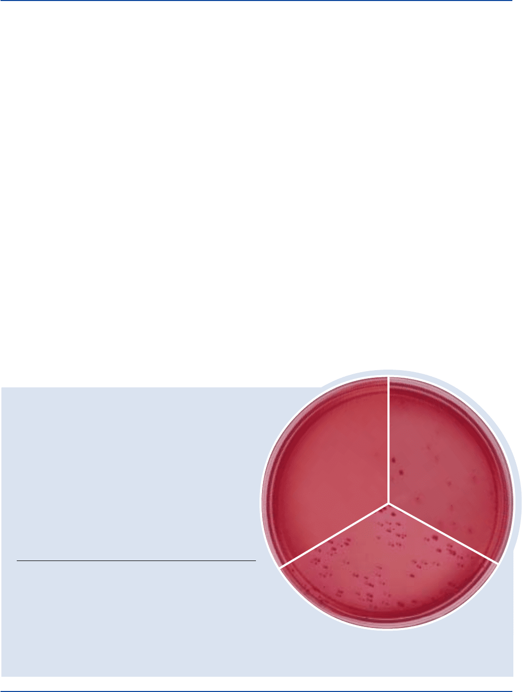
Enterobacter 13048* 30-300 good red, may have
aerogenes slight red precipitate
around colonies
Escherichia 25922* 30-300 good deep red
coli with red precipitate
around colonies
Staphylococcus 25923* 1,000-2,000 markedly to
aureus completely inhibited
Enterobacter aerogenes
ATCC
®
13048
Uninoculated
plate
Escherichia coli
ATCC
®
25922
The cultures listed are the minimum that should be used for performance testing.
*These cultures are available as Bactrol™ Disks and should be used as directed in Bactrol Disks Technical Information.
Principles of the Procedure
Violet Red Bile Agar (VRBA) contains Bacto Peptone to provide
carbon and nitrogen sources for general growth requirements.
Yeast Extract supplies B-complex vitamins which stimulate
bacterial growth. Bile Salts No. 3 and Crystal Violet inhibit most
gram-positive microorganisms. Lactose is the carbohydrate source
and Neutral Red is the pH indicator. Bacto Agar is the solidifying agent.
Formula
Violet Red Bile Agar
Formula Per Liter
Bacto Yeast Extract . . . . . . . . . . . . . . . . . . . . . . . . . . . . . . . . 3 g
Bacto Peptone . . . . . . . . . . . . . . . . . . . . . . . . . . . . . . . . . . . . 7 g
Bacto Bile Salts No. 3 . . . . . . . . . . . . . . . . . . . . . . . . . . . . 1.5 g
Bacto Lactose . . . . . . . . . . . . . . . . . . . . . . . . . . . . . . . . . . . 10 g
Sodium Chloride . . . . . . . . . . . . . . . . . . . . . . . . . . . . . . . . . . 5 g
Bacto Agar . . . . . . . . . . . . . . . . . . . . . . . . . . . . . . . . . . . . . 15 g
Neutral Red . . . . . . . . . . . . . . . . . . . . . . . . . . . . . . . . . . . . 0.03 g
Bacto Crystal Violet . . . . . . . . . . . . . . . . . . . . . . . . . . . . 0.002 g
Final pH 7.4 ± 0.2 at 25°C
Precautions
1. For Laboratory Use.
2. Follow proper established laboratory procedure in handling and
disposing of infectious materials.
Storage
Store the dehydrated medium below 30°C. The dehydrated medium is
very hygroscopic. Keep container tightly closed.
Expiration Date
The expiration date applies to the product in its intact container when
stored as directed. Do not use product if it fails to meet specifications
for identity and performance.
Procedure
Materials Provided
Violet Red Bile Agar
Materials Required but not Provided
Flask with closure
Distilled or deionized water
Autoclave
Incubator (35°C or 32°C for dairy products)
Method of Preparation
1. Suspend 41.5 grams in 1 liter distilled or deionized water.
2. Heat to boiling to dissolve completely. Do not boil for more than
2 minutes. DO NOT AUTOCLAVE.
Specimen Collection and Preparation
Refer to appropriate references for specimen collection and preparation.
Test Procedure
Presumptive test for coliforms using solid medium:
1. Transfer a 1 ml aliquot of test sample to a Petri dish.
2. Add 10 ml of Violet Red Bile Agar (at 48°C) and swirl to mix.
3. Allow medium to solidify before incubating at 35°C for 18 to
24 hours; use 32°C for dairy products.
4. Examine for purple-red colonies, 0.5 mm in diameter (or larger),
surrounded by a zone of precipitated bile acids.
5. Continue with confirmatory testing of typical coliform colonies.
1.2,3
