BD Diagnostic Systems (publ.). Difco Manual (Manual of Microbiological Culture)
Подождите немного. Документ загружается.


506 The Difco Manual
Thioglycollate Media Section II
3. For Fluid Thioglycollate Medium, Fluid Thioglycollate Medium
w/Beef Extract and Fluid Thioglycollate Medium w/K Agar,
if more than 30% of the medium is pink prior to use, reheat once
(100°C) to drive off absorbed oxygen.
For Brewer Thioglycollate Medium, if more than 20% of the
medium is green prior to use, reheat once (100°C).
After prolonged storage, reheat Thioglycollate Medium w/o
Indicator, Thioglycollate Medium w/o Dextrose or Indicator
and Thioglycollate Medium w/o Dextrose only once in flowing
steam or a boiling water bath to drive off dissolved gases.
Expiration Date
The expiration date applies to the product in its intact container when
stored as directed. Do not use a product if it fails to meet specifications
for identity and performance.
Procedure
Materials Provided
Fluid Thioglycollate Medium
NIH Thioglycollate Broth
Brewer Thioglycollate Medium
Fluid Thioglycollate Medium w/Beef Extract
Fluid Thioglycollate Medium w/K Agar
Thioglycollate Medium w/o Dextrose
Thioglycollate Medium w/o Dextrose or Indicator
Thioglycollate Medium w/o Indicator
Materials Required But Not Provided
Glassware
Autoclave
Incubator (35°C)
Waterbath
Sterile tubes
Method of Preparation
1. Suspend the appropriate amount of medium in 1 liter distilled or
deionized water:
Fluid Thioglycollate Medium 29.8 g
Brewer Thioglycollate Medium 40.5 g
NIH Thioglycollate Broth 29 g
Fluid Thioglycollate Medium w/Beef Extract 34.7 g
Fluid Thioglycollate Medium w/K Agar 29 g
Thioglycollate Medium w/o Dextrose 24 g
Thioglycollate Medium w/o Indicator 29 g
Thioglycollate Medium w/o Dextrose or Indicator 24 g
2. Heat to boiling to dissolve completely. Avoid overheating.
3. Dispense as desired, using only clean, rust-free equipment.
4. Autoclave at 121°C for 15 minutes. Cool to room temperature.
Specimen Collection and Preparation
Obtain and process specimens according to the techniques and procedures
established by laboratory policy.
Test Procedure
For a complete discussion on the isolation and identification of bacteria
and yeasts, refer to appropriate procedures outlined in the references.
Results
Typically growth is visually observed in the media. Gram negative
bacilli tend to grow diffusely, gram positive cocci exhibit puff-ball
type growth and strict aerobes, such as pseudomonads and yeast, tend
to grow in a thin layer on the surface of the broth.
References
1. The United States Pharmacopeial Convention. 1995. The United
States pharmacopeia, 23rd ed., p.1686-1690. The United States
Pharmacopeial Convention Inc. Rockville, MD.
2. Federal Register. 1992. General biological products standards.
Fed. Regist. 21:610.12.
3. Federal Register. 1992. Detection of viable bacteria and fungi
except in live vaccines. Fed. Regist. 21:113.26.
4. Quastel and Stephenson. 1926. J. Biochem. 20:1125.
5. Falk, C. R., H. Bucca, and M. P. Simmons. 1939. A comparative
study of the use of varying concentrations of agar in the test
medium used to detect contaminants in biologic products.
J. Bacteriol. 37:121-131.
6. Brewer, J. H. 1940. Clear liquid mediums for the “aerobic” culti-
vation of anaerobes. J. Amer. Med. Assoc. 115:598-600.
7. Marshall, M. S., J. B. Ginnish, and M. P. Luxen. 1940. Test for
the sterility of biologic products. Proc. Soc. Exp. Biol. Med.
43:672.
8. Nungester, W. J., M. N. Hood, and M. K. Warren. 1943. Use
of thioglycollate media for testing disinfectants. Proc. Soc. Exp.
Biol. Med. 52:287.
9. Portwood, L. 1944. Observations of the failure of sterility test
media to support the growth of laboratory contaminants.
J. Bacteriol. 48:255-256.
10. Malin, B., and R. K. Finn. 1957. The use of a synthetic resin in
anaerobic media. J. Bacteriol. 62:349-350.
11. Linden. 1941. NIH fluid thioglycollate medium for the sterility test.
12. MacFaddin, J. F. 1985. Media for isolation-cultivation-
identification maintenance of medical bacteria, vol.1, p. 755-762.
Williams & Wilkins, Baltimore, MD.
13. Harmon, S. M., D. A. Kautter, D. A. Golden, and E. J.
Rhodehamel. 1995. Clostridium perfringens, p.16.01-16.06. In
Bacteriological analytical manual, 8th ed. AOAC International,
Gaithersburg, MD.
14. Association of Official Analytical Chemists. 1995. Official
Methods of Analysis of AOAC International, 16th ed. AOAC
International, Arlington, VA.
15. Federal Register. 1992. Additional standard for human blood and
blood products. Fed Regist. 21:640.2.17.
16. Forbes, B. A., and P. A. Granato. 1995. Processing specimens
for bacteria, p. 267. In Murray, P. R., Baron E. J., Pfaller,
M. A., Tenover, F .C., and R. H. Yolken (ed.), Manual of clinical
microbiology, 6th ed. American Society for Microbiology,
Washington D.C.
17. Isenberg, H. D. (ed.) 1992. Processing and interpretation of blood
cultures, p.1.7.1 - 1.7.2. Clinical microbiology procedures handbook,
vol.1, American Society for Microbiology, Washington, D.C.

The Difco Manual 507
Section II Thiol Medium & Thiol Broth
18. Baron, E. J., L. R. Peterson, and S. M. Finegold. 1994. Cultivation
and isolation of viable pathogens, p. 91,. Bailey & Scott’s diagnostic
microbiology, 9th ed. Mosby-Year Book, Inc. St. Louis, MO.
Packaging
Fluid Thioglycollate Medium 100 g 0256-15
500 g 0256-17
2 kg 0256-07
10 kg 0256-08
NIH Thioglycollate Broth 500 g 0257-17
Brewer Thioglycollate Medium 500 g 0236-17
10 kg 0236-08
Fluid Thioglycollate Medium
w/ Beef Extract 500 g 0697-17
10 kg 0697-08
Fluid Thioglycollate Medium
w/K Agar 500 g 0607-17
2 kg 0607-07
Thioglycollate Medium w/o Dextrose 500 g 0363-17
Thioglycollate Medium w/o Dextrose
or Indicator 500 g 0432-17
Thioglycollate Medium w/o Indicator 500 g 0430-17
Bacto
®
Thiol Medium
Bacto Thiol Broth
Intended Use
Bacto Thiol Medium is used for cultivating organisms from body
fluids and other materials containing penicillin, streptomycin or
sulfonamides.
Bacto Thiol Broth is used for cultivating organisms from body
fluids and other materials containing penicillin, streptomycin or
sulfonamides.
Summary and Explanation
While studying Vibrio fetus cultivation, Huddleson
1
found that vibrios
remained viable in Thiol Medium at room temperature for at least 150
days without transfer. Christensen
2
tested Thiol Medium for the ability
to neutralize penicillin and streptomycin. Ten milliliters (10 ml) of
Thiol Medium can inactivate up to 100 units of penicillin and up to
1,000 micrograms of streptomycin, producing luxuriant growth of
staphylococci and other organisms from dilute inocula in 24 hours.
Szawatkowski
3
and Shanson and Barnicoat
4
reported Thiol Broth to
be superior in supporting the growth of Bacteroides species in blood
cultures. Thiol Broth was used to study the optimum incubation
period of blood culture broths.
5
Media containing thiol and
thioglycollate are recommended for recovery of nutritionally variant
streptococci (NVS).
6
Thiol Broth has the same formulation as Thiol Medium, omitting the
agar. Thiol is cited in Clinical Microbiology Procedures Handbook
7
as
a medium specific for anaerobic bacteria in blood cultures.
Principles of the Procedure
Proteose Peptone No. 3 and Yeast Extract provide nitrogen, vitamins
and amino acids in Thiol media. Dextrose is a carbon source. Sodium
Chloride maintains osmotic balance. Para-aminobenzoic Acid is a
preservative. Thiol Complex is rich in sulfhydryl (-SH) groups, which
neutralize the bacteriostatic and bactericidal effects of penicillin,
streptomycin and sulfonamides. Thiol Medium contains 0.1% Bacto
Agar to maintain an Eh potential that facilities anaerobic growth
and aids in dispersion of reducing substances and CO
2
formed in the
environment.
8
Formula
Thiol Medium
Formula Per Liter
Bacto Proteose Peptone No.3 . . . . . . . . . . . . . . . . . . . . . . . 10 g
Bacto Yeast Extract . . . . . . . . . . . . . . . . . . . . . . . . . . . . . . . . 5 g
Bacto Dextrose . . . . . . . . . . . . . . . . . . . . . . . . . . . . . . . . . . . 1 g
Sodium Chloride . . . . . . . . . . . . . . . . . . . . . . . . . . . . . . . . . . 5 g
Thiol Complex . . . . . . . . . . . . . . . . . . . . . . . . . . . . . . . . . . . 8 g
Bacto Agar . . . . . . . . . . . . . . . . . . . . . . . . . . . . . . . . . . . . . . 1 g
p-Aminobenzoic Acid. . . . . . . . . . . . . . . . . . . . . . . . . . . . 0.05 g
Final pH 7.1 ± 0.2 at 25°C
Thiol Broth
Formula Per Liter
Bacto Proteose Peptone No.3 . . . . . . . . . . . . . . . . . . . . . . . 10 g
Bacto Yeast Extract . . . . . . . . . . . . . . . . . . . . . . . . . . . . . . . . 5 g
Bacto Dextrose . . . . . . . . . . . . . . . . . . . . . . . . . . . . . . . . . . . 1 g
Sodium Chloride . . . . . . . . . . . . . . . . . . . . . . . . . . . . . . . . . . 5 g
Thiol Complex . . . . . . . . . . . . . . . . . . . . . . . . . . . . . . . . . . . 8 g
p-Aminobenzoic Acid. . . . . . . . . . . . . . . . . . . . . . . . . . . . 0.05 g
Final pH 7.1 ± 0.2 at 25°C
Precautions
1. For Laboratory Use.
2. Follow proper established laboratory procedures in handling and
disposing of infectious materials.
Storage
Store the dehydrated medium below 30%C. The dehydrated medium
is very hygroscopic. Keep container tightly closed.
Expiration Date
The expiration date applies to the product in its intact container when
stored as directed. Do not use a product if it fails to meet specifications
for identity and performance.
Use Thiol media within four days of preparation.
Procedure
Materials Provided
Thiol Medium
Thiol Broth

508 The Difco Manual
Materials Required But Not Provided
Glassware
Autoclave
Incubator (35°C)
Waterbath (45-50°C) (optional)
Method of Preparation
1. Suspend the medium in 1 liter distilled or deionized water:
Thiol Medium - 30 grams;
Thiol Broth - 29 grams.
2. Heat to boiling to dissolve completely.
3. Dispense as desired, using clean, rust-free equipment to prevent
precipitate formation.
4. Autoclave at 121°C for 15 minutes. Cool to room temperature.
Specimen Collection and Preparation
Obtain and process specimens according to the techniques and proce-
dures established by laboratory policy.
Test Procedure
For a complete discussion on processing and interpretation of blood
cultures and body fluids from clinical specimens, refer to appropriate
references.
7,9
Results
Refer to appropriate references and procedures for results.
Limitations of the Procedure
1. Since the nutritional requirements of organisms vary, some strains may
be encountered that fail to grow or grow poorly on this medium.
2. Strict reliance on blood culture bottles containing Thiol Broth is not
recommended for aerobic microorganisms. Always use an aerobic
medium, for example, a vented Tryptic Soy Broth, for optimum
isolation of the broad spectrum of microorganisms that can cause
bacteremia or septicemia.
References
1. Huddleson, I. F. 1948. A satisfactory medium for the isolation,
cultivation, and maintenance of viability of Vibrio fetus (bovine).
J. Bacteriol. 56:508.
2. Christensen, C. W. 1947. Presented at the Michigan Branch, Society
of American Bacteriologists, Detroit, MI., December 12, 1947.
3. Szawatkowski, M. V. 1976. A comparison of three readily available
types of anaerobic blood culture media. Med. Lab. Sci. 33:5-12.
4. Shanson, D. C., and M. Barnicoat. 1975. An experimental
comparison of Thiol broth with Brewer’s thioglycollate for anaerobic
blood cultures. J. Clin. Pathol. 28:407-409.
5. Murray, P. R. 1985. Determination of the optimum incubation
period of blood culture broths for the detection of clinically
significant septicemia. J. Clin. Microbiol. 21:481-485.
6. Donnelly, J. P. 1994. Nutritionally variant streptococci and B
6
.
Infect. Dis. Alert 6:109-112.
7. Isenberg, H. D. (ed.). 1992. Clinical microbiology procedures
handbook. American Society for Microbiology, Washington, D.C.
8. MacFaddin, J. D. 1985. Media for isolation-cultivation-
identification-maintenance medical bacteria, vol. 1, p. 802-804.
Williams & Wilkins, Baltimore, MD.
9. Murray, P. R., E. J. Baron, M. A. Pfaller, F. C. Tenover, and
R. H. Yolken (ed.). 1995. Manual of clinical microbiology, 6th ed.
American Society for Microbiology, Washington, D.C.
Packaging
Thiol Medium 500 g 0307-17
Thiol Broth 500 g 0434-17
10 kg 0434-08
User Quality Control
Identity Specifications
Thiol Medium
Dehydrated Appearance: Light beige, free-flowing,
homogeneous.
Solution: 3% solution, soluble in distilled or
deionized water on boiling. Very
light to light amber, clear to very
slightly opalescent when hot,
opalescent after cooling.
Prepared Medium: Very light amber; very slightly to
slightly opalescent when hot,
opalescent after cooling.
Reaction of 3%
Solution at 25°C: pH 7.1 ± 0.2
Thiol Broth
Dehydrated Appearance: Light beige, free-flowing,
homogeneous.
Solution: 2.9% solution, soluble in distilled or
deionized water on boiling; very
light to light amber, clear to slightly
opalescent.
Prepared Medium: Very light amber, clear to slightly
opalescent.
Reaction of 2.9%
Solution at 25°C: pH 7.1 ± 0.2
Cultural Response
Prepare Thiol Medium and Thiol Broth per label directions.
Test 5 unit, 100 unit and 1,000 unit concentrations of
penicillin and 100 µg, 1,000 µg and 10,000 µg concentrations
of streptomycin. Inoculate and incubate at 35 ± 2°C for
18-48 hours.
INOCULUM GROWTH GROWTH
ORGANISM ATCC
®
CFU w/o ANTIBIOTICS w/ANTIBIOTICS
Staphylococcus 25923* 100-1,000 good good
†
aureus
Streptococcus 19615* 100-1,000 good good
†
pyogenes
The cultures listed are the minimum that should be used for
performance testing.
*These cultures are available as Bactrol
™
Disks and should be
used as directed in Bactrol Disks Technical Information.
†Antibiotic concentrations up to 100 units of penicillin or 1000 µg
of streptomycin.
Thiol Medium & Thiol Broth Section II

The Difco Manual 509
Section II Tinsdale Agar
Tinsdale Agar
Bacto
®
Tinsdale Base
.
Bacto Tinsdale Enrichment Desiccated
User Quality Control
Identity Specifications
Tinsdale Base
Dehydrated Appearance: Light beige, free flowing, homogeneous.
Solution: 4.5% solution, soluble in distilled or
deionized water upon boiling. Solution
is light to medium amber, slightly
opalescent to opalescent, without
significant precipitate.
Prepared Medium: Light to medium amber, slightly
opalescent to opalescent without
precipitate.
Reaction of 4.5%
Solution at 25°C: pH 7.4 ± 0.2
Tinsdale Enrichment Desiccated
Lyophilized Appearance: Light to dark tan cake; variations may occur.
Solution: Soluble in distilled or deionized water. Solution
is light to dark amber, clear to opalescent, may have
a slight precipitate.
Cultural Response
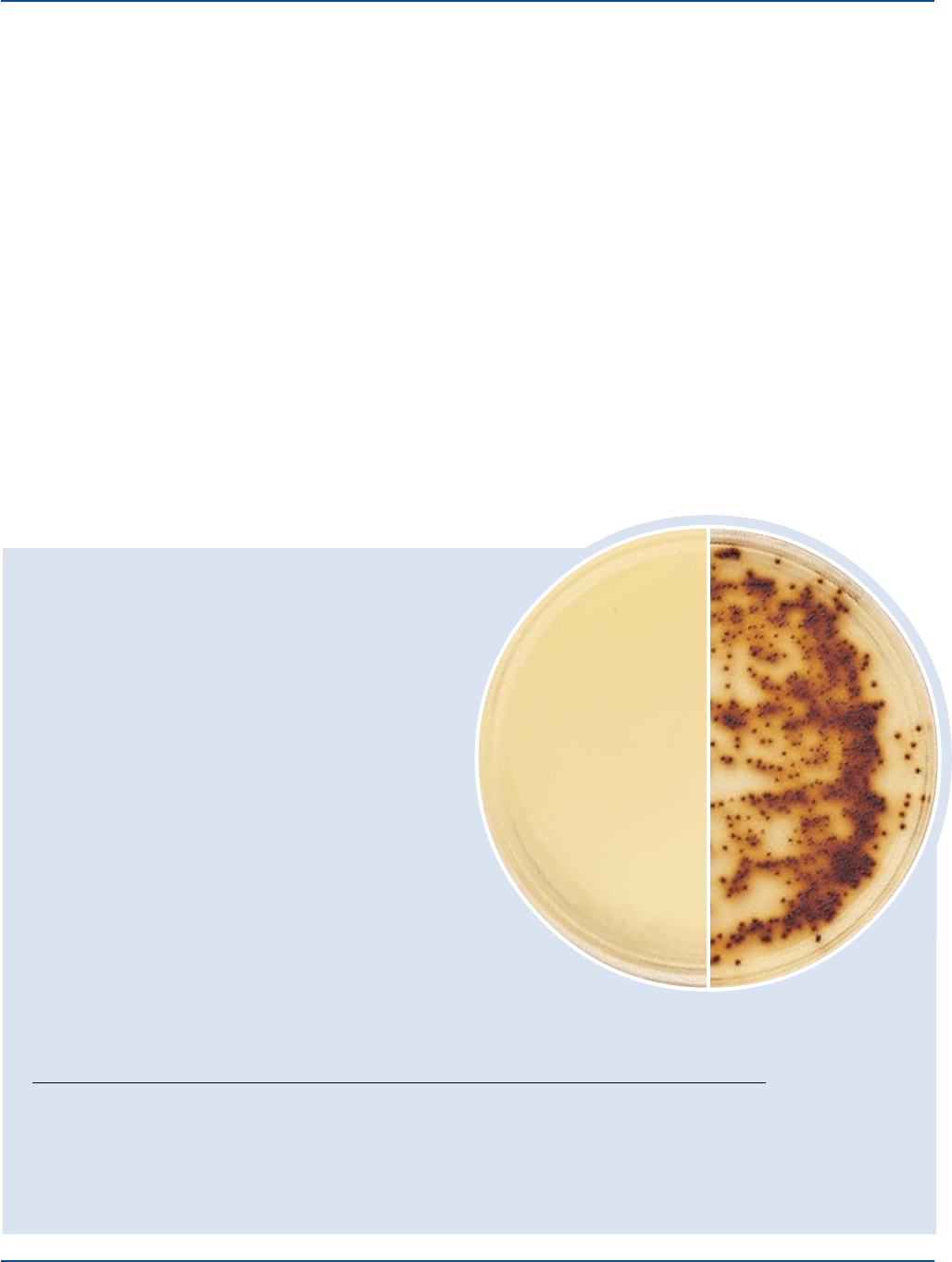
Prepare Tinsdale Agar per label directions. Inoculate and incubate at
35 ± 2°C for 18-48 hours.
Corynebacterium diphtheriae
ATCC
®
8028
Uninoculated
plate
INOCULUM
ORGANISM ATCC
®
CFU GROWTH APPEARANCE
Corynebacterium diphtheriae type gravis 8028 100-1,000 good brown with halos
Corynebacterium diphtheriae type mitis 8024 100-1,000 good brown with halos
Klebsiella pneumoniae 13883* 100-1,000 marked to complete inhibition –
Streptococcus pyogenes 19615* 100-1,000 fair brown to black without halos
The cultures listed are the minimum that should be used for performance testing.
*These cultures are available as Bactrol
™
Disks and should be used as directed in Bactrol Disks Technical Information.
Intended Use
Bacto Tinsdale Base is used with Bacto Tinsdale Enrichment Desiccated
in isolating and differentiating Corynebacterium diphtheriae.
Also Known As
Tinsdale Base (TIN) is also called Tinsdale Selective Medium, Tinsdale
Tellurite Medium and Tellurite Agar.
Summary and Explanation
Tinsdale Base, supplemented with Tinsdale Enrichment, is employed
in the cultural diagnosis of diphtheria. Diphtheria, an acute infectious
disease primarily of the upper respiratory tract but occasionally of the
skin,
1
is caused by toxigenic strains of Corynebacterium diphtheriae.
The three biotypes are mitis, intermedius and gravis.
1
The signs and
symptoms of the disease are a pharyngeal membrane, sore throat,
malaise, headache and nausea.
2
Death can result from respiratory
obstruction by the membrane or myocarditis caused by the toxin.
2
Tinsdale
3
developed a serum-cystine-thiosulfate-tellurite agar medium
for the primary isolation and differentiation of C. diphtheriae. This
formulation distinguished between C. diphtheriae and diphtheroids
which exhibited similar characteristics. The differential principle is
based on the capacity of C. diphtheriae to produce a brown or black
halo around the colonies.
Billings
4
simplified Tinsdale Basal Medium by using Proteose
Peptone No. 3 as a nutrient source. This modification improved the
differential qualities and recovery of Corynebacterium diphtheriae.
Tinsdale Base and Tinsdale Enrichment are prepared according to
the Billings
4
modification. Moore and Parsons
5
confirmed the halo
formation of C. diphtheriae with one exception; C. ulcerans occasionally
produced colonies similar to C. diphtheriae and required biochemical
identification.

510 The Difco Manual
Tinsdale Enrichment Desiccated contains Bovine Serum, Sodium
Hydroxide, L-Cystine, Sodium Thiosulfate and Potassium Tellurite in
the quantity and proportion described by Billings.
4
Principles of the Procedure
Proteose Peptone No. 3 provides the nitrogen, vitamins, carbon and
amino acids in Tinsdale Base. Sodium Chloride maintains the osmotic
balance of the medium. Bacto Agar is the solidifying agent.
Tinsdale Enrichment contains Bovine Serum, which provides essential
growth factors. Sodium Hydroxide maintains the pH. L-Cystine and
Sodium Thiosulfate are H
2
S indicators. Potassium Tellurite is a selective
agent. The formation of black to brown halos surrounding the colony
result from the reduction of potassium tellurite to metallic tellurite.
Stabbing the medium with an inoculating needle accentuates darkening
of the medium by C. diphtheriae.
Formula
Tinsdale Base
Formula Per Liter
Bacto Proteose Peptone No. 3 . . . . . . . . . . . . . . . . . . . . . . 20 g
Sodium Chloride . . . . . . . . . . . . . . . . . . . . . . . . . . . . . . . . . . 5 g
Bacto Agar . . . . . . . . . . . . . . . . . . . . . . . . . . . . . . . . . . . . . 20 g
pH 7.4 ± 0.2 at 25°C
Tinsdale Enrichment Desiccated
Contains Bovine Serum, L-Cystine, Sodium Hydroxide, Sodium
Thiosulfate and Potassium Tellurite at pH 8.0-10.0 at 25°C.
Precautions
1. For Laboratory Use.
2. Follow proper established laboratory procedures in handling and
disposing of infectious materials.
Storage
Store the dehydrated medium below 30°C. The dehydrated medium is
very hygroscopic. Keep container tightly closed.
Store Tinsdale Enrichment Desiccated at 2-8°C.
Expiration Date
The expiration date applies to the product in its intact container when
stored as directed. Do not use a product if it fails to meet specifications
for identity and performance.
Procedure
Materials Provided
Tinsdale Base
Tinsdale Enrichment Desiccated
Materials Required But Not Provided
Glassware
Autoclave
Incubator (35°C)
Waterbath (45-50°C)
Sterile Petri dishes
Method of Preparation
1. Suspend 45 grams of Tinsdale Base in 1 liter distilled or deionized
water.
2. Heat to boiling to dissolve completely.
3. Dispense 100 ml amounts into flasks.
4. Autoclave at 121°C for 15 minutes.
5. Rehydrate Tinsdale Enrichment with 15 ml sterile distilled or
deionized water and rotate in an end-over-end motion to dissolve
completely.
6. Aseptically add 15 ml rehydrated Tinsdale Enrichment to each 100
ml of Tinsdale Base at 50-55°C. Mix well.
7. Dispense into sterile Petri dishes.
Specimen Collection and Preparation
1
1. Both throat and nasopharyngeal specimens are necessary in cases
of respiratory illness. If cutaneous diphtheria is suspected, collect
skin, throat and nasopharyngeal specimens. Sterile silica gel is rec-
ommended for shipping when clinical specimens are not cultured
locally.
Test Procedure
1. For a complete discussion on the collection, isolation and identifi-
cation of Corynebacterium diphtheriae and other Corynebacterium
species, refer to the appropriate procedures outlined in the references.
2. Inoculate plates with the test organisms in a manner to obtain discrete
colonies and stab the medium several times with an inoculating needle.
3. Definitive identification of a strain of C. diphtheriae as a true
pathogen requires demonstration of toxin production.
6
Characteristic
colonies of C. diphtheriae may be inoculated directly onto KL
Virulence Agar enriched with KL Virulence Enrichment and
containing KL Antitoxin Strips for toxigenicity tests.
Limitations of the Procedure
1. Since the nutritional requirements of organisms vary, some strains
may be encountered that fail to grow or grow poorly on this medium.
2. Tinsdale Agar is not suitable as a primary plating medium, since it
may not support the growth of some strains of C. diphtheriae.
1
3. Corynebacterium ulcerans, Corynebacterium pseudotuberculosis
and (rarely) Staphylococcus species may produce a characteristic
halo on Tinsdale Agar.
1
4. Do not read Tinsdale Agar early because several organisms may
exhibit slight browning on this medium in 18 hours.
1
5. Incubation at 5-10% CO
2
retards the development of halos on
Tinsdale Agar.
1
6. On media containing tellurite, diphtheria bacilli are shorter and
stain more uniformly; however, granules are less readily observed
than when grown on Loeffler’s medium.
7
7. Further biochemical tests may be necessary to distinguish
between C. diphtheriae and C. ulcerans due to similar reactions on
this medium.
References
1. Isenberg, H. D. (ed.). 1992. Clinical microbiology procedures
handbook. American Society for Microbiology, Washington, D.C.
Tinsdale Agar Section II

The Difco Manual 511
2. Clarridge, J. E., and C. A. Spiegel. 1995. Corynebacterium and
miscellaneous irregular gram-positive rod, Erysipelothrix, and
Gardnerella, p. 357-377. In P. R. Murray, E. J. Baron, M. A.
Pfaller, F. C. Tenover, and R. H. Yolken (ed.), Manual of clinical
microbiology, 6th ed. American Society for Microbiology,
Washington, D.C.
3. Tinsdale, G. F. W. 1947. A new medium for the isolation and
identification of C. diphtheriae based on the production of hydrogen
sulphide. J. Pathol. Bacteriol. 59:461-466.
4. Billings, E. 1956. An investigation of Tinsdale Tellurite medium:
its usefulness and mechanisms of halo-formation. M.S. thesis.
University of Michigan, Ann Arbor, MI.
Section II Todd Hewitt Broth
5. Moore, M. S., and E. I. Parsons. 1958. A study of a modified
Tinsdale’s medium for the primary isolation of Corynebacterium
diphtheriae. J. Infect. Dis. 102:88.
6. Baron, E. J., L. R. Peterson, and S. M. Finegold. 1994. Bailey &
Scott’s diagnostic microbiology, 9th ed. Mosby-Year Book, Inc.,
St. Louis, MO.
7. Bailey, R. W., and E. G. Scott. 1966. Diagnostic microbiology,
2nd ed., p. 213. The C. V. Mosby Company, St. Louis, MO.
Packaging
Tinsdale Base 500 g 0786-17
Tinsdale Enrichment Desiccated 6 x 15 ml 0342-33
Bacto
®
Todd Hewitt Broth
User Quality Control
Identity Specifications
Dehydrated Appearance: Light beige, free-flowing,
homogeneous.
Solution: 3.0% solution, soluble in distilled
or deionized water; light to
medium amber, clear with no
significant precipitate.
Prepared Medium: Light to medium amber, clear with
no significant precipitate.
Reaction of 3.0%
Solution at 25°C: pH 7.8 ± 0.2
Cultural Response
Prepare Todd Hewitt Broth per label directions. Incubate
inoculated tubes at 35 ± 2°C for 18-48 hours.
INOCULUM
ORGANISM ATCC
®
CFU GROWTH
Neisseria meningitidis 13090* 100-1,000 good
Staphylococcus aureus 25923* 100-1,000 good
Streptococcus pneumoniae 6303* 100-1,000 good
Streptococcus pyogenes 19615* 100-1,000 good
The cultures listed are the minimum that should be used for
performance testing.
*These cultures are available as Bactrol
™
Disks and should
be used as directed in Bactrol Disks Technical Information.
Intended Use
Bacto Todd Hewitt Broth is used for cultivating streptococci,
pneumococci and other fastidious organisms; for cultivating group A
streptococci prior to serological typing.
Also Known As
Todd Hewitt Broth can be abbreviated as THB.
Summary and Explanation
Todd Hewitt Broth was originally developed for the production of
antigenic streptococcal hemolysin.
1
Todd Hewitt Broth is prepared
according to the formula described by Updyke and Nickle
2
who
compared media for type specific extract production of group A
streptococci. This study was performed using Todd Hewitt Broth
prepared with infusion of fresh beef heart as a control. Results showed
Todd Hewitt Broth was particularly satisfactory for growth of group
A streptococci for serological typing.
Elliott
3
reported that Todd Hewitt Broth prepared with neopeptone was
excellent for growing group A streptococci for the production of
type specific M substance. This is possible because proteinase is not
produced in this medium.
Moody, et al.
4
used Todd Hewitt Broth in the fluorescent-antibody
identification of group A streptococci from throat cultures. Todd Hewitt
Broth is recommended as an enrichment medium for the growth of
streptococcal cells in the identification of groups A and B by IF staining.
5
Todd Hewitt Broth was used as an enrichment broth for group A
streptococci in a comparison study of a rapid antigen test.
6
Principles of the Procedures
Infusion from Beef Heart and Neopeptone provides the nitrogen,
vitamins and amino acids in Todd Hewitt Broth. Dextrose is the carbon
source, and a stimulant for hemolysin production.
7
Disodium
Phosphate and Sodium Carbonate act as buffers to aid in neutralizing
acid production from dextrose fermentation, and protect hemolysin
from inactivation.
7
Sodium Chloride maintains the osmotic balance of
the medium.
Formula
Todd Hewitt Broth
Formula Per Liter
Beef Heart, Infusion from . . . . . . . . . . . . . . . . . . . . . . . . 500 g
Bacto Neopeptone. . . . . . . . . . . . . . . . . . . . . . . . . . . . . . . . 20 g
Bacto Dextrose . . . . . . . . . . . . . . . . . . . . . . . . . . . . . . . . . . . 2 g
Sodium Chloride . . . . . . . . . . . . . . . . . . . . . . . . . . . . . . . . . . 2 g
Disodium Phosphate . . . . . . . . . . . . . . . . . . . . . . . . . . . . . . 0.4 g
Sodium Carbonate . . . . . . . . . . . . . . . . . . . . . . . . . . . . . . . 2.5 g
Final pH 7.8 ± 0.2 at 25°C
Precautions
1. For Laboratory Use.
2. Follow proper established laboratory procedures in handling and
disposing of infectious materials.

512 The Difco Manual
Storage
Store the dehydrated medium below 30°C. The dehydrated medium is
very hygroscopic. Keep container tightly closed.
Expiration Date
The expiration date applies to the product in its intact container when
stored as directed. Do not use a product if it fails to meet specifications
for identity and performance.
Procedure
Materials Provided
Todd Hewitt Broth
Materials Required But Not Provided
Glassware
Autoclave
Incubator (35°C)
Waterbath (45-50°C) (optional)
Sterile tubes
Method of Preparation
1. Suspend 30 grams in 1 liter distilled or deionized water.
2. Autoclave at 121°C for 15 minutes.
3. Cool to room temperature.
Specimen Collection and Preparation
Obtain and process specimens according to the techniques and procedures
established by laboratory policy.
Test Procedure
For a complete discussion on the isolation, identification and serological
procedures of fastidious microorganisms, refer to the procedures
described in appropriate references.
4,5,8,9
Results
Refer to appropriate references and procedures for results.
Limitations of the Procedure
1. Since the nutritional requirements of organisms vary, some strains may
be encountered that fail to grow or grow poorly on this medium.
2. Todd Hewitt Broth cannot be used unbuffered for bile solubility
testing
10
References
1. Todd, E. W., and L. F. Hewitt. 1932. A new culture medium for
the production of antigenic streptococcal haemolysin. J. Pathol.
Bacteriol. 35:973.
2. Updyke, E. L., and M. I. Nickle. 1954. A dehydrated medium for
the preparation of type specific extracts of group A
streptococci. Appl. Microbiol. 2:117.
3. Elliott. 1945. J. Exp. Med. 81:573.
4. Moody, M. D., A. C. Siegel, B. Pittman, and C. C. Winter. 1963.
Fluorescent- antibody identification of group A streptococci from
throat swabs. Am. J. Public Health, 53:1083.
5. Facklam, R. R., and R. B. Carey. 1985. Streptococci and
Aerococci, p. 154-175. In, E. H.Lennette, A. Balows, W. J.
Hausler, Jr., and H. J. Shadomy (ed.), Manual of clinical
microbiology, 4th ed. American Society for Microbiology,
Washington, D.C.
6. Bourbeau, P. P., B. J. Heiter, J. P. Anhalt, and D. W. Naumovitz.
1993. Comparison of direct specimen testing utilizing testpack
strep A with testing of specimens following a two-hour broth
enrichment. Diagn. Microbiol. Infect. Dis. 17:93-96.
7. MacFaddin, J. D. 1985. Media for isolation-cultivation-
identification-maintenance medical bacteria, p.772-775. vol. 1.
Williams & Wilkins, Baltimore, MD.
8. Murray, P. R., E. J. Baron, M. A. Pfaller, F. C. Tenover, and
R. H. Yolken (ed.). 1995. Manual of clinical microbiology, 6th
ed. American Society for Microbiology, Washington, D.C.
9. Isenberg, H. D. (ed.). 1992. Clinical microbiology procedures
handbook, American Society for Microbiology, Washington, D.C.
Packaging
Todd Hewitt Broth 100 g 0492-15
500 g 0492-17
2 kg 0492-07
10 kg 0492-08
Tomato Juice Media Section II
Tomato Juice Media
Bacto
®
Tomato Juice Agar
.
Bacto Tomato Juice Agar Special
Bacto Tomato Juice Broth
Intended Use
Bacto Tomato Juice Agar is used for cultivating and enumerating
Lactobacillus species.
Bacto Tomato Juice Agar Special is used for cultivating and enumerat-
ing lactobacilli and other acidophilic microorganisms from
saliva and other specimens.
Bacto Tomato Juice Broth is used for cultivating yeasts and other
aciduric microorganisms.
Summary and Explanation
In 1925, Mickle and Breed
1
reported the use of tomato juice in culture
media used for cultivating lactobacilli. Kulp
2
investigated the use
of tomato juice on bacterial development and found that the growth
of L. acidophilus was enhanced. Tomato Juice Agar, prepared according
to Kulp and White’s
3
modification, is especially useful in cultivating
L. acidophilus from clinical specimens and foodstuffs.
4

The Difco Manual 513
Section II Tomato Juice Media
Tomato Juice Agar Special is recommended for the direct plate count
of lactobacilli from saliva and for cultivation of other acidophilic
microorganisms. The number of lactobacilli in saliva is an index of
a predisposition to dental caries as described by Jay.
5, 6
Many dentists
use the direct count of lactobacilli for the diagnosis of caries.
The acidic pH of Tomato Juice Agar Special encourages growth of
lactobacilli while inhibiting growth of accompanying bacteria. This
medium is more selective for lactobacilli than Tomato Juice Agar.
Tomato Juice Broth is recommended for use in cultivating and
isolating yeasts, lactobacilli and other aciduric microorganisms from
clinical specimens and foods.
User Quality Control
Identity Specifications
Tomato Juice Agar
Dehydrated Appearance: Tan, free-flowing, homogeneous.
Solution: 5.1% solution, soluble in distilled or
deionized water on boiling. Solution is
medium to dark amber, very slightly
opalescent without precipitate.
Reaction of 5.1%
Solution at 25°C: pH 6.1 ± 0.2
Tomato Juice Agar Special
Dehydrated Appearance: Tan, free-flowing, homogeneous.
Solution: 6.0% solution, soluble in distilled or
deionized water on boiling. Solution is
medium to dark amber, slightly opalescent.
Reaction of 6.0%
Solution at 25°C: pH 5.0 ± 0.2
Tomato Juice Broth
Dehydrated Appearance: Tan, free-flowing, homogeneous.
Solution: 4.1% solution, soluble in distilled or deionized water.
Solution is dark amber, clear without significant precipitate.
Reaction of 4.1%
Solution at 25°C: pH 6.7 ± 0.2
Tomato Juice Broth
Prepare Tomato Juice Broth per label directions. Inoculate and
incubate at 35 ± 2°C for 18-72 hours.
INOCULUM
ORGANISM ATCC
®
CFU GROWTH
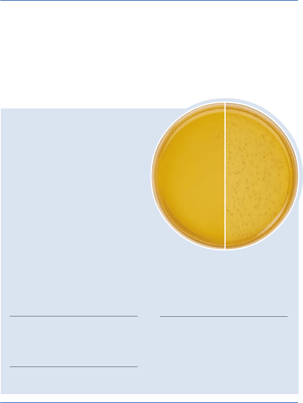
Lactobacillus casei 9595 100-1,000 good
Lactobacillus delbrueckii 4797 100-1,000 good
Saccharomyces carlsbergensis 9080 100-1,000 good
Saccharomyces cerevisiae 9763 100-1,000 good
Cultural Response
Tomato Juice Agar
Prepare Tomato Juice Agar per label directions. Inoculate using
the pour plate technique and incubate at 35 ± 2°C for 40-48 hours.
INOCULUM
ORGANISM ATCC
®
CFU GROWTH
Lactobacillus acidophilus 4356 100-1,000 good
Lactobacillus casei 9595 100-1,000 good
Lactobacillus delbrueckii 4797 100-1,000 good
Tomato Juice Agar Special
Prepare Tomato Juice Agar Special per label directions. Inoculate
and incubate at 35 ± 2°C for 18-48 hours (72 hours if necessary).
INOCULUM
ORGANISM ATCC
®
CFU GROWTH
Lactobacillus acidophilus 4356 100-1,000 good
Lactobacillus casei 9595 100-1,000 good
Lactobacillus delbrueckii 4797 100-1,000 good
Principles of the Procedure
Tomato Juice Agar and Tomato Juice Agar Special
Tomato Juice is a source of carbon, protein and nutrients. Peptone
provides a source of nitrogen, amino acids and carbon. Peptonized Milk
contains lactose as an energy source. Bacto Agar is a solidifying agent.
Tomato Juice Broth
Tomato Juice is a source of carbon, protein and nutrients. Yeast Extract
is a source of trace elements, vitamins and amino acids. Dipotassium
Phosphate and Monopotassium Phosphate provide buffering
capability. Magnesium Sulfate, Ferrous Sulfate and Manganese
The cultures listed are the minimum that should be used for
performance testing.
Lactobacillus casei
ATCC
®
9595
Uninoculated
plate

514 The Difco Manual
Sulfate provide inorganic ions. Sodium Chloride is a source of
essential ions that maintain the osmotic balance of the medium.
Formula
Tomato Juice Agar
Formula Per Liter
Tomato Juice (400 ml) . . . . . . . . . . . . . . . . . . . . . . . . . . . . 20 g
Bacto Peptone . . . . . . . . . . . . . . . . . . . . . . . . . . . . . . . . . . . 10 g
Peptonized Milk . . . . . . . . . . . . . . . . . . . . . . . . . . . . . . . . . 10 g
Bacto Agar . . . . . . . . . . . . . . . . . . . . . . . . . . . . . . . . . . . . . 11 g
Final pH 6.1 ± 0.2 at 25°C
Tomato Juice Agar Special
Formula Per Liter
Tomato Juice (400 ml) . . . . . . . . . . . . . . . . . . . . . . . . . . . . 20 g
Bacto Peptone . . . . . . . . . . . . . . . . . . . . . . . . . . . . . . . . . . . 10 g
Peptonized Milk . . . . . . . . . . . . . . . . . . . . . . . . . . . . . . . . . 10 g
Bacto Agar . . . . . . . . . . . . . . . . . . . . . . . . . . . . . . . . . . . . . 20 g
Final pH 5.0 ± 0.2 at 25°C
Tomato Juice Broth
Formula Per Liter
Tomato Juice (400 ml) . . . . . . . . . . . . . . . . . . . . . . . . . . . . 20 g
Bacto Yeast Extract . . . . . . . . . . . . . . . . . . . . . . . . . . . . . . . 10 g
Bacto Dextrose . . . . . . . . . . . . . . . . . . . . . . . . . . . . . . . . . . 10 g
Dipotassium Phosphate . . . . . . . . . . . . . . . . . . . . . . . . . . . 0.5 g
Monopotassium Phosphate. . . . . . . . . . . . . . . . . . . . . . . . . 0.5 g
Magnesium Sulfate . . . . . . . . . . . . . . . . . . . . . . . . . . . . . . . 0.2 g
Sodium Chloride . . . . . . . . . . . . . . . . . . . . . . . . . . . . . . . . 0.01 g
Ferrous Sulfate . . . . . . . . . . . . . . . . . . . . . . . . . . . . . . . . . 0.01 g
Manganese Sulfate . . . . . . . . . . . . . . . . . . . . . . . . . . . . . . 0.01 g
Final pH 6.7 ± 0.2 at 25°C
Precautions
1. Tomato Juice Agar: For Laboratory Use.
Tomato Juice Agar Special: For Laboratory Use.
Tomato Juice Broth: For Laboratory Use.
2. Follow proper established laboratory procedure in handling and
disposing of infectious materials.
Storage
Store the dehydrated medium at 2-8°C. The dehydrated medium is very
hygroscopic. Keep container tightly closed.
Expiration Date
The expiration date applies to the product in its intact container when
stored as directed. Do not use a product if it fails to meet specifications
for identity and performance.
Procedure
Materials Provided
Tomato Juice Agar
Tomato Juice Agar Special
Tomato Juice Broth
Materials Required but not Provided
Glassware
Autoclave
Distilled or deionized water
Method of Preparation
Tomato Juice Agar
1. Equilibrate the medium to room temperature before opening.
2. Suspend 51 grams in 1 liter distilled or deionized water.
3. Heat to boiling to dissolve completely.
4. Autoclave at 121°C for 15 minutes.
Tomato Juice Agar Special
1. Equilibrate the medium to room temperature before opening.
2. Suspend 60 grams in 1 liter distilled or deionized water.
3. Heat to boiling to dissolve completely.
4. Autoclave at 121°C for 15 minutes. Avoid overheating which could
cause a softer medium.
Tomato Juice Broth
1. Dissolve 41 grams in 1 liter distilled or deionized water.
2. Autoclave at 121°C for 15 minutes.
Specimen Collection and Preparation
Refer to appropriate references for specimen collection and preparation.
Test Procedure
See appropriate references for specific procedures.
Results
Refer to appropriate references and procedures for results.
References
1. Mickle and Breed. 1925. Technical Bulletin 110. NY State
Agriculture Exp. Station.
2. Kulp, W. L. 1927. Scientific apparatus and laboratory methods.
An agar medium for plating L. acidophilus and L. bulgaricus.
Science 66:512-513.
3. Kulp, J. W. L., and V. White. 1932. Modified medium for plating
L. acidophilus. Science 76:17-18.
4. MacFaddin, J. D. 1985. Media for isolation-cultivation-
identification- maintenance of medical bacteria, vol 1, p. 776-778.
Williams & Wilkins, Baltimore, MD.
5. Jay, P., and S. Gordon (ed). 1938. Bacteriology and immunology
of dental caries and dental science and dental art. Lea
and Febiger, Philadelphia, PA.
6. Jay, P., W. J. Pelton, and J. M. Wisan. 1949. Dentistry in public
health. W. B. Saunders Company, Philadelphia, PA.
Packaging
Tomato Juice Agar 500 g 0031-17
Tomato Juice Agar Special 500 g 0389-17
Tomato Juice Broth 500 g 0517-17
10 kg 0517-08
Tomato Juice Media Section II

The Difco Manual 515
Section II Transport Media
Transport Media
Bacto
®
Transport Medium Amies
.
Bacto Transport Medium
Amies w/o Charcoal
.
Bacto Transport Medium Stuart
.
Bacto
Cary-Blair Transport Medium
Intended Use
Bacto Transport Medium Amies, Transport Medium Amies w/o Charcoal
and Transport Medium Stuart are used for collecting, transporting and
preserving microbiological specimens.
Bacto Cary-Blair Transport Medium is used for collecting, transporting
and preserving microbiological specimens, particularly those containing
Vibrio cholerae.
Summary and Explanation
Transport media are chemically defined, semisolid, non-nutritive,
phosphate buffered media that provide a reduced environment.
Transport media are formulated to maintain the viability of micro-
organisms without significant increase in growth.
In 1948, Moffett, Young and Stuart described a medium for transporting
gonococcal specimens to the laboratory.
1
Stuart, Toshach and Patsula
improved this formulation, introducing what is now known as Stuart’s
Transport Medium.
2
The ability of Stuart’s medium to maintain the
viability of gonococci during transport
3,4
led other researchers to explore
its use with a variety of specimens. This medium is currently recom-
mended for throat, vaginal, and wound samples.
In 1964, Cary and Blair modified Stuart’s medium by substituting
inorganic phosphates for glycerophosphate and raising the pH to 8.4.
5
The modified medium was effective in maintaining the viability of
Salmonella and Shigella
6,7
in fecal samples. Due to its high pH,
Cary-Blair Transport Medium is also effective in maintaining the
viability of Vibrio cultures for up to four weeks.
8
Cary-Blair Transport
Medium is currently recommended for fecal and rectal samples.
Amies
9
confirmed Cary and Blair’s observations that an inorganic salt
buffer was superior to the glycerophosphate. He further modified the
formulation by using a balanced salt solution containing inorganic
phosphate buffer, omitting the methylene blue, and adding charcoal.
This modified medium yielded a higher percentage of positive
cultures than the transport medium of Stuart. Transport Medium Amies,
available with and without charcoal, is recommended for throat,
vaginal, and wound samples.
Principles of the Procedure
In the formulations, potassium chloride, calcium chloride, magnesium
chloride and sodium chloride provide essential ions that help maintain
osmotic balance while controlling permeability of bacterial cells.
Monopotassium phosphate and Disodium phosphate provide buffering
capabilities. Sodium thioglycollate suppresses oxidative changes and
provides a reduced environment. Sodium glycerophosphate is a buffer
for use with calcium chloride. Methylene blue is a colorimetric
pH indicator of the oxidation-reduction state. Charcoal neutralizes fatty
acids that are toxic to microorganisms. Bacto Agar is a solidifying agent.
Formula
Transport Medium Amies
Formula Per Liter
Sodium Chloride . . . . . . . . . . . . . . . . . . . . . . . . . . . . . . . . . . 3 g
Potassium Chloride. . . . . . . . . . . . . . . . . . . . . . . . . . . . . . . 0.2 g
Calcium Chloride . . . . . . . . . . . . . . . . . . . . . . . . . . . . . . . . 0.1 g
Magnesium Chloride . . . . . . . . . . . . . . . . . . . . . . . . . . . . . 0.1 g
Monopotassium Phosphate. . . . . . . . . . . . . . . . . . . . . . . . . 0.2 g
Disodium Phosphate . . . . . . . . . . . . . . . . . . . . . . . . . . . . . 1.15 g
Sodium Thioglycollate . . . . . . . . . . . . . . . . . . . . . . . . . . . . . 1 g
Charcoal . . . . . . . . . . . . . . . . . . . . . . . . . . . . . . . . . . . . . . . 10 g
Bacto Agar . . . . . . . . . . . . . . . . . . . . . . . . . . . . . . . . . . . . . . 4 g
Final pH 7.3 ± 0.2 at 25°C
User Quality Control
Identity Specifications
Transport Medium Amies
Dehydrated Appearance: Black, free-flowing, homogeneous.
Solution: 2.0% solution, soluble in distilled or
deionized water on boiling. Solution
is black, opaque.
Prepared Vials: Black, opaque, semi-solid.
Reaction of 2.0%
Solution at 25°C: pH 7.3 ± 0.2
Transport Medium Amies w/o Charcoal
Dehydrated Appearance: Beige, free-flowing, homogeneous.
Solution: 1.0% solution, soluble in distilled or
deionized water on boiling. Solution
is colorless to very light amber,
opalescent with precipitate.
Prepared Vials: Colorless to very light amber,
opalescent with precipitate, semi-solid.
Reaction of 1.0%
Solution at 25°C: pH 7.3 ± 0.2
Transport Medium Stuart
Dehydrated Appearance: Bluish white, free-flowing,
homogeneous.
Solution: 1.41% solution, soluble in distilled or
deionized water on boiling. Solution
is light amber, opalescent with a
bluish upper layer.
Prepared Vials: Light amber, opalescent with blue
upper layer, without precipitate,
semi-solid.
Reaction of 1.41%
Solution at 25°C: pH 7.4 ± 0.1
continued on following page
